Physical Therapists Help You Overcome Barriers to Physical Activity

Enjoying Physical Activity_GettyImages-200464866-001.jpg

According to the Department of Health and Human Services' Facts & Statistics on physical activity, more than 80% of American adults do not get enough physical activity despite the proven benefits, such as a reduced risk of some cancers and chronic diseases, as well as improved bone health, cognitive function, weight control, and quality of life. As a result, half of adults — 117 million people — have one or more chronic diseases. The good news is that regular physical activity can help prevent and improve many chronic conditions.

Barriers to movement and physical activity can be small or large, real or perceived. Whatever barriers may be preventing you from enjoying the many important health benefits of physical activity, physical therapists will partner with you to create a safe and effective program to get you moving.

Physical therapists are movement experts who improve quality of life through hands-on care, patient education, and prescribed movement. Using the latest evidence, physical therapists design physical activity plans for people of all ages and abilities specific to your needs, challenges, and goals.

Physical therapists and physical therapist assistants work together and collaborate with other members of your health care team to maximize your movement and empower you to be an active participant in your care.

You can contact a physical therapist directly for an evaluation.

11 Barriers to Physical Activity, and How to Overcome Them

1. It's too late to start, I'm too old, or I've been physically inactive for a long time.

It's never too late to get moving. According to a recent JAMA Network Open study, adding physical activity at any age has benefits. In addition to an increased life span, adding the recommended amount of physical activity for your age and ability to your daily routine can help you manage stress, improve memory and brain function, avoid chronic disease, and much more.

2. It hurts when I …

Movement is crucial to a person's health, quality of life, and independence. For some people, pain makes movement a challenge. Pain is one of the most common reasons people seek health care. A physical therapist can help you move better and safely manage your pain.

3. I don't have time.

Some physical activity is better than nothing. Try to fit in a few short bursts of physical activity a few times a day for a total of 30 minutes. Make sure that the activity increases your heart rate.

Parents should make physical activity part of their family's daily routine to establish a lifelong commitment to health for their children. Play an outdoor game like hopscotch or tag with the kids (playing is for adults too!). If you're a caretaker, maintaining your health is vital to being there for those you love. Determine when it makes the most sense to fit small amounts of physical activity into your daily routine. If possible, include movement as part of the care you provide your loved ones. They need to move, too, and you'll both benefit.

4. I don't have access to a gym or equipment.

You don't need a gym membership or fancy equipment to enjoy the benefits of physical activity. You can get plenty in and around your home. Dancing, jogging, walking, climbing stairs, and gardening are all examples of physical activity that you can do without any equipment. To improve your balance, flexibility, and strength, try doing body-weight exercises at least two days a week. Use household objects, e.g., cans, milk jugs, to strengthen muscles.

Try one of these physical therapist- and physical therapist assistant-led home exercise videos.

5. I don't like to exercise.

Competitive sports and hour-long fitness classes are not for everyone. Physical activity doesn't have to involve things you don't like doing to be effective. Discover the types of activities that you enjoy and make them part of your daily physical activity routine.

6. I can't get motivated, it's too big of a hurdle, or I don't know where to begin.

Physical activity releases endorphins, and the feeling of well-being you get after a good workout will become its own reward. To help you get started, offer yourself a small reward each time you are physically active until it becomes a habit. Perhaps looking forward to a special reward will help you reach the recommended physical activity guidelines for your age and ability. Resolve not to watch your favorite TV show unless you have met your daily movement goal. Break down long-term goals into small goals and work on achieving them one at a time. Keep a journal of how you feel after you've been physically active and each time you reach a goal.

Set yourself up for success with these tips:

  • Make it convenient (walking shoes, hand weights, or resistance bands within easy reach of your desk or where you spend the most time).

  • Schedule time for a daily physical activity break and set a calendar reminder.

  • Track your steps daily. Increase your step count goal each week.

7. I have a chronic disease, condition, or disability.

Movement is essential for everyone. Whether you use a wheelchair or other assistive device to get around or have mobility challenges due to a chronic condition or a prior injury, there are activities that you can do to challenge your muscles and lungs and improve your health and quality of life. Physical activity can even improve some chronic conditions and prevent others.

8. I'm afraid of hurting myself.

The right activity for you depends on your age, ability, and goals. A physical therapist can help you identify a safe and effective physical activity plan for your age and ability that addresses your fears and helps you reach your goals.

9. I feel out of breath when I move/walk/exercise even a little bit.

It is normal to feel a bit winded when doing physical activities in which you exert yourself more than usual.

If you worry for any reason that physical activity will be unsafe, contact a physical therapist before you begin. After an evaluation, a physical therapist can work with you to find the right duration and type of physical activity to improve your stamina and overall health.

10. I'm tired all the time; I have no energy to exercise.

Research shows that exercise boosts energy levels. Physical activity helps deliver oxygen and nutrients to our tissues and vascular system, and other body functions work more efficiently. Physical activity also improves brain function and mental health, lowers anxiety, promotes better sleep, and aids in weight management. All of these enhance our energy and lead to feelings of well-being.

11. I work out all the time but can't reach my goal.

Finding the right plan for you is essential to your success. If you are having trouble meeting strength and conditioning goals, despite your best effort, a physical therapist can work with you to identify any issues and design a program to maximize your movement and enhance performance.

Physical Therapy Guide to Anterior Cruciate Ligament (ACL) Tear

ACLAttachment_Small.jpg

An anterior cruciate ligament (ACL) tear is an injury to the knee commonly affecting athletes, such as soccer players, basketball players, skiers, and gymnasts. Nonathletes can also experience an ACL tear due to injury or accident. Approximately 200,000 ACL injuries are diagnosed in the United States each year. It is estimated that there are 95,000 ruptures of the ACL and 100,000 ACL reconstructions performed per year in the United States. Approximately 70% of ACL tears in sports are the result of noncontact injuries, and 30% are the result of direct contact (player-to-player, player-to-object). Women are more likely than men to experience an ACL tear. Physical therapists are trained to help individuals with ACL tears reduce pain and swelling, regain strength and movement, and return to desired activities.

What is an ACL Tear?

The ACL is one of the major bands of tissue (ligaments) connecting the thigh bone (femur) to the shin bone (tibia) at the knee joint. It can tear if you:

  • Twist your knee while keeping your foot planted on the ground.

  • Stop suddenly while running.

  • Suddenly shift your weight from one leg to the other.

  • Jump and land on an extended (straightened) knee.

  • Stretch the knee farther than its usual range of movement.

  • Experience a direct hit to the knee.

ACL Attachment: See More Detail

How Does It Feel?

When you tear the ACL, you may feel a sharp, intense pain or hear a loud "pop" or snap. You might not be able to walk on the injured leg because you can’t support your weight through your knee joint. Usually, the knee will swell immediately (within minutes to a few hours), and you might feel that your knee "gives way" when you walk or put weight on it.

How Is It Diagnosed?

Immediately following an injury, you may be examined by a physical therapist, athletic trainer, or orthopedic surgeon. If you see your physical therapist first, your therapist will conduct a thorough evaluation that includes reviewing your health history. Your physical therapist will ask:

  • What you were doing when the injury occurred.

  • If you felt pain or heard a "pop" when the injury occurred.

  • If you experienced swelling around the knee in the first 2 to 3 hours following the injury.

  • If you felt your knee buckle or give out when you tried to get up from a chair, walk up or down stairs, or change direction while walking.

Your physical therapist may perform gentle "hands-on" tests to determine the likelihood that you have an ACL tear, and may use additional tests to assess possible damage to other parts of your knee.

An orthopedic surgeon may order further tests, including magnetic resonance imaging (MRI), to confirm the diagnosis and rule out other possible damage to the knee.

Surgery

Most people who sustain an ACL tear will undergo surgery to repair the tear; however, some people may avoid surgery by modifying their physical activity to relieve stress on the knee. A select group can actually return to vigorous physical activity following rehabilitation without having surgery.

Your physical therapist, together with your surgeon, can help you determine if nonoperative treatment (rehabilitation without surgery) is a reasonable option for you. If you elect to have surgery, your physical therapist will help you prepare both for surgery and to recover your strength and movement following surgery.


How Can a Physical Therapist Help?

Once an ACL tear has been diagnosed, you will work with your surgeon and physical therapist to decide if you should have surgery, or if you can recover without surgery. If you don’t have surgery, your physical therapist will work with you to restore your muscle strength, agility, and balance, so you can return to your regular activities. Your physical therapist may teach you ways to modify your physical activity in order to put less stress on your knee. If you decide to have surgery your physical therapist can help you before and after the procedure.

Treatment Without Surgery

Current research has identified a specific group of patients (called "copers") who have the potential for healing without surgery following an ACL tear. These patients have injured only the ACL, and have experienced no episodes of the knee "giving out" following the initial injury. If you fall into this category, based on the specific tests your physical therapist will conduct, your therapist will design an individualized physical therapy treatment program for you. It may include treatments such as gentle electrical stimulation applied to the quadriceps muscle, muscle strengthening, and balance training.

Treatment Before Surgery

If your orthopedic surgeon determines that surgery is necessary, your physical therapist can work with you before and after your surgery. Some surgeons refer their patients to a physical therapist for a short course of rehabilitation before surgery. Your physical therapist will help you decrease your swelling, increase the range of movement of your knee, and strengthen your thigh muscles (quadriceps).

Treatment After Surgery

Your orthopedic surgeon will provide postsurgery instructions to your physical therapist, who will design an individualized treatment program based on your specific needs and goals. Your treatment program may include:

Bearing weight. Following surgery, you will use crutches to walk. The amount of weight you are allowed to put on your leg and how long you use the crutches will depend on the type of reconstructive surgery you have received. Your physical therapist will design a treatment program to meet your needs and gently guide you toward full weight bearing.

Icing and compression. Immediately following surgery, your physical therapist will control your swelling with a cold application, such as an ice sleeve, that fits around your knee and compresses it.

Bracing. Some surgeons will give you a brace to limit your knee movement (range of motion) following surgery. Your physical therapist will fit you with the brace and teach you how to use it safely. Some athletes will be fitted for braces as they recover and begin to return to their sports activities.

Movement exercises. During your first week following surgery, your physical therapist will help you begin to regain motion in the knee area, and teach you gentle exercises you can do at home. The focus will be on regaining full movement of your knee. The early exercises help with increasing blood flow, which also helps reduce swelling.

Electrical stimulation. Your physical therapist may use electrical stimulation to help restore your thigh muscle strength, and help you achieve those last few degrees of knee motion.

Strengthening exercises. In the first 4 weeks after surgery, your physical therapist will help you increase your ability to put weight on your knee, using a combination of weight-bearing and non-weight-bearing exercises. The exercises will focus on your thigh muscles (quadriceps and hamstrings) and might be limited to a specific range of motion to protect the new ACL. During subsequent weeks, your physical therapist may increase the intensity of your exercises and add balance exercises to your program.

Balance exercises. Your physical therapist will guide you through exercises on varied surfaces to help restore your balance. Initially, the exercises will help you gently shift your weight on to the surgery leg. These activities will progress to standing on the surgery leg, while on firm and unsteady surfaces to challenge your balance.

Return to sport or activities. As athletes regain strength and balance, they may begin running, jumping, hopping, and other exercises specific to their individual sport. This phase varies greatly from person-to-person. Physical therapists design return-to-sport treatment programs to fit individual needs and goals.

Can This Injury or Condition Be Prevented?

Much of the research on ACL tears has been conducted with female collegiate athletes, because women are 4 to 6 times more likely to experience the injury. Preventive physical therapy programs have proven to lower ACL injury rates by 41% for female soccer players. Researchers have made the following recommendations for a preventive exercise program:

  • The program should be designed to improve balance, strength, and sports performance. Strengthening your core (abdominal) muscles is key to preventing injury, in addition to strengthening your thigh and leg muscles.

  • Exercises should be performed 2 or 3 times per week and should include sport-specific exercises.

  • The program should last no fewer than 6 weeks.

Although most exercise studies have been conducted with female athletes, the findings may benefit male athletes as well.

Real Life Experiences

Anita is a 20-year-old student at a local university, and a star basketball player. Her team is off to a great start this year; the buzz around campus is that this could be a dream team!

But tonight, when Anita goes up for a rebound and lands off-balance, she hears a "pop" in her left knee and feels a sharp pain. When she tries to walk, she realizes that she can't put weight on her left leg. She's led back to the training room, where the school physical therapist conducts an evaluation. The test results indicate injury, and the physical therapist notices an increase in swelling around the knee just 30 minutes after the incident. She suspects an ACL tear, and refers Anita to an orthopedic surgeon. The next day, the surgeon confirms the diagnosis of an ACL tear, and tells Anita that her injury requires surgery.

After a short course of treatment by her new local physical therapist, including pain and swelling management, manual (hands-on) therapy, and knee range-of-motion and strengthening exercises, Anita has surgery the following month. Her surgeon schedules her to receive physical therapy 3 days after her surgery. She is advised to ice and elevate the knee several times per day.

Three days after surgery, Anita returns to her local physical therapist to begin her rehabilitation. He shows her how to use her crutches properly to gently begin to put weight on the operative knee. He guides her to contract/tighten the quadriceps muscle, and gently performs manual (hands-on) stretches for her to straighten the knee.

Over the next few weeks, Anita is able to gradually stop using her crutches, and begins to put her full weight on her left leg. She can also fully straighten her knee and tighten her quadriceps muscle without help from her physical therapist. She learns exercises she can safely perform at home.

After 5 weeks, Anita is able to walk normally, fully extending her knee with no pain or feelings of instability. During the next 2 months, she and her physical therapist work on her strength and balance. She finds the hardest exercises are the balance exercises, which require her to balance on a piece of foam or a rocker board while throwing a ball.

About 4 months after surgery, Anita's physical therapist designs a gentle jogging program for her. At 5 months, he allows her to begin a running program. He also adds exercises during Anita's physical therapy sessions that mimic basketball activities such as rebounding or taking a jump shot. During these activities, Anita’s physical therapist teaches her proper landing techniques to lessen the chance of reinjuring her knee when she returns to play.

After 8 months, Anita is allowed to practice with her team. They are thrilled and excited to see their star player is back. Last year was a good year for the team, but it ended in the first round of the playoffs.

Anita and her team begin a new year of full competition 11 months after her surgery. With Anita back in top form, they make the playoffs, blast through to the finals – and bring home the trophy!

This story was based on a real-life case. Your case may be different. Your physical therapist will tailor a treatment program to your specific case.

What Kind of Physical Therapist Do I Need?

Although all physical therapists are prepared through education and experience to treat a variety of conditions or injuries, you may want to consider:

  • A physical therapist who is experienced in treating people with orthopedic (musculoskeletal) problems.

  • A physical therapist who is a board-certified clinical specialist or who has completed a residency or fellowship in orthopedic physical therapy and has advanced knowledge, experience, and skills that may apply to your condition.

You can find physical therapists with these and other credentials by using Find a PT, the online tool built by the American Physical Therapy Association to help you search for physical therapists with specific clinical expertise in your geographic area.

General tips when you're looking for a physical therapist:

  • Get recommendations from family and friends or from other health care providers.

  • When you contact a physical therapy clinic for an appointment, ask about the physical therapist's experience in helping people with ACL tears.

During your first visit with the physical therapist, be prepared to describe your symptoms in as much detail as possible, and say what makes your symptoms worse.

Further Reading

The American Physical Therapy Association (APTA) believes that consumers should have access to information that could help them make health care decisions and also prepare them for their visit with their health care provider.

The following articles provide some of the best scientific evidence related to physical therapy treatment of ACL tears. The articles report recent research and give an overview of the standards of practice for treatment both in the United States and internationally. The article titles are listed by year and are linked either to a PubMed* abstract of the article or to free access of the full article, so that you can read it or print out a copy to bring with you to your health care provider.

Nyland J, Mattocks A, Kibbe S, Kalloub A, Greene JW, Caborn DN. Anterior cruciate ligament reconstruction, rehabilitation, and return to play: 2015 update. Open Access J Sports Med. 2016;7:21–32. Free Article.

Anderson MJ, Browning WM III, Urband CE, Kluczynski MA, Bisson LJ. A systematic summary of the systematic reviews on the topic of the anterior cruciate ligament. Orthop J Sports Med. 2016;4:2325967116634074. Free Article.

Anterior cruciate ligament injury. Medscape website. Accessed June 16, 2016.

Logerstedt DS, Snyder-Mackler L, Ritter RC, Axe MJ, Godges JJ; Orthopaedic Section of the American Physical Therapy Association. Knee stability and movement coordination impairments: knee ligament sprain. J Orthop Sports Phys Ther. 2010;40:A1–A37. Free Article.

Eitzen I, Moksnes H, Snyder-Mackler L, Risberg MA. A progressive 5-week exercise therapy program leads to significant improvement in knee function early after anterior cruciate ligament injury. J Orthop Sports Phys Ther. 2010;40:705-721. Free Article.

Nyland J, Brand E, Fisher B. Update on rehabilitation following ACL reconstruction. Open Access J Sports Med. 2010;1:151–166. Free Article.

Risberg MA, Holm I. The long-term effect of 2 postoperative rehabilitation programs after anterior cruciate ligament reconstruction: a randomized controlled clinical trial with 2 years of follow-up. Am J Sports Med. 2009;37:1958–1966. Free Article.

Gilchrist J, Mandelbaum BR, Melancon H, et al. A randomized controlled trial to prevent noncontact anterior cruciate ligament injury in female collegiate soccer players. Am J Sports Med. 2008;36:1476–1483. Article Summary on PubMed.

Hurd WJ, Axe MJ, Snyder-Mackler L. A 10-year prospective trial of a patient management algorithm and screening examination for highly active individuals with anterior cruciate ligament injury: Part 1, outcomes. Am J Sports Med. 2008;36:40-47. Free Article.

Benjaminse A, Gokeler A, van der Schans CP. Clinical diagnosis of an anterior cruciate ligament rupture: a meta-analysis. J Orthop Sports Phys Ther. 2006;36:267–288. Article Summary on PubMed.

Hewett TE, Ford KR, Myer GD. Anterior cruciate ligament injuries in female athletes: part 2, a meta-analysis of neuromuscular interventions aimed at injury prevention. Am J Sports Med. 2006;34:490–498. Article Summary on PubMed.

Beynnon BD, Johnson RJ, Abate JA, Fleming BC, Nichol CE. Treatment of anterior cruciate ligament injuries, part 2. Am J Sports Med. 2005;33:1751–1767. Article Summary on PubMed.

Fitzgerald GK, Piva SR, Irrgang JJ. A modified neuromuscular electrical stimulation protocol for quadriceps strength training following anterior cruciate ligament reconstruction. J Orthop Sports Phys Ther. 2003;33:492–501. Article Summary on PubMed.

*PubMed is a free online resource developed by the National Center for Biotechnology Information (NCBI). PubMed contains millions of citations to biomedical literature, including citations in the National Library of Medicine’s MEDLINE database.

Authored by Christopher Bise, PT, DPT, MS. Revised by Julie Mulcahy, PT. Reviewed by the editorial board.


Hip Labral Tears

What is a Hip Labral Tear?

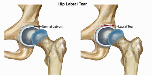
A hip labral tear occurs when there is damage to the labrum (ring of cartilage) within the hip joint. The hip joint is where the thigh bone (femur) meets the pelvis (ilium). It is described as a ball-and-socket joint. This design allows the hip to move in several directions. The bony hip socket is surrounded by the labrum, which provides additional stability and shock absorption to the hip joint.

A labral tear results when a part of the labrum separates or is pulled away from the socket. Most commonly, a labral tear is the result of repetitive stress (loading) causing irritation to the hip, often due to long-distance running or performing repeated, sharp, sports movements, such as twisting and cutting.

Repetitive loading is more likely to result in injury to the labrum when there are bony abnormalities at the hip joint. For example, hip impingement is a condition resulting in hip pain due to abnormal bony contact between the ball and socket. As the hip is moved into specific positions, this bony contact can place greater stress on the labrum.

Hip labral tears may result from a combination of several different variables, including:

  • Bony abnormalities in the hip joint (hip impingement)

  • Hip muscle tightness

  • Hip muscle weakness

  • An unstable hip joint

  • Improper technique when performing repetitive activities

  • Participation in sports that require distance running, or repetitive twisting and cutting

  • Typical wear-and-tear over time

Once torn, the labral tissue in the hip does not have the ability to heal on its own. There are surgical procedures to remove or repair torn labral tissue; however, treatment for a labral tear often begins with a course of physical therapy.

Nonsurgical treatment efforts are focused on addressing symptoms by maximizing the strength and mobility of the hip to minimize the stress placed on the injured area. In some cases, patients are able to achieve a satisfactory level of activity without surgery.

Surgical interventions are available to clean out the hip joint, and repair or reconstruct the torn labral tissue. Following surgery, patients will complete several months of physical therapy to regain function of the hip.


HipLabralTear-SM.jpg


How Does it Feel?

Many people have labral tears in the hip and do not experience symptoms; however, some labral tears can result in significant pain or limitations. Pain in the front of the hip or in the groin resulting from a hip labral tear can cause an individual to have limited ability to stand, walk, climb stairs, squat, or participate in recreational activities.

With a labral tear, you may experience:

  • A deep ache in the front of your hip or groin, often described by the "C sign." (People make a "C" with the thumb and hand, and place it on the fold at the front and side of the hip to locate their pain.)

  • Painful clicking or "catching" with hip movements; the feeling of something painful stuck in the hip or blocking hip motion.

  • Pain that increases with prolonged sitting or walking.

  • A sharp pain in the hip or groin when squatting.

  • Pain that comes on gradually rather than with one specific episode.

  • Weakness in the muscles surrounding the hip, or a feeling of the hip “giving way.”

  • Stiffness in the hip.


How Is It Diagnosed?

Your physical therapist will begin your evaluation by gathering information about your condition and medical history. Although a hip labral tear may be the result of a single injury, it most likely is a condition that develops as a consequence of repetitive irritation in the hip. Your physical therapist may ask you to describe:

  • Your current symptoms and how they affect your activities in a typical day

  • Any pain you are experiencing, its intensity and location, and how it may vary during the day

  • What activities you may be unable to do or have difficulty completing

  • What activities aggravate your symptoms, and how you reduce the level of your discomfort

  • Prior injury occurrences before your symptoms began

  • Other health care professional visits and any tests received


Your physical examination will focus on the region where your symptoms are occurring, but also include other areas that may have been affected as your body adjusted to pain. Your physical therapist may watch you walk, step onto a stair, squat, or balance on one leg.

Your physical therapist will gently but skillfully palpate (touch) the front, side, and back of your hip to determine exactly where it is most painful. The therapist will assess the mobility and strength of your hip and other regions of the body to determine the areas that require treatment.

Following the interview and physical examination, your physical therapist will discuss the findings with you and, through mutual collaboration, develop an individualized treatment program to begin your recovery.

Your physical therapist also may refer you to an orthopedic physician who specializes in hip injuries for diagnostic imaging (ie, X-ray, MRI). An X-ray helps to identify any bony abnormalities, such as those that occur with hip impingement, which may be contributing to your pain. An MRI helps to identify a labral tear.


How Can a Physical Therapist Help?

When you have been diagnosed with a hip labral tear, your physical therapist will work with you to develop a plan to help achieve your specific goals. To do so, your therapist will select treatment strategies in any or all of the following areas:

Education. Your physical therapist will work with you to identify and change any external factors causing your pain, such as exercise selection, footwear, or the amount of exercises you perform.

Pain management. Many pain-relief strategies may be implemented; the most beneficial strategy to alleviate hip pain is to apply ice to the area and to decrease or eliminate specific activities causing your symptoms. Your physical therapist will identify specific movements that aggravate the inside of your hip joint, and design an individualized treatment plan for you, beginning with a period of rest, and gradually adding a return to certain activities as appropriate. Physical therapists are experts in prescribing pain-management techniques that reduce or eliminate the need for medication, including opioids.

Manual therapy. Your physical therapist may apply hands-on treatments to gently move your muscles and joints to decrease your pain and improve motion and strength. These techniques often address areas that are difficult to treat on your own.

Movement reeducation. Your back and hip may be moving improperly, causing increased tension at the hip joint. Your physical therapist may teach you self-stretching techniques for the lower body to decrease tension and help restore normal motion in the back, hip, and leg. There are, however, certain hip motions to avoid following an injury to the hip labrum. Your physical therapist will carefully prescribe exercises that improve your range of motion while protecting the area that has the labral tear.

Muscle strengthening. Muscle weaknesses or imbalances can be the cause or the result of hip pain. Based on your specific condition, your physical therapist will design a safe, individualized, progressive resistance program for you, likely including your core (midsection) and lower extremity. You may begin by performing strengthening exercises while lying down, and advance to performing exercises in a standing position. Your physical therapist will choose what exercises are right for you.

Functional training. Once your pain, strength, and motion improve you will be able to safely transition back into more demanding activities. To minimize tension on the hip, it is important to teach your body safe, controlled movements. Based on your own unique movement assessment and goals, your physical therapist will create a series of activities to help you learn how to use and move your body correctly and safely. Your therapist also will discuss specific positions and activities that should be avoided or modified to protect your hip.


Can this Injury or Condition be Prevented?

Repetitive motion, such as sports or long-distance running, can create the risk of sustaining a labral injury. It is imperative to be aware of any hip pain that you experience, particularly with sitting and squatting, as these are signs of a potential hip injury. Identifying and addressing these injuries early is helpful in their treatment. A physical therapist can help an active individual learn proper body movements to lessen the possibility of injury.

After recovering from a hip labral tear, it is important to continue the lower-extremity mobility and muscle strengthening practices taught to you by your physical therapist, to help reduce the risk of further irritation or injury. In some cases, complete avoidance of the activity that contributed to the symptoms may be recommended.


Real Life Experiences

Erin is a 27-year-old accountant who is training for an upcoming half-marathon. She runs 5 days a week and also enjoys performing weight training and strengthening exercises 2 to 3 days a week. Over the past 2 weeks, Erin has begun to experience an achy pain in the front of her right hip. Her pain is worse after running, and while sitting in her car and at her desk. She also experiences occasional "catching" in her hip when reaching forward to pick up her 1-year-old daughter.

Erin is concerned about the pain she feels between runs and her inability to sit without discomfort. She is worried about her ability to perform daily activities, care for her daughter, and train for her upcoming race. She consults her physical therapist.

Erin’s physical therapist conducts a comprehensive assessment of her current symptoms and her health history. She assesses Erin’s motion, strength, balance, movement, and running mechanics. She skillfully palpates (touches) the front, side, and back of Erin’s hip to determine the precise location of her pain. Erin describes her typical daily running routine, her stretching routine, and her footwear. Based on these findings, her physical therapist suspects an injury to her labrum within her hip joint.

Because Erin’s hip is so tender, her physical therapist refers her to an orthopedic surgeon. The surgeon confirms the diagnosis of a hip labral tear. Erin and her surgeon discuss treatment options; the decision is made for nonoperative management of the condition, with a 2-month period of physical therapy.

Erin and her physical therapist work together to establish short- and long-term goals and identify immediate treatment priorities, including icing and activity modification to decrease her pain as well as gentle hip-strengthening exercises. Her physical therapist also teaches her a home-exercise program to perform daily to help speed her recovery.

Together, they outline a 4-week rehabilitation program. Erin sees her physical therapist 1 to 2 times each week; she assesses Erin’s progress, performs manual therapy techniques, and advances her exercise program as appropriate. She advises Erin on exercise and activity modifications that will enhance her recovery. Erin maintains her daily exercise routine at home.

After 6 weeks, Erin's hip no longer "catches" when she bends forward, and she only experiences periodic mild discomfort when sitting or running.

On the day of the half-marathon, Erin runs pain free—and is proud to high five her husband and her little daughter at the finish line!


What Kind of Physical Therapist Do I Need?

All physical therapists are prepared through education and experience to treat a labral injury in the hip. However, you may want to consider:

  • A physical therapist who is experienced in treating people with hip labral injuries or tears, and hip impingement. Some physical therapists have a practice with an orthopedic or musculoskeletal focus.

  • A physical therapist who is a board-certified clinical specialist, or who completed a residency or fellowship in orthopedic or sports physical therapy. This physical therapist has advanced knowledge, experience, and skills that may apply to your condition.

You can find physical therapists who have these and other credentials by using Find a PT, the online tool built by the American Physical Therapy Association to help you search for physical therapists with specific clinical expertise in your geographic area.

General tips when you're looking for a physical therapist (or any other health care provider):

  • Get recommendations from family and friends or from other health care providers.

  • When you contact a physical therapy clinic for an appointment, ask about the physical therapists' experience in helping people who have hip labral injury or hip impingement.

  • During your first visit with the physical therapist, be prepared to describe your symptoms in as much detail as possible, and describe what makes your symptoms worse.


Further Reading

The American Physical Therapy Association (APTA) believes that consumers should have access to information that could help them make health care decisions and also prepare them for a visit with their health care provider.

The following articles provide some of the best scientific evidence related to physical therapy treatment of labral tears in the hip. The articles report recent research and give an overview of the standards of practice both in the United States and internationally. The article titles are linked either to a PubMed* abstract of the article or to free full text, so that you can read it or print out a copy to bring with you to your health care provider.

McGovern RP, Martin RR, Kivlan BR, Christoforetti JJ. Non-operative management of individuals with non-arthritic hip pain: a literature review. Int J Sport Phys Ther. 2019;14(1): 135–147. Free Article.

Pennock AT, Bomar AD, Johnson KP, Randich K, Upasani W. Nonoperative management of femoroacetabular impingement: a prospective study. Am J Sports Med. 2018;46(14):3415–3422. Article Summary in PubMed.

Griffin DR, Dickenson EJ, O’Donnell J, et al. The Warwick Agreement on femoroacetabular impingement syndrome (FAI syndrome): an international consensus statement. Br J Sports Med. 2016;50:1169–1176. Free Article.

*PubMed is a free online resource developed by the National Center for Biotechnology Information (NCBI). PubMed contains millions of citations to biomedical literature, including citations in the National Library of Medicine’s MEDLINE database

Authored by Allison Mumbleau, PT, DPT, SCS. Revised by Jennifer Bagwell, PT, PhD, DPT, member of APTA's Academy of Orthopaedic Physical Therapy. Reviewed by an APTA section liaison. 


Guide to Anterior Cruciate Ligament (ACL) Tear

An anterior cruciate ligament (ACL) tear is an injury to the knee commonly affecting athletes, such as soccer players, basketball players, skiers, and gymnasts. Nonathletes can also experience an ACL tear due to injury or accident. Approximately 200,000 ACL injuries are diagnosed in the United States each year. It is estimated that there are 95,000 ruptures of the ACL and 100,000 ACL reconstructions performed per year in the United States. Approximately 70% of ACL tears in sports are the result of noncontact injuries, and 30% are the result of direct contact (player-to-player, player-to-object). Women are more likely than men to experience an ACL tear. Physical therapists are trained to help individuals with ACL tears reduce pain and swelling, regain strength and movement, and return to desired activities.

What is an ACL Tear?

The ACL is one of the major bands of tissue (ligaments) connecting the thigh bone (femur) to the shin bone (tibia) at the knee joint. It can tear if you:

  • Twist your knee while keeping your foot planted on the ground.

  • Stop suddenly while running.

  • Suddenly shift your weight from one leg to the other.

  • Jump and land on an extended (straightened) knee.

  • Stretch the knee farther than its usual range of movement.

  • Experience a direct hit to the knee.

ACLAttachment_Small.jpg

ACL Attachment: See More Detail

Back to Top

How Does it Feel?

When you tear the ACL, you may feel a sharp, intense pain or hear a loud "pop" or snap. You might not be able to walk on the injured leg because you can’t support your weight through your knee joint. Usually, the knee will swell immediately (within minutes to a few hours), and you might feel that your knee "gives way" when you walk or put weight on it.

Back to Top

How Is It Diagnosed?

Immediately following an injury, you may be examined by a physical therapist, athletic trainer, or orthopedic surgeon. If you see your physical therapist first, your therapist will conduct a thorough evaluation that includes reviewing your health history. Your physical therapist will ask:

  • What you were doing when the injury occurred.

  • If you felt pain or heard a "pop" when the injury occurred.

  • If you experienced swelling around the knee in the first 2 to 3 hours following the injury.

  • If you felt your knee buckle or give out when you tried to get up from a chair, walk up or down stairs, or change direction while walking.

Your physical therapist may perform gentle "hands-on" tests to determine the likelihood that you have an ACL tear, and may use additional tests to assess possible damage to other parts of your knee.

An orthopedic surgeon may order further tests, including magnetic resonance imaging (MRI), to confirm the diagnosis and rule out other possible damage to the knee.

Surgery

Most people who sustain an ACL tear will undergo surgery to repair the tear; however, some people may avoid surgery by modifying their physical activity to relieve stress on the knee. A select group can actually return to vigorous physical activity following rehabilitation without having surgery.

Your physical therapist, together with your surgeon, can help you determine if nonoperative treatment (rehabilitation without surgery) is a reasonable option for you. If you elect to have surgery, your physical therapist will help you prepare both for surgery and to recover your strength and movement following surgery.


Back to Top

How Can a Physical Therapist Help?

Once an ACL tear has been diagnosed, you will work with your surgeon and physical therapist to decide if you should have surgery, or if you can recover without surgery. If you don’t have surgery, your physical therapist will work with you to restore your muscle strength, agility, and balance, so you can return to your regular activities. Your physical therapist may teach you ways to modify your physical activity in order to put less stress on your knee. If you decide to have surgery your physical therapist can help you before and after the procedure.

Treatment Without Surgery

Current research has identified a specific group of patients (called "copers") who have the potential for healing without surgery following an ACL tear. These patients have injured only the ACL, and have experienced no episodes of the knee "giving out" following the initial injury. If you fall into this category, based on the specific tests your physical therapist will conduct, your therapist will design an individualized physical therapy treatment program for you. It may include treatments such as gentle electrical stimulation applied to the quadriceps muscle, muscle strengthening, and balance training.

Treatment Before Surgery

If your orthopedic surgeon determines that surgery is necessary, your physical therapist can work with you before and after your surgery. Some surgeons refer their patients to a physical therapist for a short course of rehabilitation before surgery. Your physical therapist will help you decrease your swelling, increase the range of movement of your knee, and strengthen your thigh muscles (quadriceps).

Treatment After Surgery

Your orthopedic surgeon will provide postsurgery instructions to your physical therapist, who will design an individualized treatment program based on your specific needs and goals. Your treatment program may include:

Bearing weight. Following surgery, you will use crutches to walk. The amount of weight you are allowed to put on your leg and how long you use the crutches will depend on the type of reconstructive surgery you have received. Your physical therapist will design a treatment program to meet your needs and gently guide you toward full weight bearing.

Icing and compression. Immediately following surgery, your physical therapist will control your swelling with a cold application, such as an ice sleeve, that fits around your knee and compresses it.

Bracing. Some surgeons will give you a brace to limit your knee movement (range of motion) following surgery. Your physical therapist will fit you with the brace and teach you how to use it safely. Some athletes will be fitted for braces as they recover and begin to return to their sports activities.

Movement exercises. During your first week following surgery, your physical therapist will help you begin to regain motion in the knee area, and teach you gentle exercises you can do at home. The focus will be on regaining full movement of your knee. The early exercises help with increasing blood flow, which also helps reduce swelling.

Electrical stimulation. Your physical therapist may use electrical stimulation to help restore your thigh muscle strength, and help you achieve those last few degrees of knee motion.

Strengthening exercises. In the first 4 weeks after surgery, your physical therapist will help you increase your ability to put weight on your knee, using a combination of weight-bearing and non-weight-bearing exercises. The exercises will focus on your thigh muscles (quadriceps and hamstrings) and might be limited to a specific range of motion to protect the new ACL. During subsequent weeks, your physical therapist may increase the intensity of your exercises and add balance exercises to your program.

Balance exercises. Your physical therapist will guide you through exercises on varied surfaces to help restore your balance. Initially, the exercises will help you gently shift your weight on to the surgery leg. These activities will progress to standing on the surgery leg, while on firm and unsteady surfaces to challenge your balance.

Return to sport or activities. As athletes regain strength and balance, they may begin running, jumping, hopping, and other exercises specific to their individual sport. This phase varies greatly from person-to-person. Physical therapists design return-to-sport treatment programs to fit individual needs and goals.

Back to Top

Can this Injury or Condition be Prevented?

Much of the research on ACL tears has been conducted with female collegiate athletes, because women are 4 to 6 times more likely to experience the injury. Preventive physical therapy programs have proven to lower ACL injury rates by 41% for female soccer players. Researchers have made the following recommendations for a preventive exercise program:

  • The program should be designed to improve balance, strength, and sports performance. Strengthening your core (abdominal) muscles is key to preventing injury, in addition to strengthening your thigh and leg muscles.

  • Exercises should be performed 2 or 3 times per week and should include sport-specific exercises.

  • The program should last no fewer than 6 weeks.

Although most exercise studies have been conducted with female athletes, the findings may benefit male athletes as well.

Back to Top

Real Life Experiences

Anita is a 20-year-old student at a local university, and a star basketball player. Her team is off to a great start this year; the buzz around campus is that this could be a dream team!

But tonight, when Anita goes up for a rebound and lands off-balance, she hears a "pop" in her left knee and feels a sharp pain. When she tries to walk, she realizes that she can't put weight on her left leg. She's led back to the training room, where the school physical therapist conducts an evaluation. The test results indicate injury, and the physical therapist notices an increase in swelling around the knee just 30 minutes after the incident. She suspects an ACL tear, and refers Anita to an orthopedic surgeon. The next day, the surgeon confirms the diagnosis of an ACL tear, and tells Anita that her injury requires surgery.

After a short course of treatment by her new local physical therapist, including pain and swelling management, manual (hands-on) therapy, and knee range-of-motion and strengthening exercises, Anita has surgery the following month. Her surgeon schedules her to receive physical therapy 3 days after her surgery. She is advised to ice and elevate the knee several times per day.

Three days after surgery, Anita returns to her local physical therapist to begin her rehabilitation. He shows her how to use her crutches properly to gently begin to put weight on the operative knee. He guides her to contract/tighten the quadriceps muscle, and gently performs manual (hands-on) stretches for her to straighten the knee.

Over the next few weeks, Anita is able to gradually stop using her crutches, and begins to put her full weight on her left leg. She can also fully straighten her knee and tighten her quadriceps muscle without help from her physical therapist. She learns exercises she can safely perform at home.

After 5 weeks, Anita is able to walk normally, fully extending her knee with no pain or feelings of instability. During the next 2 months, she and her physical therapist work on her strength and balance. She finds the hardest exercises are the balance exercises, which require her to balance on a piece of foam or a rocker board while throwing a ball.

About 4 months after surgery, Anita's physical therapist designs a gentle jogging program for her. At 5 months, he allows her to begin a running program. He also adds exercises during Anita's physical therapy sessions that mimic basketball activities such as rebounding or taking a jump shot. During these activities, Anita’s physical therapist teaches her proper landing techniques to lessen the chance of reinjuring her knee when she returns to play.

After 8 months, Anita is allowed to practice with her team. They are thrilled and excited to see their star player is back. Last year was a good year for the team, but it ended in the first round of the playoffs.

Anita and her team begin a new year of full competition 11 months after her surgery. With Anita back in top form, they make the playoffs, blast through to the finals – and bring home the trophy!

This story was based on a real-life case. Your case may be different. Your physical therapist will tailor a treatment program to your specific case.

Back to Top

What Kind of Physical Therapist Do I Need?

Although all physical therapists are prepared through education and experience to treat a variety of conditions or injuries, you may want to consider:

  • A physical therapist who is experienced in treating people with orthopedic (musculoskeletal) problems.

  • A physical therapist who is a board-certified clinical specialist or who has completed a residency or fellowship in orthopedic physical therapy and has advanced knowledge, experience, and skills that may apply to your condition.

You can find physical therapists with these and other credentials by using Find a PT, the online tool built by the American Physical Therapy Association to help you search for physical therapists with specific clinical expertise in your geographic area.

General tips when you're looking for a physical therapist:

  • Get recommendations from family and friends or from other health care providers.

  • When you contact a physical therapy clinic for an appointment, ask about the physical therapist's experience in helping people with ACL tears.

During your first visit with the physical therapist, be prepared to describe your symptoms in as much detail as possible, and say what makes your symptoms worse.

Back to Top

Further Reading

The American Physical Therapy Association (APTA) believes that consumers should have access to information that could help them make health care decisions and also prepare them for their visit with their health care provider.

The following articles provide some of the best scientific evidence related to physical therapy treatment of ACL tears. The articles report recent research and give an overview of the standards of practice for treatment both in the United States and internationally. The article titles are listed by year and are linked either to a PubMed* abstract of the article or to free access of the full article, so that you can read it or print out a copy to bring with you to your health care provider.

Nyland J, Mattocks A, Kibbe S, Kalloub A, Greene JW, Caborn DN. Anterior cruciate ligament reconstruction, rehabilitation, and return to play: 2015 update. Open Access J Sports Med. 2016;7:21–32. Free Article.

Anderson MJ, Browning WM III, Urband CE, Kluczynski MA, Bisson LJ. A systematic summary of the systematic reviews on the topic of the anterior cruciate ligament. Orthop J Sports Med. 2016;4:2325967116634074. Free Article.

Anterior cruciate ligament injury. Medscape website. Accessed June 16, 2016.

Logerstedt DS, Snyder-Mackler L, Ritter RC, Axe MJ, Godges JJ; Orthopaedic Section of the American Physical Therapy Association. Knee stability and movement coordination impairments: knee ligament sprain. J Orthop Sports Phys Ther. 2010;40:A1–A37. Free Article.

Eitzen I, Moksnes H, Snyder-Mackler L, Risberg MA. A progressive 5-week exercise therapy program leads to significant improvement in knee function early after anterior cruciate ligament injury. J Orthop Sports Phys Ther. 2010;40:705-721. Free Article.

Nyland J, Brand E, Fisher B. Update on rehabilitation following ACL reconstruction. Open Access J Sports Med. 2010;1:151–166. Free Article.

Risberg MA, Holm I. The long-term effect of 2 postoperative rehabilitation programs after anterior cruciate ligament reconstruction: a randomized controlled clinical trial with 2 years of follow-up. Am J Sports Med. 2009;37:1958–1966. Free Article.

Gilchrist J, Mandelbaum BR, Melancon H, et al. A randomized controlled trial to prevent noncontact anterior cruciate ligament injury in female collegiate soccer players. Am J Sports Med. 2008;36:1476–1483. Article Summary on PubMed.

Hurd WJ, Axe MJ, Snyder-Mackler L. A 10-year prospective trial of a patient management algorithm and screening examination for highly active individuals with anterior cruciate ligament injury: Part 1, outcomes. Am J Sports Med. 2008;36:40-47. Free Article.

Benjaminse A, Gokeler A, van der Schans CP. Clinical diagnosis of an anterior cruciate ligament rupture: a meta-analysis. J Orthop Sports Phys Ther. 2006;36:267–288. Article Summary on PubMed.

Hewett TE, Ford KR, Myer GD. Anterior cruciate ligament injuries in female athletes: part 2, a meta-analysis of neuromuscular interventions aimed at injury prevention. Am J Sports Med. 2006;34:490–498. Article Summary on PubMed.

Beynnon BD, Johnson RJ, Abate JA, Fleming BC, Nichol CE. Treatment of anterior cruciate ligament injuries, part 2. Am J Sports Med. 2005;33:1751–1767. Article Summary on PubMed.

Fitzgerald GK, Piva SR, Irrgang JJ. A modified neuromuscular electrical stimulation protocol for quadriceps strength training following anterior cruciate ligament reconstruction. J Orthop Sports Phys Ther. 2003;33:492–501. Article Summary on PubMed.

*PubMed is a free online resource developed by the National Center for Biotechnology Information (NCBI). PubMed contains millions of citations to biomedical literature, including citations in the National Library of Medicine’s MEDLINE database.

Authored by Christopher Bise, PT, DPT, MS. Revised by Julie Mulcahy, PT. Reviewed by the editorial board.
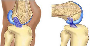
Posterior Cruciate Ligament (PCL) Injury

The posterior cruciate ligament (PCL) is a thick band of tissue deep inside the knee that connects the thighbone to the shinbone. The PCL prevents the shinbone from sliding too far backward under the thighbone. Any force that pushes the shinbone backward under the thighbone can cause a PCL injury. The PCL may be stretched, partially torn, or completely torn.

Knee injuries that tear the PCL often damage other ligaments or cartilage in the knee. A PCL injury can also break a piece of bone loose within the knee. PCL injuries can occur quickly—with a blunt force injury to the knee—or slowly, as the ligament is stressed over time.

download.jpg


How Does it Feel?

With an injured PCL, you may experience:

  • Sharp or dull pain deep inside the knee joint or the back of the knee

  • Pain in the knee when lifting a heavy load

  • Pain when walking longer distances

  • Swelling throughout the knee

  • Stiffness in the knee

  • A wobbly feeling in the knee

  • Difficulty walking on the injured leg

  • Difficulty going up or down stairs

  • Difficulty when starting to run

PCL injuries sometimes do not cause a "popping" sound or sensation when they first occur, and for that reason, people may not be sure of the exact time of their injury.

How Is It Diagnosed?

If you see your physical therapist first, the therapist will conduct a thorough evaluation that includes taking your health history. Your physical therapist will also ask you detailed questions about your injury, such as:

  • How and when did you notice the pain?

  • Did you feel pain or hear a "pop" when you injured your knee?

  • Did your knee straighten out very quickly, past its normal position?

  • Did your knee get forcefully bent, past its normal position?

  • Did you receive a direct hit to the leg while the knee was bent?

  • Did you fall on a bent knee with the foot pointed downward?

  • Did you see swelling around the knee in the first 2 to 3 hours following the injury?

  • Does your knee feel like it is wobbling, locking, catching, buckling, or giving away when you try to use it?

Your physical therapist also will perform special tests to help determine the likelihood that you have a PCL injury. Your physical therapist will gently press on the front of your knee to determine if the ligament feels loose, and may perform additional tests to determine if other parts of your knee are injured. Your physical therapist will also observe how you are walking.

To provide a definitive diagnosis, your physical therapist may collaborate with an orthopedic physician or other health care provider, who may order further tests, such as an x-ray, to confirm the diagnosis and to rule out other damage to the knee, including fracture.

How Can a Physical Therapist Help?

Your physical therapist will work with you to design a specific treatment program that will speed your recovery, including exercises you can do at home. Physical therapy will help you return to your normal lifestyle and activities. The time it takes to heal the condition varies, but improvement is generally noted in 2 to 12 weeks.

During the first 24 to 48 hours following your injury, your physical therapist may advise you to:

  • Rest your knee by using crutches or a brace, reducing the amount of weight you put on your injured leg, and avoiding any activity that causes pain.

  • Apply ice packs to the area for 15– to 20 minutes every 2 hours.

  • Consult with a physician for further services such as medication or diagnostic tests.

Your physical therapist will work with you over time to:

Reduce Pain and Swelling

Your physical therapist may use a variety of treatments and technologies to control and reduce your pain and swelling, which may include ice, heat, ultrasound, electrical stimulation, taping, exercises, and hands-on therapy, such as massage.

Improve Motion

Your physical therapist will choose specific activities and treatments to help restore normal movement in the knee and leg. These might begin with passive motions that your physical therapist performs for you to gently move your leg and knee joint, and progress to active exercises and stretches that you do yourself.

Improve Flexibility

Your physical therapist will determine if any of your leg muscles are tight, and teach you how to stretch them with gentle exercise.

Improve Strength

Certain exercises will aid healing at each stage of recovery; your physical therapist will choose and teach you the correct exercises and equipment to steadily restore your strength and agility. These may include the use of cuff weights, stretch bands, weight-lifting equipment, and cardio-exercise equipment, such as treadmills or stationary bicycles.

Improve Balance

Regaining your sense of balance is important after an injury. Your physical therapist will teach you exercises to improve your balance skills.

Restore Agility

Speed and accuracy of leg movement is important in athletic activities. Your physical therapist will help you regain these skills in preparation for a return to sports activities.

Speed Recovery Time

Your physical therapist is trained and experienced in choosing the best treatments and exercises to help you heal, return to your normal lifestyle, and reach your goals faster than you are likely to do on your own.

Your physical therapist will work with you to set your work, sport, and home-life recovery goals. Your treatment program will help you reach those goals in the safest, fastest, and most effective way possible. Your physical therapist will teach you exercises, work retraining activities, and sport-specific techniques and drills to help you return to your regular activities.

If Surgery Is Necessary

Surgery is not usually required to treat a mild PCL injury. However, it may be needed if:

  • The PCL is completely torn

  • A piece of bone has broken loose

  • There are other ligament injuries

  • You constantly feel like your knee is going to buckle beneath you

Athletes may elect to undergo surgical replacement of the PCL to improve the stability of the knee during sports activities. If other parts of the knee are injured at the same time as the PCL, you may need different treatment for those injuries, including surgery. If surgery is needed, you will follow a recovery program over several weeks guided by your physical therapist. Your physical therapist will help you minimize pain, regain motion and strength, and return to normal activities in the speediest manner possible.

Can this Injury or Condition be Prevented?

Your physical therapist can recommend a home-exercise program to strengthen and stretch the muscles around your knee, upper leg, and abdomen to help prevent future injury. These may include strength and flexibility exercises for the leg, knee, and core muscles.

To help prevent a recurrence of the injury, your physical therapist may advise you to:

  • Always use a seat belt to help prevent injury during a car accident.

  • Position your car seat so it is not too close to the dashboard.

  • Avoid intentionally landing on the front of your shinbone or on your knees.

  • Always warm up before starting a sport or heavy physical activity.

  • Maintain or improve sport-specific conditioning and techniques that are right for your level of sport activity.

  • Wear shoes that are in good condition and fit well.

  • Maintain a healthy weight.

Real Life Experiences

Steven is a 35-year-old nurse. While driving home one day, he is involved in an accident. His knees hit his car's dashboard very hard. His left knee is bruised but otherwise fine;  the right knee feels painful and swells overnight. After a few days, the right knee does not feel better and starts to feel wobbly. He is not able work. He calls his physical therapist. 

Steven's physical therapist is able to see him immediately, and thoroughly examines the knee. She can see there is swelling throughout the knee-joint area. The bruised areas are painful when she gently touches them. She tests the ligaments of the knee and finds that the PCL is noticeably looser than the PCL on the other leg. She determines that the PCL is moderately sprained—overstretched, but not torn. She applies an ice pack and electrical stimulation to help reduce the pain and swelling, and a brace to support the knee and limit bending. She teaches Steven how to use crutches, and advises him to consult with his personal physician to rule out a bone fracture. His physician confirms the diagnosis of PCL sprain.

When Steven returns for his next session, his physical therapist teaches him gentle motion and strengthening exercises. Over the next few weeks, she helps him progress through a rehabilitation program that includes strengthening, stretching, and balance exercises. The physical therapy program reduces the swelling and restores the knee's motion and strength, so that Steven no longer needs crutches after 2 weeks, and he is able to stop using the brace after 3 weeks. After about 6 weeks, Steven is able to resume all of his normal daily activities, including his full-time duties as a nurse.

What Kind of Physical Therapist Do I Need?

All physical therapists are prepared through education and experience to treat PCL sprains. However, you may want to consider:

  • A physical therapist who is experienced in treating people with orthopedic injuries. Some physical therapists have a practice with an orthopedic focus.

  • A physical therapist who is a board-certified clinical specialist or who completed a residency or fellowship in orthopedic or sports physical therapy. This therapist has advanced knowledge, experience, and skills that may apply to your condition.

You can find physical therapists that have these and other credentials by using Find a PT, the online tool built by the American Physical Therapy Association to help you search for physical therapists with specific clinical expertise in your geographic area.

General tips when you're looking for a physical therapist (or any other health care provider):

  • Get recommendations from family and friends or from other health care providers.

  • When you contact a physical therapy clinic for an appointment, ask about the physical therapists' experience in helping people who have your type of injury.

  • During your first visit with the physical therapist, be prepared to describe your symptoms in as much detail as possible, and say what makes your symptoms worse.

Further Reading

The American Physical Therapy Association (APTA) believes that consumers should have access to information that could help them make health care decisions and also prepare them for their visit with their health care provider.

The following articles provide some of the best scientific evidence related to physical therapy treatment of your injury. The articles report recent research and give an overview of the standards of practice both in the United States and internationally. The article titles are linked either to a PubMed* abstract of the article or to free full text, so that you can read it or print out a copy to bring with you to your health care provider.

Kopkow C, Freiberg A, Kirschner S, et al. Physical examination tests for the diagnosis of posterior cruciate ligament rupture: a systematic review. J Orthop Sports PhysTher. 2013;43:804-813. Article Summary on PubMed.

Kim JG, Lee YS, Yang BS, et al. Rehabilitation after posterior cruciate ligament reconstruction: a review of the literature and theoretical support. Arch Orthop Trauma Surg. 2013;133:1687-1695. Article Summary on PubMed.

Jansson KS, Costello KE, O'Brien L, et al. A historical perspective of PCL bracing. Knee Surg Sports Traumatol Arthrosc. 2013;21:1064-1070. Article Summary on PubMed.

Pierce CM, O'Brien L, Griffin LW, Laprade RF. Posterior cruciate ligament tears: functional and postoperative rehabilitation. Knee Surg Sports Traumatol Arthrosc. 2013;21:1071-1084. Article Summary on PubMed.

Rosenthal MD, Rainey CE, Tognoni A, Worms R. Evaluation and management of posterior cruciate ligament injuries. Phys Ther Sport. 2012;13:196-208. Article Summary on PubMed.

Chandrasekaran S, Ma D, Scarvell JM, et al. A review of the anatomical, biomechanical and kinematic findings of posterior cruciate ligament injury with respect to non-operative management. Knee. 2012;19:738-745. Article Summary on PubMed.

*PubMed is a free online resource developed by the National Center for Biotechnology Information (NCBI). PubMed contains millions of citations to biomedical literature, including citations in the National Library of Medicine’s MEDLINE database.

Authored by Andrea Avruskin, PT, DPT. Reviewed by the editorial board.
Pes Anserine Bursitis

Pes anserine bursitis is a condition that produces pain on the inside of the knee and lower leg. It occurs most commonly in young people involved in sports (eg, running or swimming the breaststroke), middle-aged women who are overweight, and people aged 50 to 80 years who have osteoarthritis of the knee. Up to 75% of people who have osteoarthritis of the knee have symptoms of pes anserine bursitis. The condition is also commonly associated with type 2 diabetes; 24% to 34% of patients with type 2 diabetes who report knee pain are found to have pes anserine bursitis. However, in some cases no direct cause can be identified. Physical therapists treat people with pes anserine bursitis to reduce pain, swelling, stiffness, and weakness, as well as identify and treat the underlying cause of the condition.

download.jpg

What is Pes Anserine Bursitis?

The pes anserine bursa is a small, fluid-filled sac located 2 to 3 inches below the knee joint on the inside of the lower leg. It lies beneath 3 tendons that attach to muscles of the thigh, and prevents the tendons from rubbing on the shinbone (tibia). The term “bursitis” describes a condition where the bursa has become irritated and inflamed. This condition is usually painful and associated with some swelling in the affected area. Certain positions, motions, or disease processes can cause increased friction or stress on the bursa, leading to the development of bursitis.

Pes anserine bursitis can be caused by:

  • Repetitive activities, like squatting, stair climbing, and other work or household activities that are often repeated

  • Incorrect sports training techniques, such as a lack of stretching, sudden increases in run distances, or too much uphill running

  • Obesity

  • Osteoarthritis of the knee

  • Valgus positioning of the knee (ie, a “knock-knee” position where the knees angle inward)

  • Turning the leg sharply with the foot planted on the ground

  • Injury, such as a direct hit to the leg

  • Tight hamstring (back of the thigh) muscles

  • A tear in the cartilage of the knee

  • Flat feet

How Does it Feel?

With pes anserine bursitis, you may experience:

  • Pain and swelling on the inside of the lower leg, 2 to 3 inches below the knee joint; this pain may also extend to the front of the knee and down the lower leg

  • Pain when touching the inside of the lower leg, 2 to 3 inches below the knee joint

  • Pain when bending or straightening the knee

  • Pain or difficulty walking, sitting down, rising from a chair, or climbing stairs

How Is It Diagnosed?

Your physical therapist will conduct a thorough examination that includes taking your health history as well as asking you detailed questions about your injury, such as:

  • How and when did you notice the pain?

  • Did you feel pain or hear a "pop" when you injured your leg?

  • Did you turn your leg with your foot planted on the ground?

  • Did you change direction quickly while running?

  • Did you receive a direct hit to the leg while your foot was planted on the ground?

  • Did you see swelling around the knee in the first 2 to 3 hours following the injury?

  • Does your knee feel like it is buckling or “giving way” when you try to use it?

Your physical therapist also will perform special tests to help determine the likelihood that you have pes anserine bursitis. Your therapist may:

  • Gently press on the inner side of your knee to see if it is painful to the touch

  • Assess the range of motion you have at the knee and hip, as well as the strength of some of the muscles at these joints

  • Observe how you are walking, squatting, and performing other functional and sports-specific tasks as appropriate

To provide a definitive diagnosis, your physical therapist may collaborate with an orthopedic physician or other health care provider, who may order further tests, such as an x-ray, to confirm the diagnosis and to rule out other damage to the knee.

How Can a Physical Therapist Help?

Your physical therapist will work with you to design a specific treatment program that will speed your recovery, including exercises and treatments that you can do at home. Physical therapy will help you return to your normal lifestyle and activities.

The First 24 to 48 Hours

If you see a physical therapist within 24 to 48 hours of your injury, your therapist may advise you to:

  • Rest the area by avoiding walking or any activity that causes pain.

  • Apply ice packs to the area for 15 to 20 minutes every 2 hours.

  • Consult with a physician for further services, such as medication or diagnostic tests.

Individualized Treatment

Depending on your condition and goals, your individualized rehabilitation plan may include treatments to:

Reduce pain and swelling. Your physical therapist may use different types of treatments to control and reduce your pain and swelling, including ice, heat, ultrasound, electrical stimulation, taping, exercises, and hands-on therapy, such as massage.

Improve motion. Your physical therapist will choose specific activities and treatments to help restore normal movement in the knee and leg. These might begin with "passive" motions that the physical therapist performs for you to gently move your leg and knee joint, and then progress to active exercises that you do yourself.

Improve flexibility. Pes anserine bursitis is often related to tight hamstring (back of the thigh) muscles. Your physical therapist will determine if your hamstring muscles or any other leg muscles are tight, and teach you how to stretch them.

Improve strength. Certain exercises will aid healing at each stage of recovery. Your physical therapist will choose and teach you the correct exercises and equipment to steadily restore your muscle strength and power. These may include using cuff weights, stretch bands, weight-lifting equipment, and cardio-exercise equipment, such as treadmills or stationary bicycles.

Improve balance. Regaining your sense of balance is important after an injury. Your physical therapist may teach you exercises to improve your balance skills.

Speed recovery time. Your physical therapist is trained and experienced in choosing the best treatments and exercises to help you heal, return to a normal lifestyle, and reach your goals faster than you are likely to do on your own.

Return to activities. Initially, your physical therapist may recommend that you reduce or eliminate activities that aggravate your condition for a period of time. Your physical therapist will discuss your goals with you and set up a treatment program to help you meet them in the safest, fastest, and most effective way possible. You may learn specific exercises, work retraining activities, and sport-specific techniques and drills to help you achieve your own unique goals.

Other Treatment Options

Studies have shown that some patients who do not respond to conservative treatment, such as physical therapy, may benefit from medical therapy. Your physical therapist may recommend that you discuss other treatment options with your physician, including surgery. Although surgery is rarely prescribed for pes anserine bursitis, it sometimes is needed. If surgery is required for your condition, you will follow a recovery program over several weeks guided by your physical therapist. Your physical therapist will help you minimize pain, regain motion and strength, and return to normal activities in the safest and speediest manner possible.

Can this Injury or Condition be Prevented?

Your physical therapist can recommend a home-exercise program to strengthen and stretch the muscles around your knees, upper legs, and abdomen to help prevent the onset or recurrence of pes anserine bursitis. These may include strength and flexibility exercises for the legs, knees, and core muscles.

To help prevent a recurrence of the injury, or prevent its onset if you seek guidance before injury, your physical therapist may advise you to:

  • Learn correct knee positioning when participating in athletic activities.

  • Follow a consistent flexibility and strength exercise program, especially for the leg and hip muscles, to maintain good physical conditioning.

  • Practice balance and agility exercises and drills.

  • Always warm up before starting a sport or heavy physical activity.

  • Avoid sudden increases in running mileage or uphill running.

  • Wear shoes that are in good condition and fit well.

  • Maintain a healthy weight.

  • Treat and manage diabetes very closely.

  • Wear orthotics to reduce flat feet and valgus (knock-knee) positioning of the lower extremities.

  • Wear a knee brace to support the knee and reduce strain on the inside of the joint.

Real Life Experiences

Martha is a 40-year-old secretary who has become obese. Her goal is to lose 100 pounds with diet and exercise. To reach that goal, Martha recently joined a gym, and decided to try the latest craze—a Zumba class. Martha enjoyed the first week of classes, but when leaving the gym after the fourth class, she felt a sharp pain in the inner, lower side of her right knee. It got worse when she bent and straightened her knee and when she walked upstairs to go to bed that night. The next day, the pain was still there, making it hard for her to get to work. She contacted her physical therapist.

Martha’s physical therapist performed special tests on the tendons and muscles around the knee, and found that her hamstring (back of the thigh) muscles were extremely tight and her quadriceps (front of the thigh) muscles were weak. Martha’s knee was tender to the touch, and mildly swollen 2 to 3 inches below the knee joint on the inner side of the leg, where the pes anserine bursa is located.

Martha's physical therapist explained that her pes anserine bursa was irritated and swollen. He applied ice and electrical stimulation to the area for 20 minutes. He also applied some tape to gently support Martha's hamstring muscles and alleviate the swelling and pain. He showed her how to stretch her hamstring muscles at home, and how to apply ice every few hours. He recommended that she not attend her Zumba class until her symptoms cleared up.

When Martha returned for her next visit, her physical therapist taught her some exercises to improve the strength of the muscles of her legs and “core,” and to improve her balance. Martha and her physical therapist worked together consistently over the next few weeks. Her treatment program, both in the clinic and at home, as well as her return to activity, were carefully adjusted to help ensure her safe and effective recovery.

Martha received physical therapy treatments for 6 weeks, at which time she felt almost 100% pain free—and much stronger. Martha returned to the gym to perform the exercises and stretches she learned in physical therapy as well as a modified fitness program. By the fourth week, she was able to participate in half of the Zumba class and by the fifth week, to finish the full class.

Martha has continued to do the stretches and exercises she learned from her physical therapist, and is proud to report to her friends and family that she is now pain free—and losing weight!

What Kind of Physical Therapist Do I Need?

All physical therapists are prepared through education and experience to treat pes anserine bursitis. However, you may want to consider:

  • A physical therapist who is experienced in treating people with orthopedic injuries. Some physical therapists have a practice with an orthopedic focus.

  • A physical therapist who is a board-certified clinical specialist or who has completed a residency or fellowship in orthopedic or sports physical therapy. This physical therapist has advanced knowledge, experience, and skills that may apply to your condition.

You can find physical therapists who have these and other credentials by using Find a PT, the online tool built by the American Physical Therapy Association to help you search for physical therapists with specific clinical expertise in your geographic area.

General tips when you're looking for a physical therapist (or any other health care provider):

  • Get recommendations from family and friends, or from other health care providers.

  • When you contact a physical therapy clinic for an appointment, ask about the physical therapists' experience in helping people who have your type of injury.

  • During your first visit with the physical therapist, be prepared to describe your symptoms in as much detail as possible, and say what makes your symptoms worse.

Further Reading

The American Physical Therapy Association (APTA) believes that consumers should have access to information that could help them make health care decisions and also prepare them for a visit with their health care provider.

The following articles provide some of the best scientific evidence related to physical therapy treatment of pes anserine bursitis. The articles report recent research and give an overview of the standards of practice both in the United States and internationally. The article titles are linked either to a PubMed* abstract of the article or to free full text, so that you can read it or print out a copy to bring with you to your health care provider.

Uysal F, Akbal A, Gökmen F, Adam G, Reşorlu M. Prevalence of pes anserine bursitis in symptomatic osteoarthritis patients: an ultrasonographic prospective study. Clin Rheumatol. 2015;34(3):529–533. Article Summary in PubMed.

Klontzas ME, Akoumianakis ID, Vagios I, Karantanas AH. MR imaging findings of medial tibial crest friction. Eur J Radiol. 2013;82(11):e703–e706. Article Summary in PubMed.

Chatra PS. Bursae around the knee joints. Indian J Radiol Imaging. 2012;22(1):27-30. Free Article.

Helfenstein M Jr, Kuromoto J. Anserine syndrome [article in English and Portuguese]. Rev Bras Reumatol. 2010;50(3):313–327. Free Article.

Alvarez-Nemegyei J. Risk factors for pes anserinus tendinitis/bursitis syndrome: a case control study. J Clin Rheumatol. 2007;13(2):63–65. Article Summary in PubMed

Rennie WJ, Saifuddin A. Pes anserine bursitis: incidence in symptomatic knees and clinical presentation. Skeletal Radiol. 2005;34:395–398. Article Summary in PubMed

Handy JR. Anserine bursitis: a brief review. South Med J. 1997;90(4):376–377. Article Summary on PubMed.

Butcher JD, Salzman KL, Lillegard WA. Lower extremity bursitis [published correction in: Am Fam Physician. 1996;54(2):468]. Am Fam Physician. 1996;53(7):2317-2324. Article Summary on PubMed.

Hemler DE, Ward WK, Karstetter KW, Bryant PM. Saphenous nerve entrapment caused by pes anserine bursitis mimicking stress fracture of the tibia. Arch Phys Med Rehabil. 1991;72(5):336–337. Article Summary on PubMed.

*PubMed is a free online resource developed by the National Center for Biotechnology Information (NCBI).  PubMed contains millions of citations to biomedical literature, including citations in the National Library of Medicine’s MEDLINE database.

Revised by Daniel Farwell, PT, DPT, a board-certified sports clinical specialist. Authored by Andrea Avruskin, PT. Reviewed by the editorial board.

Osteoporosis

What is Osteoporosis?

Osteoporosis is a bone disease characterized by low bone density (thickness of the bone), decreased bone strength, and a change in the bone structure, which can lead to an increased risk of fracture. The normal bone structure becomes thinned out and porous with poor nutrition, aging, or when osteoporosis develops, lessening the ability of the bone to withstand the typical forces that are applied in everyday living. Fractures from low bone density and osteoporosis can be serious, causing pain and affecting quality of life.

Bone is living tissue. Normally, one type of cell removes bone and another type of cell adds bone in a balanced, ongoing process. In osteoporosis, bones weaken when not enough new bone is formed and/or too much bone is lost. This imbalance commonly begins in women during the first 5 years of menopause. However, it can also occur in men and in children, often due to diseases that affect bone development, such as celiac disease, inflammatory bowel disease, rheumatoid arthritis, spina bifida, cystic fibrosis, or kidney disease. Some medicines, such as steroids, may increase the risk of developing osteoporosis. Athletes who are underweight during the time of peak bone development are also susceptible.

There are many factors that can cause a person to be at risk for developing osteoporosis. It is important to know your risks so that you can be diagnosed and proactive in your treatment.


Unknown.jpeg

Risk Factors for Osteoporosis

Noncontrollable risks

  • Female gender

  • Small frame

  • Advanced age

  • Hormone levels

  • Genetics

  • Predisposing medical conditions

Controllable risks

  • Cigarette smoking

  • Excessive alcohol intake

  • Inactive lifestyle

  • Excessive caffeine intake

  • Lack of weight-bearing exercise

  • Drugs (eg, steroids, heparin)

  • Poor health

  • Low weight

  • Calcium-poor diet

  • Low vitamin D levels


How Does it Feel?

Osteoporosis is a disease that can be "silent." There may be no outward symptoms until a fracture occurs. If you are middle-aged or older, you may notice a loss of height or the appearance of a humpback. You may also begin to experience pain between your shoulder blades or above the crest of the pelvis.

People with low bone density may experience fractures in everyday situations that would not occur in persons with healthy bones, such as breaking a hip or a wrist with a fall from a standing height, breaking a rib when opening a window or when receiving a hug, or breaking an ankle after stepping off a curb. These are called fragility fractures and are a red flag for bone disease. Spinal compression fractures, particularly those in the upper back or thoracic spine (area between the neck and the lower back), are the most common fractures, followed by hip and wrist fractures.


How Is It Diagnosed?

If you are seeing a physical therapist for back pain or other rehabilitation issues, the therapist will review your medical, family, medication, exercise, dietary, and hormonal history, conduct a complete physical examination, and determine your risk factors for osteoporosis. The assessment may lead the physical therapist to recommend further testing.

Osteoporosis is best diagnosed through a quick and painless specialized X-ray called the DXA, which measures bone density. The results are reported using T-scores and Z-scores.

  • The T-score compares your score to that of healthy 30-year-old adults. If you have a T-score of -1 or less, you have a greater risk of having a fracture.

  • If the T-score is -2.5 or less you will receive the diagnosis of osteoporosis.

  • The Z-score compares your bone mineral density to those of the same sex, weight, and age. It is used for those whose bone mass has not yet peaked, premenopausal women, and men older than 50.

Other methods of measuring bone density include X-ray, ultrasound, and CT scan. 


How Can a Physical Therapist Help?

Your physical therapist can develop a specific program based on your individual needs to help improve your overall bone health, keep your bones healthy, and help you avoid fracture. Your physical therapist may teach you:

  • Specific exercises to build bone or decrease the amount of bone loss

  • Proper posture to protect your spine from fracture

  • Proper alignment during activities of daily living

  • How to improve your balance so as to reduce your risk of falling

  • How to adjust your environment to protect your bone health

Healthy bone is built and maintained through a healthy lifestyle. Your physical therapist will teach you specific exercises to meet your particular needs.

The exercise component for bone building or slowing bone loss is very specific and similar for all ages. Bone grows when it is sufficiently and properly stressed, just as muscle grows when challenged by more than usual weight. Two types of exercise are optimal for bone health: weight-bearing and resistance.

It is best for a physical therapist to provide your individual bone-building prescription to ensure that you are neither overexercising nor underexercising. Typically, exercises are performed 2 to 3 times a week as part of an overall fitness program.

Weight-bearing exercises

  • Dancing

  • Jogging (if your bone density is higher than -3.0)

  • Racquet sports

  • Heel drops

  • Stomping

Resistance exercises

  • Weight lifting in proper spine and lower-extremity alignment

  • Use of exercise bands

  • Gravity resistance (eg, push-ups, prone trunk extension with cushion to protect lowest ribs, single-leg heel raises, squats, lunges, sustained standing yoga poses in neutral spine position)

  • Exercises that reduce or stabilize kyphosis (hunchback)

  • Balance exercises

If you are diagnosed with osteoporosis or low bone density, your physical therapist will work with you to:

  • Build bone or lessen the amount of bone loss at areas most vulnerable to fracture through exercise—hip, spine, shoulder, arms.

  • Improve your dynamic balance to avoid falls.

  • Improve your posture.

  • Adjust your work and living environments to limit risk.

  • Help you avoid exercises and movements that may contribute to spinal fracture, including any type of sit-up or crunch, and excessive spinal or hip twisting.

Conservative treatment of a fracture includes bed rest and appropriate pain treatment. Your physical therapist will work with you to:

  • Decrease your pain through positioning and other pain-relieving modalities. Individualized physical therapist regimens can help reduce pain without the need for medications, such as opioids.

  • Provide appropriate external devices, such as bracing, to promote healing and improve posture.

  • Decrease your risk of a fall, strengthen your muscles, and improve your postural alignment.

  • Avoid exercises that involve too much forward or side bending or twisting.

  • Avoid water or endurance exercises, as they have been shown to negatively affect bone density.

If your pain lasts longer than 6 weeks following a spinal fracture, you can discuss surgical options, such as vertebroplasty or kyphoplasty, with your physical therapist, primary care physician, and surgeon.

Children and adolescents. Physical therapists can educate families and youth groups on proper exercise and posture, and about the need to move daily to build bone strength and prevent bone loss. Children with health issues such as spina bifida, diabetes, Crohn's disease, and cerebral palsy are at a greater risk for bone disease and can particularly benefit from the guidance of a physical therapist. Proper physical conditioning is crucial for children and adolescents: the majority of bone is built during adolescence and peaks by the third decade of life.

Middle-aged and older adults. As people age, they may begin to notice postural, balance, and strength changes. Physical therapists work with middle-aged and older adults to:

  • Develop individualized exercise programs to promote bone growth or lessen bone loss

  • Improve dynamic balance to avoid falls

  • Improve posture

  • Improve the strength of back muscles

  • Improve hip strength and mobility


Can this Injury or Condition be Prevented?

Osteoporosis can be prevented by building adequate bone density through childhood, adolescence, and early adulthood. Building strong bones requires an adequate intake of calcium and vitamin D, and regular exercise.

There are steps to take to improve bone health at any age. An active lifestyle that includes resistance and weight-bearing exercise is important to maintain healthy bone. It is also important to avoid habits that promote bone loss, such as smoking, excessive alcohol consumption, and an inadequate intake of calcium in your diet. Maintaining good body mechanics and posture also contribute to good bone health. We have no control over the genetic tendencies we have inherited, but we can choose to manage osteoporosis through proper medication, diet, and appropriate exercise.

As with any health issue, an overall healthy lifestyle is important for staying well.


Real Life Experiences

Anna is a 69-year-old retired legal secretary. She has enjoyed her early years of retirement, taking long walks in beautiful settings across the United States. Two years into her retirement, however, she began having knee pain during some of her walks, which gradually grew worse. Last year, she had a total knee replacement due to arthritis. She now walks with a cane because of chronic knee and ankle pain, and has experienced a loss of balance. She also has developed a rounded upper back, and low back pain. She seeks the help of a physical therapist.

Anna's physical therapist performs an assessment that includes a medical review for osteoporosis risk factors and for other health issues. He evaluates her range of motion and strength, testing her arms, legs, and trunk—especially her upper back. He tests the flexibility of her spine and her balance, her walking ability, and her risk of falling. Anna's walking style is uneven and she leans heavily on her cane. A DXA scan reveals that Anna has lost bone density in her spine and both hips. A vertebral fracture assessment X-ray shows that she has painless compression fractures of her spine. Her physical therapist diagnoses osteoporosis of the spine.

Anna first works with her physical therapist to improve her posture and knee function through flexibility and strengthening exercises, so she can walk more normally while working on her balance to lower her fall risk. She tells him her main goal is to be able to take walks in the park again.

Anna’s physical therapist teaches her safe trunk movement to avoid spinal fracture. Anna agrees to wear a dynamic trunk brace 2 hours a day to help make her posture more upright. She practices weight-bearing exercises with considerations for her arthritis, and learns resistive strengthening exercises for her spine and hip. Anna's physical therapist designs a gentle home-exercise program for her as well.

By her last visit, the flexibility and strength of Anna’s trunk and legs and her tolerance of physical activity have improved. The quality of her walking and dynamic balance are measurably improved, and her risk of falling has decreased. Anna feels much more confident about managing her condition.

Just this past week, Anna joined a therapeutic senior walking group that meets at the local botanic garden twice a week. She is thrilled to be enjoying gentle walks in nature again, and looks forward to coordinating other activities with her new group of friends!

This story was based on a real-life case. Your case may be different. Your physical therapist will tailor a treatment program to your specific case.


What Kind of Physical Therapist Do I Need?

All physical therapists are prepared through education and experience to treat those with osteoporosis. However, if you have a diagnosis of osteoporosis or low bone density, you may want to consider:

  • A physical therapist who is a board-certified clinical specialist or who completed a residency or fellowship in orthopedic physical therapy or geriatric physical therapy. This physical therapist has advanced knowledge, experience, and skills that may apply to your condition.

  • A physical therapist who specializes in the treatment of osteoporosis.

You can find physical therapists with these and other credentials by using Find a PT, the online tool by the American Physical Therapy Association that can help you search for physical therapists with specific clinical expertise in your geographic area.

General tips when you're looking for a physical therapist (or any other health care provider):

  • Get recommendations from family and friends or from other health care providers.

  • When you contact a physical therapy clinic for an appointment, ask about the physical therapists' experience in helping people who have osteoporosis.


Further Reading

The American Physical Therapy Association (APTA) believes that consumers should have access to information that can help them make health care decisions and also prepare them for a visit with their health care provider.

The following websites are important and reputable resources to obtain more information about improving your bone health:

National Osteoporosis Foundation. Accessed March 28, 2018.   

American Bone Health. Accessed March 28, 2018.

American Bone Health. FORE fracture risk calculator. Accessed March 28, 2018.

Osteoporosis Canada. Accessed March 28, 2018.

Osteoporosis Canada. Too fit to fracture series. Accessed March 28, 2018.

National Bone Health Alliance. Accessed March 28, 2018.

Own the Bone. Accessed March 28, 2018.

National Osteoporosis Foundation and Pilates Anytime. Safe movement video series. Accessed March 28, 2018.

MedBridge. Osteoporosis education courses for physical therapists. Accessed March 28, 2018.

Office of the US Surgeon General. The 2004 Surgeon General’s report on bone health and osteoporosis. Accessed March 28, 2018. 

Physical Activity Guidelines Advisory Committee, US Dept of Health and Human Services. Physical Activity Guidelines Advisory Committee Report, 2008Washington, DC: US Department of Health and Human Services. Published June 2008. Accessed March 28, 2018. 

The following articles provide some of the best scientific evidence related to physical therapy treatment of osteoporosis and fracture prevention. The articles report recent research and give an overview of the standards of practice both in the United States and internationally. The article titles are linked either to a Pub Med* abstract of the article or to free full text, so that you can read it or print out a copy to bring with you to your health care provider.

Watson SL, Weks BK, Weis LJ, Harding AT, Horan SA, Beck BR. High-intensity resistance and impact training improves bone mineral density and physical function in postmenopausal women with osteopenia and osteoporosis: the LIFTMOR randomized controlled trial. J Bone Miner Res. 2018;33(2):211–220. Article Summary in PubMed.

Beck BR, Daly RM, Singh MA, Taaffe DR. Exercise and Sports Science Australia (ESSA) position statement on exercise prescription for the prevention and management of osteoporosis. J Sci Med Sport. 2017;20(5):438–445. Article Summary in PubMed.

Sözen T, Özışık L, Başaran NÇ. An overview and management of osteoporosis. Eur J Rheumatol. 2017;4(1):46–56. Free Article.

Giangregorio LM, McGill S, Wark JD, et al. Too fit to fracture: outcomes of a Delphi consensus process on physical activity and exercise recommendations for adults with osteoporosis with or without vertebral fractures. Osteoporos Int. 2015;26(3):891–910. Free Article.

Bansal S, Katzman WB, Giangregorio LM. Exercise for improving age-related hyperkyphotic posture: a systematic review. Arch Phys Med Rehabil. 2014;95(1):129–140. Free Article.

Clark EM, Carter L, Gould VC, Morrison L, Tobias JH. Vertebral fracture assessment (VFA) by lateral DXA scanning may be cost-effective when used as part of fracture liaison services or primary care screening. Osteoporos Int. 2014;25(3):953–964. Article Summary in PubMed.

Siris ES, Adler R, Bilezikian J, et al. The clinical diagnosis of osteoporosis: a position statement from the National Bone Health Alliance Working Group. Osteoporos Int. 2014;25(5):1439–1443. Free Article.

Silva BC, Boutroy S, Zhang C, et al. Trabecular bone score (TBS): a novel method to evaluate bone microarchitectural texture in patients with primary hyperparathyroidism. J Clin Endocrinol Metab. 2013;98(5):1963–1970. Free Article.

Cheung AM, Giangregorio L. Mechanical stimuli and bone health: what is the evidence? Curr Opin Rheumatol. 2012;24:561–566. Article Summary in PubMed.

Pfeifer M, Kohlwey L, Begerow B, Minne HW. Effects of two newly developed spinal orthoses on trunk muscle strength, posture, and quality-of-life in women with postmenopausal osteoporosis: a randomized trial. Am J Phys Med Rehabil. 2011;90:805–815. Article Summary on PubMed.

Kasukawa Y, Miyakoshi N, Hongo M, et al. Relationships between falls, spinal curvature, spinal mobility and back extensor strength in elderly people. J Bone Miner Metab. 2010;28:82–87. Article Summary in PubMed.

Nikander R, Kannus P, Dastidar M, et al. Targeted exercises against hip fragility. Osteoporos Int. 2009;20:1321–1328. Article Summary in PubMed.

Hongo M, Itoi E, Sinaki M, et al. Effect of low-intensity back exercise on quality of life and back extensor strength in patients with osteoporosis: a randomized controlled trial. Osteoporos Int. 2007;18:1389–1395. Article Summary in PubMed.

Vainionpaa A, Korpelainen R, Leppaluoto J, Jamsa T. Effects of high-impact exercise on bone mineral density: a randomized controlled trial in premenopausal women. Osteoporos Int. 2005;16:191–197. Article Summary in PubMed.

*PubMed is a free online resource developed by the National Center for Biotechnology Information (NCBI). PubMed contains millions of citations to biomedical literature, including citations in the National Library of Medicine's MEDLINE database.

Authored by Mary Saloka Morrison, PT, DScPT, MHS. Reviewed by the editorial board.


Greater Trochanteric Bursitis

Greater trochanteric bursitis (GTB) is an irritation of the bursa, a fluid-filled sac that sits on top of the greater trochanter, a bony prominence on the outside of the hip bone (femur). The bursa acts as a cushion to decrease friction between the outside of the hip bone and muscles attaching to the bone; bursitis results when the bursa on the outside of the hip bone becomes irritated. Greater trochanteric pain syndrome is the term used when the condition also includes irritation to the tendons of the gluteal muscles that sit beneath the bursa. 

Most often, GTB is the result of repetitive friction to the bursa due to a combination of muscle weakness and tightness affecting the outside of the hip. The condition is most often treated with physical therapy to restore normal function.

GTB may result from a combination of several different variables, including:

  • Gluteal muscle weakness

  • Iliotibial (IT) band (a thick band of tissue that runs along the outside of the leg from the pelvis to the knee) tightness

  • Hip muscle tightness

  • Abnormal hip or knee structure

  • Abnormal hip or knee mechanics (movement)

  • Improper movement technique with repetitive activities

  • Change in an exercise routine or sport activity

  • Improper footwear


images-1.jpeg


How Does it Feel?

People with GTB may experience:

  • Tenderness to touch on the outside of the hip

  • Pain that can vary from sharp to dull, and can radiate to the buttock, groin, thigh, or knee

  • Pain that is intermittent and symptomatic for a prolonged period

  • Pain when lying on the involved side

  • Pain and stiffness with prolonged sitting, walking (worst with the first few steps), negotiating stairs, or squatting

  • Pain that may increase during prolonged activity


How Is It Diagnosed?

The goals of the initial examination are to assess the degree of the injury, and determine the cause and contributing factors to it. GTB is a condition that develops as a consequence of repetitive irritation in the hip; it seldom results from a single injury. Your physical therapist will begin by gathering information about your condition, including your health history and your current symptoms. Your therapist will then examine your hip and thigh region to determine the presence of GTB. Your physical therapist may ask you questions about:

  • Your health history

  • Your current symptoms and how they may affect your typical day

  • The location and intensity of your pain, and how it may vary during the day

  • How the pain affects your activity level, and what you do to reduce the pain

  • How any injury may have occurred prior to your symptoms developing

  • How you have sought treatment, such as seeing other health care practitioners or having imaging or other tests done

Your physical examination will focus on the region of your symptoms, but also include other areas that may have been affected as your body has adjusted to pain. Your physical therapist may watch you walk, step onto a stair, squat, or balance on one leg. Following the interview and physical examination, your physical therapist will assess the results and develop an individualized treatment program to address your specific condition and goals. 

Imaging techniques, such as X-ray or MRI, are typically not needed to diagnose GTB.


How Can a Physical Therapist Help?

You and your physical therapist will work together to develop a plan to help achieve your specific goals. To do so, your physical therapist will select treatment strategies in any or all of the following areas:

  • Patient education. Your physical therapist will work with you to identify and change any external factors causing your pain, such as the type and amount of exercises you perform, your athletic activities, or your footwear. Your therapist will recommend improvements in your daily activities, and develop a personalized exercise program to help ensure a pain-free return to your desired activity level.

  • Pain management. Your physical therapist will design a program to address your pain that includes applying ice to the affected area as well as a trial of heat, such as a hot shower or heating pad. The exercises discussed below also can have a pain-reducing component. Your physical therapist also may recommend decreasing some activities that cause pain. Physical therapists are experts in prescribing pain-management techniques that reduce or eliminate the need for medication.

  • Range-of-motion exercise. Your low back, hip, or knee joint may be moving improperly, causing increased tension at the greater trochanter. Your physical therapist may teach you self-stretching techniques to decrease tension and help restore normal motion in the back, hip, and knee.

  • Manual therapy. Your physical therapist may apply “hands-on” treatments to gently move your muscles and joints, most likely in your low back, hip, or thigh. These techniques help improve motion and strength, and often address areas that are difficult to treat on your own.

  • Muscle strength. Muscle weaknesses or imbalances can result in excessive strain at the greater trochanter. Based on your specific condition, your physical therapist will design a safe, individualized, progressive resistance program for you, likely including your core (midsection) and lower extremity. You may begin by performing strengthening exercises lying on a table or at home on the bed or floor (eg, lifting your leg up while lying in different positions). You then may advance to exercises in a standing position (eg, standing squats). Your physical therapist will choose what exercises are right for you based on your age and physical condition.

  • Functional training. Once your pain, strength, and motion improve you will need to safely transition back into more demanding activities. To minimize the tension on the hip and your risk of repeated injury, it is important to teach your body safe, controlled movements. Based on your own unique movement assessment and goals, your physical therapist will create a series of activities to help you learn how to use and move your body correctly and safely.

Physical therapy promotes recovery from GTB by addressing issues, such as pain in the body structure, that is under stress from any lack of strength, flexibility, or body control. Your physical therapist may also recommend a period of relative rest, then help you slowly resume activities and carefully guide your progression. When GTB remains untreated, however, your pain will persist and result in long-term difficulty performing your desired activities.


Can this Injury or Condition be Prevented?

GTB may be the result of changes in the body’s shape, such as one leg being longer or shorter on the involved side. This condition can occur from an injury to the lower extremity or subtle differences that occur in the body’s growth and development.

Maintaining appropriate lower-extremity mobility and muscle strength, and paying particular attention to your exercise routine—especially changes in an exercise activity, the volume of exercises performed, and your footwear—are the best methods for preventing GTB.

Your physical therapist will help guide you through a process that will progressively reintegrate more demanding activities into your routine without overstraining your hip. Keep in mind that returning to activities too soon after injury can cause the condition to be more difficult to fix, and often leads to persistent pain.


Real Life Experiences

Karen is a 47-year-old teacher who is training for her first 5K road race. She runs 3 to 4 days each week, then walks the other days. Over the past 2 weeks, she has begun to experience pain in the outside of her right hip. Her pain is worse while running and lying on her right side; she experiences hip pain and stiffness when taking her first steps in the morning and walking up stairs, and also notes a dull ache with prolonged sitting and standing. She typically performs stretches for 5 minutes before her runs. Karen had not run consistently before she began training for the 5K.

Karen is concerned about the sharp hip pain she feels when running and her inability to complete her training due to pain. She is worried about her ability to perform daily activities and train for her upcoming race. She decides to seek the help of a physical therapist.

Karen's physical therapist takes a full history of her condition. Karen describes her typical daily running routine, including distance, pace, and running surface; her stretching routine; and her footwear. Her physical therapist then assesses Karen’s motion, strength, balance, movement, and running mechanics. He skillfully palpates (gently presses on) the front, side, and back of her hip to determine the precise location of her pain. Based on these findings, he diagnoses greater trochanteric bursitis.

Karen and her physical therapist work together to establish short- and long-term goals and identify immediate treatment priorities, including icing and stretching to decrease her pain, as well as gentle hip-strengthening exercises. They also discuss temporary alternative methods for Karen to maintain her fitness without continuing to aggravate her injury and prolong her recovery, such as swimming or biking. She is also prescribed a home-exercise program consisting of a series of activities to perform daily to help speed her recovery.

Together, they outline a 4-week rehabilitation program. Karen sees her physical therapist 1 to 2 times each week. He assesses her progress, performs manual therapy techniques, and advances her exercise program as appropriate. He advises her as to when she can begin to carefully resume running, and establishes a day-by-day plan to help her safely build back up to her desired mileage. Karen performs an independent daily exercise routine at home, including stretching and strengthening activities, which her physical therapist modifies as she regains strength and movement.

After 4 weeks of patient work, Karen no longer experiences pain or stiffness in her hip, and resumes her desired training program in preparation for her upcoming 5K race.

On the day of the race, Karen runs pain free and crosses the finish line in a personal best time!


What Kind of Physical Therapist Do I Need?

All physical therapists are prepared through education and experience to treat greater trochanteric bursitis. However, you may want to consider:

  • A physical therapist who is experienced in treating people with greater trochanteric bursitis. Some physical therapists have a practice with an orthopedic or musculoskeletal focus.

  • A physical therapist who is a board-certified clinical specialist or who completed a residency or fellowship in orthopedic or sports physical therapy. This physical therapist has advanced knowledge, experience, and skills that may apply to your condition.

You can find physical therapists who have these and other credentials by using Find a PT, the online tool built by the American Physical Therapy Association to help you search for physical therapists with specific clinical expertise in your geographic area.

General tips when you're looking for a physical therapist (or any other health care provider):

  • Get recommendations from family and friends or from other health care providers.

  • When you contact a physical therapy clinic for an appointment, ask about the physical therapists' experience in helping people who have greater trochanteric bursitis.

  • During your first visit with the physical therapist, be prepared to describe your symptoms in as much detail as possible, and describe what makes your symptoms worse.


Further Reading

The American Physical Therapy Association (APTA) believes that consumers should have access to information that could help them make health care decisions and also prepare them for a visit with their health care provider.

The following articles provide some of the best scientific evidence related to physical therapy treatment of greater trochanteric bursitis. The articles report recent research and give an overview of the standards of practice both in the United States and internationally. The article titles are linked either to a PubMed* abstract of the article or to free full text, so that you can read it or print out a copy to bring with you to your health care provider.

Tan LA, Benkli B, Tuchman A, et al. High prevalence of greater trochanteric pain syndrome among patients presenting to spine clinic for evaluation of degenerative lumbar pathologies. J Clin Neurosci. 2018;53:89–91. Article Summary in PubMed.

Mulligan EP, Middleton EF, Brunette M. Evaluation and management of greater trochanter pain syndrome. Phys Ther Sport. 2015;16(3):205–214. Article Summary in PubMed.

Grumet RC, Frank RM, Slabaugh MA, et al. Lateral hip pain in an athletic population: differential diagnosis and treatment options. Sports Health. 2010;2(3):191–196. Free Article.

* PubMed is a free online resource developed by the National Center for Biotechnology Information (NCBI). PubMed contains millions of citations to biomedical literature, including citations in the National Library of Medicine’s MEDLINE database.

Authored by Allison Mumbleau, PT, DPT, SCS. Reviewed by the MoveForwardPT.com editorial board. Revsied by Caleb Pagliero, PT, of APTA's Academy of Orthopaedic Physical Therapy. Reviewed by APTA Section liaison.  


Shin Splints (Medial Tibial Stress Syndrome)

Medial tibial stress syndrome (MTSS) develops when too much stress is placed on the tibia (main shin bone). The muscles that attach to the tibia can cause an overload of stress on the bone, and strain themselves at their insertion onto the bone as well. These muscles include the posterior tibialis muscle, the soleus muscle, and the flexor digitorum longus muscle.

The most common risk factors of MTSS include:

  • Flattening of the arch of the foot (overpronation) while standing and walking/running

  • Participation in a sport that requires repetitive jumping and/or running

  • Excessive hip motion (moving the hip through a greater range than is typical)

  • A high body mass index (>20.2 BMI)

  • A previous running injury

shin splint.jpg

How Does it Feel?

If you have developed MTSS, you may feel pain in the middle or bottom third of the inside of the shin. The pain may be sharp when you touch the tender area, or occur as an ache during or after exercise. Generally, however, the pain is initially provoked with activity and lessens with rest. When MTSS is developing, the pain may be present during the beginning of exercise and less noticeable as exercise progresses. Over time, the condition can worsen and pain may be felt throughout any exercise regimen and continue after exercise.

How Is It Diagnosed?

Your physical therapist will perform a thorough examination that will include taking a full health history and observing you as you walk and perform the activity that causes your symptoms, such as running or jumping.

Your physical therapist will further perform a series of tests and measures of your musculoskeletal system that assess your strength, mobility, flexibility, and pain response. The most reliable symptom of MTSS is pain felt when pressure is applied to specific locations on the shin.

If the results of the examination suggest MTSS, your physical therapist will discuss with you the goals of treatment and develop a specialized rehabilitation program for you. If a more serious condition could be contributing to your pain, you may be referred to a physician for further tests.

How Can a Physical Therapist Help?

Your physical therapist will determine what risk factors have caused your MTSS and will teach you how to address those causes. A treatment plan will be developed that is specific to you and what your body needs to recover and to prevent reinjury.

To relieve pain, your physical therapist may prescribe:

  • Rest from the aggravating activity or exercise

  • Icing the tender area for 5 to 10 minutes, 1 to 3 times a day

  • Exercises to gently stretch the muscles around the shin

  • Taping the arch of the foot or the affected leg muscles

  • Hands-on massage of the injured tissue

To help strengthen weak muscles, your physical therapist may teach you:

  • Exercises that increase the strength of hip rotation, hip abduction (lifting the leg away from the other leg), and hip extension (lifting the leg behind your body) to decrease stress to the lower leg

  • Exercises that increase your arch and shin muscle strength to decrease the overpronation (flattening out) of the arch of the foot

Your treatment also may include:

  • Calf and foot muscle stretches

  • Single-leg exercises including squats, reaching exercises, or heel raises

  • Modified take-off and landing techniques for jumping athletes

  • Modified leg and foot control during walking and running

  • Suggestions for footwear that provides better support when walking or exercising

Your physical therapist also may prescribe orthotics or shoe inserts that support the arch of the foot if your feet flatten out too much, or if your foot muscles are weak.

Physical therapists help athletes with MTSS alter their training schedules to safely return to sport, and offer specific guidance for reducing the possibility of reinjury. 

Can this Injury or Condition be Prevented?

To prevent MTSS, physical therapists recommend that you:

  • Get an annual functional fitness examination, including strength, flexibility, mobility, and sport-specific analyses.

  • Perform dynamic stretches before exercising and static stretches after exercising.

  • Perform strength and endurance exercises for the foot, hip, and pelvic muscles.

  • Perform balance exercises on each leg.

  • Follow a recommended training program when starting or increasing an exercise program. MTSS is commonly seen in the early part of a person’s training or the beginning of an athlete’s season. All exercise programs should begin gently and progress slowly.

  • Choose appropriate footwear for the activity that is being performed.

  • Choose appropriate cross-training activities to condition the core and leg muscles.

  • Exercise on a softer, more pliable surface whenever possible.

Your physical therapist can teach you exercises to ensure maximum strength and health, and prevent MTSS.

Real Life Experiences

John is a 35-year-old high school teacher who is training for his third half marathon. Recently, John began to feel shin pain in both legs during the first mile of his runs, which went away during the remaining miles. Over the next few days, the pain lasted longer during his run. John had been to physical therapy before for treatment of a knee problem. Concerned that he might be causing a new injury, John contacted his physical therapist.

John’s physical therapist begins his session with a detailed interview concerning his shin pain. She also asks John about his general health to rule out any other condition that could be a contributing factor to his pain.

John's physical therapist conducts a thorough examination to assess his pelvic, trunk, hip, leg, foot, and ankle strength. She asks him to try to hold test positions as she applies pressure to his legs and hips. John can't hold his position when she applies pressure to the hip area. During further tests, John demonstrates excessive flattening of each of his feet, and his knees show weakness. John’s physical therapist applies pressure to the muscles surrounding the shins and reproduces pain over the muscles on the lower one-third of the inside of the shin on each leg. She diagnoses MTSS in both legs.

John’s physical therapist begins his treatment by applying gentle massage to the painful area in both shins. She shows John how to apply ice to the painful areas for 5 to 10 minutes, 1 to 3 times a day. She teaches him gentle strengthening exercises for the hip and foot muscles. She also recommends that he temporarily modify his training program to run fewer overall miles, and to stop when his symptoms reappear. She offers John suggestions for specific footwear that will provide better support and cushioning, and educates him about choosing safer surfaces to run on when he resumes his full marathon training.

Because John sought help as soon as his symptoms began, after 2 weeks of treatment, his pain is much less. He slowly rebuilds his marathon training program with the advice of his physical therapist. He continues his prescribed exercise regimen and his physical therapy treatments for a few more weeks.

The following month, feeling stronger than he has in years, John competes pain free in the half marathon, and is proud to report a personal-best finishing time!

What Kind of Physical Therapist Do I Need?

All physical therapists are prepared through education and experience to treat MTSS. However, you may want to consider:

  • A physical therapist who is experienced in treating people with MTSS, or who has experience treating patients who participate in your sport.

  • A physical therapist who is a board-certified clinical specialist or who has completed a residency or fellowship in orthopaedic or sports physical therapy. This physical therapist has advanced knowledge, experience, and skills that may apply to your condition.

You can find physical therapists who have these and other credentials by using Find a PT, the online tool built by the American Physical Therapy Association to help you search for physical therapists with specific clinical expertise in your geographic area.

General tips when you’re looking for a physical therapist (or any other health care provider):

  • Get recommendations from family and friends or from other health care providers.

  • When you contact a physical therapy clinic for an appointment, ask about the physical therapists’ experience helping people who have MTSS.

  • During your first visit with the physical therapist, be prepared to describe your symptoms in as much detail as possible, and say what makes your symptoms worse.

Further Reading

The American Physical Therapy Association (APTA) believes that consumers should have access to information that could help them make health care decisions and also prepare them for a visit with their health care provider.

The following articles provide some of the best scientific evidence related to physical therapy treatment of MTSS. The articles report recent research and give an overview of the standards of practice for treatment of it both in the United States and internationally. The article titles are linked either to a PubMed* abstract of the article or to free full text, so that you can read it or print out a copy to bring with you to your health care provider.

References

Newman P, Witchalls J, Waddington G, Adams R. Risk factors associated with medial tibial stress syndrome in runners: a systematic review and meta-analysis. Open Access J Sports Med. 2013;4:229–241. Free Article.

Moen MH, Holtslag L, Bakker E, et al. The treatment of medial tibial stress syndrome in athletes: a randomized clinical trial. Sports Med Arthrosc Rehabil Ther Technol. 2012;4:12. Free Article.

Moen MH, Bongers T, Bakker EW, et al. Risk factors and prognostic indicators for medial tibial stress syndrome. Scand J Med Sci Sports. 2012;22(1):34–39. Article Summary on PubMed.

Moen MH, Tol JL, Weir A, et al. Medial tibial stress syndrome: a critical review. Sports Med. 2009;39(7):523-546. Article Summary on PubMed.

* PubMed is a free online resource developed by the National Center for Biotechnology Information (NCBI). PubMed contains millions of citations to biomedical literature, including citations in the National Library of Medicine’s MEDLINE database.

Revised by Stephen Reischl, PT, DPT.  He is a board-certified orthopaedic clinical specialist. Authored by Kari Brown Budde, PT, DPT. She is a board-certified sports clinical specialist. Reviewed by the MoveForwardPT.com editorial board.