Benign Paroxysmal Positional Vertigo (BPPV)

Every year, millions of people in the United States develop vertigo, a sensation that you or your surroundings are spinning.The sensation can be very disturbing and may increase the risk of falling. If you've been diagnosed with benign paroxysmal positional vertigo (BPPV), you're not alone—at least 9 out of every 100 older adults are affected, making it one of the most common types of episodic vertigo. The good news is that BPPV is treatable. Your physical therapist will use unique tests to confirm vertigo, and use special exercises and maneuvers to help.

What Is BPPV?

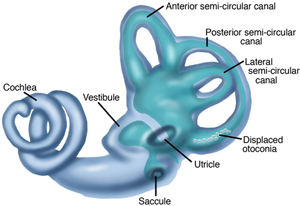
Benign paroxysmal positional vertigo is a common inner-ear problem affecting the vestibular system, a system used to maintain balance. BPPV causes short periods of dizziness when your head is moved in certain positions, relative to gravity. Benign means that this disorder is not life threatening, and generally, the disorder is not progressive. Paroxysmal means that the vertigo (spinning sensation) occurs suddenly. Positional means that the vertigo is triggered by changes in head position, most commonly when lying down, turning over in bed, or looking up. This dizzy or spinning sensation is called vertigo.

A layer of calcium carbonate material is present naturally in 1 part of your inner ear (the utricle). BPPV occurs when pieces of this material break off and move to another part of the inner ear, the semicircular canals (usually the posterior canal). These tiny calcium crystals (otoconia) are sometimes called “ear rocks.”

When you move your head a certain way, the crystals move inside the canal and stimulate the nerve endings, causing you to become dizzy. The cause of BPPV is usually not known; however, the crystals may become loose due to trauma to the head, infection, conditions, such as Meniere’s disease, or aging. BPPV is more common among females, and it may be hereditary.

InnerEar_sm.jpg

How Does it Feel?

BPPV occurs most commonly following position changing, such as lying down, turning over in bed, bending over, and looking up. A short delay, often less than 15 seconds, may follow a position change before symptoms start. This dizzy sensation, called vertigo, is brief and intense and usually lasts for about 15-45 seconds. However, symptoms may last for up to 2 minutes if the crystals become stuck to part of the inner ear. The episodes of vertigo occur frequently for weeks or months at a time. During these episodes, you may feel like the room is spinning around you, and you also may feel lightheaded, off balance, and nauseous.

Signs and Symptoms

The signs and symptoms of BPPV usually last less than a minute. The signs and symptoms may come and go or may disappear for a period of time, and then recur. Movement of the head causes most of the signs and symptoms of BPPV, which may include:

  • Dizziness

  • A sense that you or your surroundings are spinning or moving (vertigo)

  • A loss of balance or unsteadiness

  • Nausea

  • Vomiting

How Is It Diagnosed?

The diagnosis of BPPV is based on whether you have a particular kind of involuntary eye movement (called "nystagmus"), and whether you have vertigo when your head is moved into certain positions. Your physical therapist will perform tests that move your head in specific ways to see whether vertigo and involuntary eye movement results. These tests will help the therapist determine the cause and type of your dizziness, and whether you should be referred to a physician for any additional testing.

The positional tests are meant to recreate BPPV symptoms. By moving your head into certain positions and watching your eyes, your physical therapist may determine the appropriate repositioning maneuver needed to reduce or eliminate your vertigo.

Many different types and causes of dizziness exist, and dizziness is difficult for people to describe, making BPPV and other causes of dizziness more challenging to diagnose. When talking to your clinician, be as specific as possible when describing your symptoms.

For example, explain if you have lightheadedness or if you see or feel the room spinning during an episode. Also, describe how long your symptoms last (seconds, minutes, hours, or days). Do your best to describe what makes your dizziness better or worse. For example, is your dizziness made worse by movement or position changes? Is your dizziness eased by stillness or rest?

Be sure to discuss any recent illnesses or injuries, problems with your immune system, changes in medications or hormones, or headaches. These clues will be very insightful for your physical therapist and can assist in establishing an accurate diagnosis, or indicate the need for a referral to another specialist.

How Can a Physical Therapist Help?

Fortunately, most people recover from BPPV with a simple but very specific head and neck maneuver performed by a physical therapist. Your physical therapist will guide you through a series of 2-4 position changes. Each position may be held for 30 seconds to 2 minutes, as prescribed by your physical therapist. These repositioning treatments are designed to move the crystals from the semicircular canal back into the appropriate area in the inner ear (the utricle). A repositioning treatment called the Epley maneuver is used for the resolution of posterior canal BPPV, the most commonly involved canal. No medication has been found to be effective with BPPV and, in some cases, medication could cause more harm.

In a very few cases, BPPV cannot be managed with treatment maneuvers, and a surgical procedure called a “posterior canal plugging” may be considered—but, surgical intervention is rare.

Can this Injury or Condition be Prevented?

No known ways exist to prevent BPPV, especially when caused by such factors as head injury or aging. Once a person has experienced BPPV, symptoms can return if new crystals break off and get into the semicircular canal, or if you dislodge loose crystals by placing your head in a certain position. Some people report that their BPPV symptoms recur predictably, perhaps seasonally, or with changes in the weather.

Within 3 years of having BPPV, about 50% of people may have a recurrence. BPPV resulting from head trauma is more likely to recur. Once a person has experienced BPPV, symptoms can return if new crystals break. Although your BPPV might return, you'll be able to recognize the symptoms and keep yourself safe until you can get help. Your physical therapist will apply the appropriate maneuver to return the crystals to their correct position in the inner ear, and also will teach you how to do exercises that can reduce or eliminate the symptoms.

Real Life Experiences

Laura B. is a 68-year-old woman with vertigo that began one morning 2 weeks ago when she got out of bed and the world started to spin. Since then, she's been having vertigo, nausea, and problems with her balance. When she visits her physical therapist, he gives her a special questionnaire to find out exactly what brings on her dizziness and balance difficulties. Turning over in bed, bending over, or looking up cause the most severe symptoms.

The physical therapist reviews Laura's medical history to make sure that no past condition may be contributing to the vertigo. He performs an examination, explains what tests he will use, and tells Laura that she should try to keep her eyes open and stay in position. The tests show that in certain positions, Laura's eyes move when they shouldn't, and she has vertigo that lasts 5 seconds. The therapist determines that she has the "canalithiasis form" of vertigo, which means that some crystals are displaced and are flowing through her semicircular ear canals, causing vertigo.

The therapist uses "canalith repositioning" to move the crystals into a proper position, using the Epley maneuver. Afterwards, he asks Laura to wait in the waiting room for a while so that he can retest her. Laura no longer has the symptoms that she had when the therapist tested her the first time, so he shows her how to do the canalith repositioning maneuver at home. She is to perform the maneuver once every day in the morning for 1 week, and then return to the clinic to make sure that she is progressing as expected.

This story was based on a real-life case. Your case may be different. Your physical therapist will tailor a treatment program to your specific case.

What Kind of Physical Therapist Do I Need?

All physical therapists are prepared through education and experience to treat people who have dizziness and balance problems. You may want to consider:

  • A physical therapist who is experienced in treating people with neurological problems.

  • A physical therapist with specialized training and experience in vestibular rehabilitation.

  • A physical therapist who is a board-certified neurological clinical specialist, called NCS, or who completed a residency or fellowship in neurologic physical therapy, or who has advanced knowledge, experience, and skills that may apply to your condition.

You can find physical therapists who have these and other credentials by using Find a PT, the online tool built by the American Physical Therapy Association to help you search for physical therapists with specific clinical expertise in your geographic area.

General tips when you're looking for a physical therapist (or any other health care provider):

  • Get recommendations from family and friends or from other health care providers.

  • When you contact a physical therapy clinic for an appointment, ask about the physical therapists' experience in helping people with inner ear injury.

  • During your first visit with the physical therapist, be prepared to describe your symptoms in as much detail as possible, and say what makes your symptoms worse.

Further Reading

The American Physical Therapy Association (APTA) believes that consumers should have access to information that could help them make health care decisions and also prepare them for their visit with their health care provider.

The following articles provide some of the best scientific evidence about treatment of BPPV. The articles report recent research and give an overview of the standards of practice for treatment both in the United States and internationally. The article titles are linked either to a PubMed abstract of the article or to free full text, so that you can read it or print out a copy to bring with you to your health care provider.

Hilton MP, Pinder DK. The Epley (canalith repositioning) manoeuvre for benign paroxysmal positional vertigo. Cochrane Database Syst Rev. 2014;12:CD003162. Article Summary on PubMed.

Helminski JO. Effectivess of the canalith repositioning procedure in the treatment of benign paroxysmal positional vertigo. Phys Ther. 2014:94(10):1373–1382. Article Summary on PubMed.

Helminski JO, Zee DS, Janssen I, Hain TC. Effectiveness of particle repositioning maneuvers in the treatment of benign paroxysmal positional vertigo: a systematic review.  Phys Ther. 2010;90:663–678. Free Article

Cohen HS, Sangi-Haghpeykar H. Canalith repositioning variations for benign paroxysmal positional vertigo. Otolaryngol Head Neck Surg. 2010;143:405–412. Free Article.

Clinch CR, Kahill A, Klatt LA, Stewart D. Clinical inquiries: what is the best approach to benign paroxysmal positional vertigo in the elderly? J Fam Pract. 2010;59:295–297. Review. Article Summary on PubMed.

Fife TD, Iverson DJ, Lempert T, et al. Practice parameter: therapies for benign paroxysmal positional vertigo (an evidence-based review): report of the Quality Standards Subcommittee of the American Academy of Neurology. Neurology. 2008;70:2067-2074. Free Article.

Vestibular Disorders Association. Benign paroxysmal positional vertigo (BPPV). Accessed June 20, 2015.

Authored by Susan J. Herdman, PT, PhD; Shannon L.G. Hoffman, PT, DPT; Marcia Thompson, PT, DPT; Bob Wellmon, PT, PhD, NCS; and APTA’s Section on Neurology. Reviewed by the MoveForwardPT.com editorial board.


Shoulder Impingement

What is Shoulder Impingement?

Shoulder impingement syndrome is a condition that develops when the rotator-cuff tendons in the shoulder are overused or injured, causing pain and movement impairments. Shoulder impingement syndrome may also be referred to as "subacromial" impingement syndrome because the tendons, ligaments, and bursa under the "acromion" can become pinched or compressed. The shoulder is made up of 3 bones called the humerus, the scapula, and the clavicle. The acromion is a bony prominence on the top of the scapula, which can be felt as a bump at the tip of the shoulder.

The rotator cuff tendon and the bursa sit beneath the acromion. The bursa is a fluid-filled sac that provides a cushion between the bony acromion and the rotator cuff tendon, and it can become compressed underneath the acromion. Impingement symptoms can occur when compression and microtrauma harm the tendons. There are several causes to shoulder impingement syndrome including:

  • Repetitive overhead movements, such as golfing, throwing, racquet sports, and swimming, or frequent overhead reaching or lifting.

  • Injury, such as a fall, where the shoulder gets compressed.

  • Bony abnormalities of the acromion, which narrow the subacromial space.

  • Osteoarthritis in the shoulder region.

  • Poor rotator cuff and shoulder blade muscle strength, causing the humeral head to move abnormally.

  • Thickening of the bursa.

  • Thickening of the ligaments in the area.

  • Tightness of the soft tissue around the shoulder joint called the joint capsule.

How Does it Feel?

Individuals with shoulder impingement may experience:

  • Restriction in shoulder motion with associated weakness in movement patterns, such as reaching overhead, behind the body, or out to the side.

  • Pain in the shoulder when moving the arm overhead, out to the side, and beside the body.

  • Pain and discomfort when attempting to sleep on the involved side.

  • Pain with throwing motions and other dynamic movement patterns.

How Is It Diagnosed?

A physical therapist will perform an evaluation and ask you questions about the pain you are feeling, and other symptoms. Your physical therapist may perform strength and motion tests on your shoulder, ask about your job duties and hobbies, evaluate your posture, and check for any muscle imbalances and weakness that can occur between the shoulder and the scapular muscles.  

Special tests involving gentle movements of your arm and shoulder may be performed to determine exactly which tendons are involved. X-rays may also be taken to identify other conditions that could be contributing to your discomfort, such as bony spurs or abnormalities, or arthritis.

How Can a Physical Therapist Help?

It is important to get proper treatment for shoulder impingement as soon as it occurs. Secondary conditions can result from the impingement of the tissues in the shoulder, including irritation of the bursa and rotator-cuff tendinitis or tears.

Physical therapy can be very successful in treating shoulder impingement syndrome. You will work with your physical therapist to devise a treatment plan that is specific to your condition and goals. Your individual treatment program may include:

Pain Management. Your physical therapist will help you identify and avoid painful movements, as well as correct abnormal postures to reduce impingement compression. Therapeutic modalities, like iontophoresis (medication delivered through an electrically charged patch) and ultrasound may be applied. Ice may also be helpful to reduce pain.

Manual Therapy. Your physical therapist may use manual techniques, such as gentle joint movements, soft-tissue massage, and shoulder stretches to get your shoulder moving properly, so that the tendons and bursa avoid impingement.

Range-of-Motion Exercises. You will learn exercises and stretches to help your shoulder and shoulder blade move properly, so you can return to reaching and lifting without pain.

Strengthening Exercises. Your physical therapist will determine which strengthening exercises are right for you, depending on your specific condition. Often with shoulder impingement syndrome, the head of the humerus tends to drift forward and upward due to the rotator-cuff muscles becoming weak. Strengthening the rotator-cuff and scapular muscles helps position the head of the humerus bone down and back to ease the impingement. You may also perform resistance training exercises to strengthen your weaker muscles. You will receive a home-exercise program to continue your strengthening long after you have completed your formal physical therapy.

Patient Education. Learning proper posture is an important part of rehabilitation. For example, when your shoulders roll forward as you lean over a computer, the tendons in the front of the shoulder can become impinged. Your physical therapist will work with you to help improve your posture, and may suggest adjustments to your work station and work habits.

Functional Training. As your symptoms improve, your physical therapist will teach you how to correctly perform a range of functions using proper shoulder mechanics, such as lifting an object onto a shelf or throwing a ball. This training will help you return to pain-free function on the job, at home, and when playing sports.

Can this Injury or Condition be Prevented?

Shoulder impingement syndrome can be prevented by:

  • Maintaining proper strength in the shoulder and shoulder-blade muscles.

  • Regularly stretching the shoulders, neck, and middle-back region.

  • Maintaining proper posture and shoulder alignment when performing reaching and throwing motions.

  • Avoiding forward-head and rounded-shoulder postures (being hunched over) when spending long periods of time sitting at a desk or computer.

Real Life Experiences

Bob is a 33-year-old engineer who spends many hours drafting, creating spreadsheets, and writing reports on his computer at work. He enjoys playing tennis after work a few times a week.

Recently, Bob began feeling pain in his right shoulder when he reached overhead to serve a tennis ball. He felt the same kind of pain when he tried to throw a softball during his daughter's softball practice.

After a few weeks, the shoulder pain worsened and Bob began to have difficulty just reaching into a cupboard to get a glass at home. Last night, the pain in his shoulder woke him up every time he rolled onto it. He decided to call his physical therapist.

Bob's physical therapist performed a full evaluation of his shoulder. She noted he felt pain with certain shoulder movements, and had difficulty performing them due to weakness in the rotator-cuff and scapular muscles. She also saw that he had a rounded posture when sitting. She performed range-of-motion and other special tests on his shoulder. Based on her findings, she diagnosed shoulder impingement syndrome.

Bob and his physical therapist worked together to establish short- and long-term goals for his treatment. She prescribed ice to help decrease his pain, and taught him some gentle movement and strengthening exercises. She performed manual (hands-on) therapy on his shoulder to gently mobilize the joints and tissues.

Bob's physical therapist showed him how to improve his posture when sitting at his desk at work, and taught him a home-exercise program of stretching, strengthening, and postural exercises, which she modified throughout the course of his therapy as his condition improved.

After a few weeks of diligent physical therapy sessions and performing his home-exercise program, Bob reported he was able to raise his shoulder completely overhead without pain or limitation, and sleep on his side comfortably at night.

Today, Bob is back playing tennis pain-free, and is able to "throw long" for his daughter during softball practice.

What Kind of Physical Therapist Do I Need?

All physical therapists are prepared through education and experience to treat shoulder impingement syndrome. However, you may want to consider:

  • A physical therapist who is experienced in treating people with shoulder impingement syndrome. Some physical therapists have a practice with an orthopedic or musculoskeletal focus.

  • A physical therapist who is a board-certified clinical specialist, or who completed a residency or fellowship in orthopedic or sports physical therapy. This therapist has advanced knowledge, experience, and skills that may apply to your condition.

You can find physical therapists who have these and other credentials by using  Find a PT, the online tool built by the American Physical Therapy Association to help you search for physical therapists with specific clinical expertise in your geographic area.

General tips when you're looking for a physical therapist (or any other health care provider):

  • Get recommendations from family and friends or from other health care providers.

  • When you contact a physical therapy clinic for an appointment, ask about the physical therapists' experience in helping people who have shoulder impingement syndrome. During your first visit with the physical therapist, be prepared to describe your symptoms in as much detail as possible, and say what makes your symptoms worse.

Further Reading

The American Physical Therapy Association (APTA) believes that consumers should have access to information that could help them make health care decisions and also prepare them for their visit with their health care provider.

The following articles provide some of the best scientific evidence for the treatment of shoulder impingement. The articles report recent research and give an overview of the standards of practice both in the United States and internationally. The article titles are linked either to a PubMed* abstract of the article or to free full text, so that you can read it or print out a copy to bring with you to your health care provider.

Khan Y, Nagy MT, Malal J, Waseem M. The painful shoulder: shoulder impingement syndrome. Open Orthop J. 2013;7:347–351. Free Article.

Wilk KE, Hooks TR, Macrina LC. The modified sleeper stretch and modified cross-body stretch to increase shoulder internal rotation range of motion in the overhead throwing athlete. J orthop Sports Phys Ther. 2013;43(12):891–894. Free Article.

De Mey K, Danneels LA, Cagnie B, Huyghe L, Seyns E, Cools AM. Conscious correction of scapular orientation in overhead athletes performing selected shoulder rehabilitation exercises: the effect on trapezius muscle activation measured by surface electromyography. J Orthop Sports Phys Ther. 2013;43(1):3–10. Free Article.

Struyf F, Nijs J, Baeyens JP, Mottram S, Meeusen R. Scapular positioning and movement in unimpaired shoulders, shoulder impingement syndrome, and glenohumeral instability. Scand J Med Sci Sports. 2011;21(3):352–358. Article Summary on PubMed.

Castagna A, Garofalo R, Cesari E, Markopoulos N, Borroni M, Conti M. Posterior superior internal impingement: an evidence-based review [erratum in: Br J Sports Med. 2010;44(8):604.]. Br J Sports Med. 2010;44(5):382–388. Article Summary on PubMed.

Faber E, Kuiper JI, Burdorf A, Miedema HS, Verhaar JA. Treatment of impingement syndrome: a systematic review of the effects on functional limitations and return to work. J Occup Rehabil. 2006;16(1):7–25. Article Summary on PubMed.

*PubMed is a free online resource developed by the National Center for Biotechnology Information (NCBI). PubMed contains millions of citations to biomedical literature, including citations in the National Library of Medicine’s MEDLINE database.

Authored by Julie A. Mulcahy, PT, MPT. Reviewed by the MoveForwardPT.com editorial board.


Osgood-Schlatter Disease

Osgood-Schlatter disease occurs when there is irritation to the top, front portion of the shin bone (tibia) where the tendon attached to the kneecap (patella) meets the shin bone. It occurs when there is an increased amount of stress placed upon the bones where the tendons attach. This is most often the result of increased activity levels by an adolescent athlete.

Our musculoskeletal system is made up of bones and surrounding soft tissue structures, including muscles, ligaments (which connect bone to bone), and tendons (which connect muscle to bone). These structures all play a role in helping us move.

During adolescence our bodies grow at a rapid rate. As our bodies develop, our bones are growing longer. Throughout this phase, our growth plates (epiphyseal plates) are susceptible to injury. A growth plate is the site at the end of a bone where new bone tissue is made and bone growth occurs. Females typically experience the most rapid growth between approximately 11 to 12 years of age, and males typically experience this growth surge between approximately 13 and 14 years of age. Males experience OS more frequently than females, likely due to an increased rate of sports participation.

Structures in our body might become irritated if they are asked to do more than they are capable of doing. Injuries can occur in an isolated event, but OS disease is most likely the cumulative effect of repeated trauma. OS is most frequently experienced in adolescents who regularly participate in running, jumping, and "cutting" (rapid changes in direction) activities.

When too much stress is present (ie, from rapid growth) and when the body is overworked (ie, either too much overall volume of exercise, or too much repetition), the top of the shin can become painful and swollen. As this condition progresses, the body’s response to bone stress can be an increase in bone production; an adolescent may begin to develop a boney growth that feels like a bump on the front of the upper shin.

OS can start as mild soreness, but can progress to long-lasting pain and limited function, if not addressed early and appropriately.

How Does it Feel?

With Osgood-Schlatter, you may experience:

  • Gradually worsening pain below your knee, at the top of the shin bone.

  • Pain that worsens with exercise.

  • Swelling and tenderness at the top of the shin.

  • A boney growth at the top of the shin.

  • Loss of strength in the quadriceps muscle (connecting the hip to the knee).

  • Increased tightness in the quadriceps muscle.

  • Loss of knee motion.

  • Discomfort with daily activities that use your knee, like kneeling, squatting, or walking up and down stairs.

How Is It Diagnosed?

Diagnosis of OS begins with a thorough medical history, including specific questions regarding athletic participation (sports played, frequency of practices/games, positions). Your physical therapist will assess different measures, such as sensation, motion, strength, flexibility, tenderness, and swelling. Your physical therapist will perform several tests specific to the knee joint, and may ask you to briefly demonstrate the activities or positions that cause your pain, such as walking, squatting, and stepping up or down stairs.

Because the knee and hip are both involved in these aggravating activities, your physical therapist will likely examine your hip as well. Other nearby areas, such as your feet and core, will also be examined to determine whether they, too, might be contributing to your knee condition.

If your physical therapist suspects there may be a more involved injury than increased stress-related irritation (ie, if there is a recent significant loss of motion or strength, or severe pain when the knee is moved), your therapist will likely recommend a referral to an orthopedic physician for diagnostic imaging, such as ultrasound, x-ray, or MRI.

How Can a Physical Therapist Help?

Once other conditions have been ruled out and OS is diagnosed, your physical therapist will work with you to develop an individualized treatment plan tailored to your specific knee condition and your goals. The goal of physical therapy is to accelerate your recovery and return to pain-free activity. There are many physical therapy treatments that have been shown to be effective in treating OS, and among them are:

Range of Motion Therapy. Your physical therapist will assess the motion of your knee and its surrounding structures, and design gentle exercises to help you work through any stiffness and swelling to return to a normal range of motion.

Strength Training. Your physical therapist will teach you exercises to strengthen the muscles around the knee so that each muscle is able to properly perform its job, and stresses are eased so the knee joint is properly protected.

Manual Therapy.Physical therapists are trained in manual (hands-on) therapy. If needed, your physical therapist will gently move your kneecap or patellar tendon and surrounding muscles as needed to improve their motion, flexibility, and strength. These techniques can target areas that are difficult to treat on your own. 

Pain Management.Your physical therapist may recommend therapeutic modalities, such as ice and heat, or a brace to aid in pain management.

Functional Training.Physical therapists are experts at training athletes to function at their best. Your physical therapist will assess your movements and teach you to adjust them to relieve any extra stress on the front of your knee.

Education. The first step to addressing your knee pain is rest. Your physical therapist will explain why this is important and develop a plan for your complete rehabilitation.

Can this Injury or Condition be Prevented?

Fortunately, there is much that can be done to prevent the cascade of events that lead to OS. Physical therapists focus on:

  • Educating coaches, parents, and athletes on guidelines for sports participation, explaining common causes of overuse injuries, and providing strategies for prevention.

  • Educating athletes on the risks of playing through pain.

  • Scheduling adequate rest time to recover between athletic events.

  • Tracking a young athlete’s growth curves (height, weight, BMI) to identify periods of increased injury risk.

  • Developing an athlete-specific flexibility and strengthening routine to be followed throughout the athletic season.

  • Encouraging consultation with a physical therapist whenever symptoms appear.

Real Life Experiences

Caleb is a 13-year-old boy who has been playing basketball since he was in the first grade. When younger, Caleb played only during the winter season. Over the course of the last year, however, he has attended 2 basketball camps during the summer, played on his middle-school team during the winter, and is now playing AAU basketball in the spring.

Caleb also chose to join the track team this spring, competing in the high jump and sprint events to improve his basketball skills. Over the past 3 months, Caleb has grown 2 inches, and both he and his basketball coaches are excited about his recent growth.

Recently, Caleb has been busy playing in weekly AAU tournaments with 1 to 2 track meets during the week. But when he got home from track practice on Monday, he told his dad that his leg was hurting. He said that it had begun getting sore while playing basketball over the weekend, but he didn’t want to tell his coach because he wanted to continue to play. Now he feels like the top of his shin is tender to touch, and he is unable to fully bend his knee without increased pain.

His dad realizes that this is more than the expected postactivity soreness; he immediately calls their local physical therapist.

Caleb's physical therapist takes his health history and performs an extensive examination. It becomes clear that Caleb has not scheduled appropriate rest times between his athletic activities, and that he is experiencing a growth spurt. The physical examination reveals that the top of Caleb’s shin is very tender, the area around his knee is swollen, and he has lost knee motion and strength. OS is diagnosed.

Together, Caleb and his physical therapist, father, and basketball and track coaches develop a treatment plan to help him return to pain-free sport participation. It begins with a 2-week period of rest where Caleb performs only minimal running exercise, and works regularly with his physical therapist on stretching, strengthening, balance, and coordination exercises, and on improving his squatting movements.

While at basketball practice, Caleb continues to work on his ball handling and free-throw shooting, activities that won't increase his knee pain.

After 2 weeks, when his knee is less tender, Caleb, his physical therapist, and his coaches develop a plan for his gradual return to full participation in track and basketball. They help Caleb understand how important it is to be honest about his knee pain, and to communicate with his coach if it starts to bother him again.

A month later, Caleb is back participating in all his track and basketball activities. He has changed his routine to allow for adequate warm-up time before and after each practice, and sufficient rest periods between activities. He makes sure his dad or coaches are keeping track of how much time he spends at practice and at rest.

Caleb decides to spend more time during the week working on his stretching, so he can reduce any risk of pain as he continues to grow. At the end of the season, Caleb’s AAU team wins the state championship—and he sets a new personal-best record in both the high jump and the 100-meter sprint!

What Kind of Physical Therapist Do I Need?

All physical therapists are prepared through education and clinical experience to treat a variety of conditions or injuries. You may want to consider:

  • A physical therapist who is experienced in treating people with orthopedic or musculoskeletal injuries.

  • A physical therapist who is a board-certified specialist or has completed a residency in orthopedic or sports physical therapy, as the therapist will have advanced knowledge, experience, and skills that apply to an athletic population.

You can find physical therapists that have these and other credentials by using Find a PT, the online tool built by the American Physical Therapy Association to help you search for physical therapists with specific clinical expertise in your geographic area.

General tips when you're looking for a physical therapist (or any other health care provider):

  • Get recommendations from family and friends or from other health care providers.

  • When you contact a physical therapy clinic for an appointment, ask about the physical therapists' experience in helping young athletes with knee pain.

  • During your first visit with the physical therapist, be prepared to describe your symptoms in as much detail as possible, and report activities that make your symptoms worse.

Further Reading

The American Physical Therapy Association (APTA) believes that consumers should have access to information that could help them make health care decisions, and also prepare them for a visit with their health care provider.

The following articles provide some of the best scientific evidence related to physical therapy treatment of athletic injuries. The articles report recent research and give an overview of the standards of practice both in the United States and internationally. The article titles are linked either to a PubMed* abstract of the article or to free full text, so that you can read it or print out a copy to bring with you to your health care provider.

Whitmore A. Osgood-Schlatter disease. JAAPA. 2013;26(10):51–52. Article Summary on PubMed.

Maffulli N, Longo UG, Spiezia F, Denaro V. Aetiology and prevention of injuries in elite young athletes. Med Sport Sci. 2011;56:187–200. Article Summary on PubMed.

Stein CJ, Micheli LJ. Overuse injuries in youth sports. Phys Sportsmed. 2010;38(2):102–108. Article Summary on PubMed.

* PubMed is a free online resource developed by the National Center for Biotechnology Information (NCBI). PubMed contains millions of citations to biomedical literature, including citations in the National Library of Medicine’s MEDLINE database.

 Authored by Allison Mumbleau, PT, DPT, SCS. Reviewed by the MoveForwardPT.com editorial board.


Rotator Cuff Tendinitis

The rotator cuff muscles are a group of 4 muscles that attach the humerus (upper-arm bone) to the scapula (shoulder blade). The rotator cuff muscles help raise, rotate, and stabilize the upper arm. A tendon is a bundle of fibers that connect the muscles to the bone. Rotator cuff tendinitis occurs when the tendon connected to the rotator cuff muscles becomes inflamed and irritated. It can be caused by:

  • Poor posture, such as rounded shoulders caused by leaning over a computer for long periods of time.

  • Repetitive arm movements, such as those performed by a hair stylist or painter.

  • Overhead shoulder motions, such as those performed by baseball pitchers or swimmers.

  • Tight muscles and tissues around the shoulder joint.

  • Weakness and muscle imbalances in the shoulder blade and shoulder muscles.

  • Bony abnormalities of the shoulder region that cause the tendons to become pinched (shoulder impingement syndrome).

How Does it Feel?

Rotator cuff tendinitis is characterized by shoulder pain that can occur gradually over time or start quite suddenly. The pain occurs in the shoulder region and sometimes radiates into the upper arm. It does not usually radiate past the elbow region. You may be symptom free at rest or experience a mild, dull ache; however, pain can be moderate to severe with certain shoulder movements. Reaching behind the body to perform a motion, as in fastening a seat belt, can be very painful. So can overhead activities, such as throwing, swimming, reaching into a cupboard, or combing your hair. The pain can worsen at night, especially when rolling over or attempting to sleep on the painful side. You may notice weakness when lifting and reaching for household items. Holding a heavy platter or taking a pan off the stove may become difficult.

How Is It Diagnosed?

A physical therapist will perform an evaluation and ask you questions about the pain and other symptoms you are feeling. Your therapist may perform strength and motion tests on your shoulder, ask about your job duties and hobbies, evaluate your posture, and check for any muscle imbalances and weakness that can occur between the shoulder and the scapular muscles. Your physical therapist will gently touch your shoulder in specific areas to determine which tendon or tendons are inflamed, and special tests may need to be performed to determine this.

How Can a Physical Therapist Help?

It is important to get proper treatment for tendinitis as soon as it occurs. A degenerated tendon that is not treated can begin to tear causing a more serious condition. Physical therapy can be very successful in treating rotator cuff tendinitis, tendinosis, and shoulder impingement syndrome. You will work with your physical therapist to devise a treatment plan that is specific to your condition and goals. Your individual treatment program may include:

Pain management. Your physical therapist will help you identify and avoid painful movements to allow the inflamed tendon to heal. Ice, ice massage, or moist heat maybe used for pain management. Therapeutic modalities, such as iontophoresis (medication delivered through an electrically charged patch) and ultrasound may be applied.

Manual therapy. Your physical therapist may use manual techniques, such as gentle joint movements, soft-tissue massage, and shoulder stretches to get your shoulder moving again in harmony with your scapula.

Range-of-motion exercises. You will learn exercises and stretches to help your shoulder and shoulder blade move properly, so you can return to reaching and lifting without pain.

Strengthening exercises. Your physical therapist will determine which strengthening exercises are right for you, depending on your specific condition. You may use weights, medicine balls, resistance bands, and other types of resistance training to challenge your weaker muscles. You will receive a home-exercise program to continue rotator cuff and scapular strengthening, long after you have completed your formal physical therapy.

Patient education. Posture education is an important part of rehabilitation. For example, when your shoulders roll forward as you lean over a computer, the tendons in the front of the shoulder can become pinched. Your physical therapist may suggest adjustments to your workstation and work habits.

Functional training. As your symptoms improve, your physical therapist will help you return to your previous level of function that may include household chores, job duties and sports- related activities. Functional training can include working on lifting a glass into a cupboard or throwing a ball using proper shoulder mechanics. You and your physical therapist will decide what your goals are, and get you back to your prior level of functioning as soon as possible.

Can this Injury or Condition be Prevented?

Rotator cuff tendinitis can be prevented by:

  • Maintaining proper shoulder and spinal posture during daily activities, including sitting at a computer.

  • Performing daily stretches to the shoulder and upper back to maintain normal movement. Tightness in the upper back, or a rounded shoulder posture will decrease the ability to move your torso, and that makes the shoulder have to work harder to perform everyday activities, such as reaching for objects.

  • Keeping your upper body strong, including the upper back and shoulder-blade muscles will help prevent tendinitis. Many people work the muscles in their chest, arms, and shoulders, but it is also important to work the muscles around the shoulder blade and upper back. These muscles provide a strong foundation for your shoulder function. Without a strong foundation, muscle imbalances occur and put the shoulder at risk for injury.

Real Life Experiences

Mary is a 51-year-old piano teacher with 14 students. She teaches 3 days a week; each session lasts 30 minutes. Mary also plays piano for her church, and for her own enjoyment. A few weeks ago, she began to feel pain in her left shoulder when reaching her arm overhead or behind her body. Her symptoms worsened, and she began experiencing pain even when at rest. Now the pain is so severe, it wakes her up at night; she can no longer sleep on her left side. She contacts her physical therapist.

Mary's physical therapist performs a full evaluation of her shoulder, and her scapula and upper-back strength and mobility. Mary describes how long she sits at the piano each week. Her therapist gently feels all around her shoulder and finds that it is very tender over the rotator cuff region. She has pain when her therapist performs resistive-muscle testing to the rotator cuff. He also discovers that Mary has tightness in her upper back region that limits her ability to fully twist her body to the right and left. Special tests were performed on her shoulder, and the results indicate the rotator cuff is irritated. Based on these findings, he diagnoses rotator cuff tendinitis.

Mary and her physical therapist work together to establish short- and long-term goals for her treatment. He prescribes ice to help decrease her pain, and teaches her some gentle movement and strengthening exercises. He also shows Mary how to improve her posture when sitting at the piano, and teaches her a home-exercise program of stretching, strengthening, and postural exercises, which he modifies throughout the course of her therapy as her condition improves.

Mary and her physical therapist work together in a 6-week program of 2-3 rehabilitation sessions per week. He performs gentle passive movements of her shoulder, scapula, and upper back to increase her joint motion. Mary learns proper movement patterns for reaching her arm overhead. She finds that using a therapeutic chair helps improve her posture and strengthens her core during her piano lessons.

After a few weeks of diligent therapy sessions and working with her home-exercise program, Mary notices she is able to sleep on her left side again without pain, and can easily reach to get a mug from her upper kitchen shelf.

Mary is soon able to return to all of her daily activities and enjoy her life as a piano teacher—free of pain.

What Kind of Physical Therapist Do I Need?

All physical therapists are prepared through education and experience to treat rotator cuff tendinitis. However, you may want to consider:

  • A physical therapist who is experienced in treating people with rotator cuff tendinitis. Some physical therapists have a practice with an orthopedic or musculoskeletal focus.

  • A physical therapist who is a board-certified clinical specialist or who completed a residency or fellowship in orthopedic or sports physical therapy. This therapist has advanced knowledge, experience, and skills that may apply to your condition.

You can find physical therapists who have these and other credentials by using  Find a PT, the online tool built by the American Physical Therapy Association to help you search for physical therapists with specific clinical expertise in your geographic area.

General tips when you are looking for a physical therapist (or any other health care provider):

  • Get recommendations from family and friends or from other health care providers.

  • When you contact a physical therapy clinic for an appointment, ask about the physical therapists' experience in helping people who have rotator cuff tendinitis. During your first visit with the physical therapist, be prepared to describe your symptoms in as much detail as possible, and say what makes your symptoms worse.

Further Reading

The American Physical Therapy Association (APTA) believes that consumers should have access to information that could help them make health care decisions and also prepare them for their visit with their health care provider.

The following articles provide some of the best scientific evidence related to physical therapy treatment of rotator cuff tendinitis. The articles report recent research and give an overview of the standards of practice both in the United States and internationally. The article titles are linked either to a PubMed* abstract of the article or to free full text, so that you can read it or print out a copy to bring with you to your health care provider.

Thornton AL, McCarty CW, Burgess MJ. Effectiveness of low-level laser therapy combined with an exercise program to reduce pain and increase function in adults with shoulder pain: a critically appraised topic. J Sport Rehabil. 2013;22(1):72-78. Article Summary on PubMed.

Childress MA, Beutler A. Management of chronic tendon injuries. Am Fam Physician. 2013;87(7):486-490. Article Summary on PubMed.

Scott A, Docking S, Vicenzino B, et al. Sports and exercise-related tendinopathies: a review of selected topical issues by participants of the second International Scientific Tendinopathy Symposium (ISTS) Vancouver 2012 [erratum in: Br J Sports Med. 2013;47(12):744]. Br J Sports Med. 2013;47(9):536-544. Free Article.

Littlewood C, Ashton J, Chance-Larsen K, et al. Exercise for rotator cuff tendinopathy: a systematic review. Physiotherapy. 2012;98(2):101-109. Article Summary on PubMed.

Andres BM, Murrell GA. Treatment of tendinopathy: what works, what does not, and what is on the horizon. Clin Orthop Relat Res. 2008;466(7): 1539–1554. Free Article.

Senbursa G, Galtaci G, Atay A. Comparison of conservative treatment with and without manual physical therapy for patients with shoulder impingement syndrome: a prospective, randomized clincial trial. Knee Surg Sports Traumatol Arthrosc. 2007;15(7):915-921. Article Summary on PubMed.

*PubMed is a free online resource developed by the National Center for Biotechnology Information (NCBI). PubMed contains millions of citations to biomedical literature, including citations in the National Library of Medicine’s MEDLINE database.

Authored by Julie A. Mulcahy, PT, MPT. Reviewed by the MoveForwardPT.com editorial board.


Plantar Fasciitis

Plantar fasciitis is a condition causing heel pain. Supporting the arch, the plantar fascia, a thick band of tissue connecting the heel to the ball of the foot, can become inflamed or can tear. You experience pain when you put weight on your foot—particularly when taking your first steps in the morning. The pain can be felt at the heel, or along the arch and the ball of the foot.

Plantar fasciitis is a common foot condition. It occurs in as many as 2 million Americans per year and 10% of the population over their lifetimes.

Factors that contribute to the development of plantar fasciitis include:

  • Age (over 40 years)

  • A job, sport, or hobby that involves prolonged standing or other weight-bearing activity

  • Rapid increases in length or levels of activity, such as beginning a new running program or changing to a job that requires a lot more standing or walking than you are accustomed to

  • Decreased calf muscle flexibility

  • Increased body weight (Body Mass Index greater than 30)

  • Tendency to have a flat foot (pronation)

Plantar fasciitis affects people of all ages, both athletes and non-athletes. Men and women have an equal chance of developing the condition.

Treatment generally reduces pain and restores your ability to put weight on your foot again.

PlantarFasciitis_SM.jpg

What is Plantar Fasciitis?

Plantar fasciitis is a condition causing heel pain. Supporting the arch, the plantar fascia, a thick band of tissue connecting the heel to the ball of the foot, can become inflamed or can tear. The condition develops when repeated weight-bearing activities put a strain on the plantar fascia. People who are diagnosed with plantar fasciitis also may have heel spurs, a bony growth that forms on the heel bone. However, people with heel spurs may not experience pain.

Plantar fasciitis occurs most frequently in people in their 40s but can occur in all age groups.

The condition can develop in athletes who run a great deal and in non-athletes who are on their feet most of the day, such as police officers, cashiers, or restaurant workers.

Signs and Symptoms

The onset of symptoms of plantar fasciitis frequently occurs with a sudden increase in activity. You might feel a stabbing pain on the underside of your heel, and a sensation of tightness and/or tenderness along your arch.

People with plantar fasciitis may experience pain:

  • In the morning, when stepping out of bed and taking the first steps of the day

  • With prolonged standing

  • When standing up after sitting for awhile

  • After an intense weight-bearing activity such as running

  • When climbing stairs

  • When walking barefoot or in shoes with poor support

As your body warms up, your pain may actually decrease during the day but then worsen again toward the end of the day because of extended walking. Severe symptoms may cause you to limp.

How Is It Diagnosed?

The physical therapist’s diagnosis is based on your health and activity history and a clinical evaluation. Your therapist also will take a medical history to make sure that you do not have other possible conditions that may be causing the pain. Sharing information about the relationship of your symptoms to your work and recreation, and reporting any lifestyle changes, will help the physical therapist diagnose your condition and tailor a treatment program for your specific needs.

To diagnose plantar fasciitis, your therapist may conduct the following physical tests to see if symptoms occur:

  • Massaging and pressing on the heel area (palpation)

  • Gently stretching the ankle to bend the top of the foot toward the leg (dorsiflexion)

  • Gently pressing the toes toward the ankle

How Can a Physical Therapist Help?

Physical therapists are trained to evaluate and treat plantar fasciitis.

When you are diagnosed with plantar fasciitis, your physical therapist will work with you to develop a program to decrease your symptoms that may include:

  • Stretching exercises to improve the flexibility of your ankle and the plantar fascia

  • Use of a night splint to maintain correct ankle and toe positions

  • Selection of supportive footwear and/or shoe inserts that minimize foot pronation and reduce stress to the plantar fascia

  • Application of ice to decrease pain and inflammation

  • Iontophoresis (a gentle way to deliver medication through the skin)

  • Taping of the foot to provide short-term relief

Research shows that most cases of plantar fasciitis improve over time with these conservative treatments, and surgery is rarely required.

Can this Injury or Condition be Prevented?

Guidelines for the prevention or management of plantar fasciitis include:

  • Choosing shoes with good arch support

  • Replacing your shoes regularly, so that they offer arch support and provide shock absorption to your feet

  • Using a thick mat if you must stand in one place for much of the day

  • Applying good principles to your exercise program, such as including a warm-up and gradually building up the intensity and duration of your exercises to avoid straining the plantar fascia

  • Stretching your calves and feet before and after running or walking

  • Maintaining a healthy body weight

Real Life Experiences

Jason has worked as a cook in a restaurant kitchen for 5 years. He has gradually gained about 25 pounds over those years. He began to develop pain in both his heels about 2 months ago. He does not exercise.

Jason asks a friend who has received physical therapy for advice. His friend suggests he see a physical therapist to find the cause of his heel pain.

Jason's physical therapist conducts a detailed history, asking questions about his health, lifestyle, and work, and performs a thorough evaluation. Jason says that his heel pain is worst when he gets up in the morning. After a shower and walking around for a while, his pain diminishes. However, when he is cooking at the restaurant during the evening shift, his heel pain returns, extending to the balls of his feet. Due to food debris in the restaurant kitchen, Jason says he wears old beat-up sneakers to work.

After conducting a physical examination, Jason's therapist diagnoses plantar fasciitis. She teaches Jason several stretches to perform twice a day and designs a home exercise program that will fit his goals and lifestyle. The therapist recommends he choose a shoe with a good arch support and replace them when they are worn out. She also suggests an orthotic (shoe insert) to place into his new shoes. She instructs him to apply ice to the bottom of his feet several times throughout the day. The therapist does not prescribe a night splint at this time, because Jason has had symptoms for less than 3 months. The therapist recommends that for his general health, Jason begin a low-impact exercise program, including swimming and using an exercise bike. This will help him lose the excess weight he has gained without further aggravating his plantar fasciitis.

Jason follows the advice of his physical therapist. He purchases new footwear for work and performs the stretching exercises and icing as instructed. After 2 weeks, he is 90% pain-free. Jason keeps his follow-up visit with his physical therapist 1 month later to review his condition and adjust his home program.

What Kind of Physical Therapist Do I Need?

All physical therapists are prepared through education and experience to treat plantar fasciitis. However, you may want to consider:

  • A physical therapist who is experienced in treating people with orthopedic and sports injuries, particularly those with experience working with the ankle and foot

  • A physical therapist who is a board-certified clinical specialist or who completed a residency or fellowship in orthopedic or sports physical therapy, meaning that this therapist has advanced knowledge, experience, and skills that may apply to your condition

You can find physical therapists who have these and other credentials by using Find a PT, the online tool built by the American Physical Therapy Association to help you search for physical therapists with specific clinical expertise in your geographic area.

General tips when you're looking for a physical therapist (or any other health care provider):

  • Get recommendations from family and friends or from other health care providers.

  • When you contact a physical therapy clinic for an appointment, ask about the physical therapists' experience in helping people who have heel pain.

  • During your first visit with the physical therapist, be prepared to describe your symptoms in as much detail as possible, and say what makes your symptoms worse.

Further Reading

The American Physical Therapy Association (APTA) believes that consumers should have access to information that could help them make health care decisions and also prepare them for their visit with their health care provider.

The following articles provide some of the best scientific evidence related to physical therapy treatment of plantar fasciitis. The articles report recent research and give an overview of the standards of practice both in the United States and internationally. The article titles are linked either to a PubMed* abstract of the article or to free access of the full article, so that you can read it or print out a copy to bring with you to your health care provider.

Cole C, Seto C, Gazewood J. Plantar fasciitis: evidence-based review of diagnosis and therapy. Am Fam Physician. 2005;72:2237–2242. Free Article.

Irving DB, Cook JL, Young MA, Menz HB. Obesity and pronated foot type may increase the risk of chronic plantar heel pain: a matched case-control study. BMC Musculoskelet Disord. 2007;8:41. Free Article.

McPoil TG, Martin RL, Cornwall MW, et al. Heel pain—plantar fasciitis: clinical practice guildelines linked to the International Classification of Function, Disability and Health from the Orthopaedic Section of the American Physical Therapy Association [erratum in: J Orthop Sports Phys Ther. 2008;38:648]. J Orthop Sports Phys Ther. 2008;38:A1–A18. .

Riddle DL, Pulisic M, Pidcoe P, Johnson RE. Risk factors for plantar fasciitis: a matched case-control study [erratum in: J Bone Joint Surg Am. 2003;85-A:1338]. J Bone Joint Surg Am. 2003;85-A:872–877.  Article Summary on PubMed.

Riddle DL, Schappert SM. Volume of ambulatory care visits and patterns of care for patients diagnosed with plantar fasciitis: a national study of medical doctors. Foot Ankle Int. 2004;25:303–310. Article Summary on PubMed.

Scher DL, Belmont PJ Jr, Bear R, et al. The incidence of plantar fasciitis in the United States military. J Bone Joint Surg Am. 2009;91:2867–872. Article Summary on PubMed.

*PubMed is a free online resource developed by the National Center for Biotechnology Information (NCBI). PubMed contains millions of citations to biomedical literature, including citations in the National Library of Medicine's MEDLINE database.

Authored by Shaw Bronner, PT, PhD, OCS. Reviewed by the MoveForwardPT.com editorial board.

Hip Impingement

Hip impingement involves a change in the shape of the surface of the hip joint that predisposes it to damage, resulting in stiffness and pain. Hip impingement is a process that may precede hip osteoarthritis. It most often occurs in young, active people. A recent study found that 87% of teens and adults with hip pain showed evidence of hip impingement on diagnostic images taken of their hip joints. To treat hip impingement, physical therapists prescribe stretches and strengthening exercises to better balance the muscles around the hip to protect it, and use manual therapies to help restore range of motion and increase comfort.

What is Hip Impingement?

There are 2 types of hip impingement; they may occur alone or together.

Pincer-Type Impingement

  • In pincer-type impingement, the hip socket (acetabulum), which is usually angled forward, may be angled toward the back, or protruding bone may be present on the pelvis side of the hip joint making the socket a deeper recess that covers more of the ball or head of the femur bone.

  • The overgrown bone or incorrect angle of the socket causes the labrum, a rim of connective tissue around the edge of the hip socket, to be pinched. Over time, this extra pressure to the labrum when flexing (moving the leg forward) leads to wear and tear that can cause inflammation and could result in a tear. If this condition persists, eventually the cartilage that lines the hip joint can become worn and form holes.

  • This condition affects men and women equally; symptoms often begin early, appearing at any time between 15 to 50 years of age.


Cam-Type Impingement

  • In cam-type impingement, the shape of the bone around the head of the femur—the ball at the top of the bone in the thigh—is misshapen. It can vary from the normal round ball shape, or have overgrown bone formed at the top and front. The nickname “pistol grip” deformity is given to the appearance of the bony overgrowth on x-rays.

  • The overgrown or misshapen bone contacts the cartilage that lines the hip socket, and can cause it to peel away from the bone in the socket. The labrum can become worn, frayed, or torn as well.

  • This condition affects men to women at a ratio of 3 to 1; symptoms often manifest during the teen years and 20s.

HipImpingement-SM.jpg

Signs and Symptoms

Hip impingement may cause you to experience:

  • Stiffness or deep aching pain in the front or side of the hip or front of the upper thigh while resting.

  • Sharp, stabbing pain when standing up from a chair, squatting, rising from a squat, running, "cutting," jumping, twisting, pivoting, or making lateral motions.

  • Hip pain described in a specific location by making a "C" with the thumb and hand and placing it on the fold at the front and side of the hip, known as the "C-sign."

  • Pain that most often starts gradually, but can also remain after another injury resolves.

  • Pain that increases with prolonged sitting or forward leaning.

  • Feeling less flexible at the hips, including a decreased ability to turn your thigh inward on the painful side.

How Is It Diagnosed?

Your physical therapist will evaluate the range of motion (movement) of the hip and surrounding joints, and test the strength of the muscles in that area. Your therapist will feel the hip joint and surrounding muscles to evaluate their condition. The examination will include observing how you move, standing from a sitting position, walking, running, or squatting, as appropriate. Your physical therapist may perform special tests to help determine whether the hip is the source of your symptoms. For instance, the therapist may gently roll your leg in and out (the “log roll” test), or bend your hip up and in while turning the lower leg out to the side (the "FADDIR" test) to assess your condition.

If further diagnosis is needed, your doctor may order diagnostic tests to help identify any joint changes, including x-rays, magnetic resonance imaging (MRI), or diagnostic injections. Hip impingement can occur at the same time as low back, buttock, or pelvic pain, or from conditions such as bursitis or groin strain. The final diagnosis of hip impingement may take some time, especially when other conditions are present.

How Can a Physical Therapist Help?

Without Surgery

When an active person develops hip pain, but does not have severe symptoms or joint damage, the recommended treatment is physical therapy. The following interventions can help decrease pain, improve movement, and avoid the progression of hip impingement and the need for surgery:

  • Improving the strength of your hips and trunk. Strengthening of the hips and trunk can reduce abnormal forces on the already injured joint and help with strategies to compensate.

  • Improving hip muscle flexibility and joint mobility. Stretching tight muscles can reduce abnormal forces that cause pain with motion. Joint mobilization may help ease pain from the hip joint; however, these treatments do not always help range of motion, especially if the shape of the bone at the hip joint has changed.

  • Improving tolerance of daily activities. Your physical therapist can consider your job and recreational activities and offer advice regarding maintaining postures that are healthier for your hip and activity modification. Often this involves limiting the amount of bending at the hip to avoid further hip damage.


Following Surgery

Surgery for hip impingement is performed with arthroscopy. This is a minimally invasive type of surgery, where the surgeon makes small incisions in the skin and inserts pencil-sized instruments into the joint to repair damage. The surgeon may perform 1 or several techniques during your procedure as needed. The surgeon may remove or reshape the bone on the pelvis or femur side of the joint, and repair or remove the damaged labrum or cartilage of the hip joint.

Postsurgical physical therapy varies based on the procedure performed. It may include:

  • Ensuring your safety as you heal. Your physical therapist may recommend that you limit the amount of weight you put on the operated leg if there was a repair of the labrum. You may wear a brace to help limit the amount of bending at the hip. You might also use crutches to avoid overloading the leg if the bone on the femur was reshaped.

  • Improving your range of motion, strength, and balance. Your physical therapist will guide you through safe range-of-motion, strengthening, and balance activities to improve your movement as quickly as possible while allowing the surgical site to heal properly.

  • Instructions on returning to an active lifestyle. Most people return to normal daily activities about 3 months after surgery, and to high-level activities and sports 4 to 6 months after surgery. Your physical therapist will recommend a gradual return to activity based on your condition—research shows that 60% to 90% of athletes return to their previous playing ability depending on the surgical procedure performed and the sport.

Can this Injury or Condition be Prevented?

Currently there are no recommendations to prevent hip impingement. Despite a major increase in research to learn more about hip impingement, there is a great deal that is unknown. For instance, many active young people whose x-rays show hips as being abnormal do not have pain despite continuing to live active lives and participate in sports.

However, there is evidence that physical therapy interventions along with anti-inflammatory drugs can decrease pain, slow joint damage, and improve function. This is particularly important in those with mild hip impingement, those who are attempting to avoid surgery, and those who are not candidates for surgery.

Real Life Experiences

Lindsay is an active high school senior who plays shortstop for her school's softball team. Over the last several months, she has had progressively worsening pain on the front and side of her left hip. It started as an occasional sharp pain when she fielded ground balls at practice, and it eventually developed into aching and stiffness of the hip while resting. Lindsay occasionally develops hip pain while sitting in class or at the movies. In the past couple of weeks, she has found it hard to lean forward to tie her shoes. Her mom has been worrying about her pain and takes Lindsay to her physical therapist.

At the evaluation, the physical therapist finds that Lindsay has weakness around her hip and trunk muscles, decreased hip mobility, pain when flexing the hip, pain returning to a standing position after squatting, and decreased balance when standing on her affected leg. Her physical therapist diagnoses mild hip impingement in her left hip. Lindsay sees her physical therapist 1-2 times a week for the next 6 weeks.

Her treatments focus on developing a home program for strengthening her hips and trunk, and the therapist uses manual therapy for the hip to improve her comfort and allow her to perform more activities. The therapist works with Lindsay to change how she moves when standing from a seated position, and also to modify how she moves when playing the infield in softball. Lindsay also spends less time in the positions that bother her hip in the weight room and on the practice field, following recommendations from her physical therapist. After 3 weeks, the majority of her pain has subsided, and by 6 weeks, she is playing in games pain-free.

Lindsay meets her goal of finishing her senior year with the softball team. However, she is considering other ways to stay active after she graduates that don’t involve bending forward as much.

This story was based on a real-life case. Your case may be different. Your physical therapist will tailor a treatment program to your specific case.

What Kind of Physical Therapist Do I Need?

All physical therapists are prepared through education and experience to treat patients who have hip impingement. You may want to consider:

  • A physical therapist who is experienced in treating people with musculoskeletal problems. Some physical therapists have a practice with a sports or orthopaedic focus.

  • A physical therapist who is a board-certified clinical specialist or who completed a residency or fellowship in sports or orthopaedic physical therapy. This therapist has advanced knowledge, experience, and skills that may apply to your condition.

You can find physical therapists who have these and other credentials by using Find a PT, the online tool built by the American Physical Therapy Association to help you search for physical therapists with specific clinical expertise in your geographic area.

General tips when you're looking for a physical therapist (or any other health care provider):

  • Get recommendations from family and friends or from other health care providers.

  • When you contact a physical therapy clinic for an appointment, ask about the physical therapists' experience in helping people with hip impingement.

  • During your first visit with the physical therapist, be prepared to describe your symptoms in as much detail as possible, and say what makes your symptoms worse.

Further Reading

The American Physical Therapy Association (APTA) believes that consumers should have access to information that could help them make health care decisions and also prepare them for their visit with their health care provider.

The following articles provide some of the best scientific evidence related to physical therapy treatment of problems related to hip impingement. The articles report recent research and give an overview of the standards of practice for treatment both in the United States and internationally. The article titles are linked either to a PubMed* abstract of the article or to free full text, so that you can read it or print out a copy to bring with you to your health care provider.

Byrd JW. Femoroacetabular impingement in athletes, part I: cause and assessment. Sports Health. 2010;2:321-333. Free Article.

Byrd JW. Femoroacetabular impingement in athletes, part II: treatment and outcomes. Sports Health. 2010;2:403-409. Free Article.

Enseki KR, Martin RL, Draovitch P, et al. The hip joint: arthroscopic procedures and postoperative rehabilitation. J Orthop Sports Phys Ther. 2006:36:516-525. Article Summary in PubMed.

*PubMed is a free online resource developed by the National Center for Biotechnology Information (NCBI). PubMed contains millions of citations to biomedical literature, including citations in the National Library of Medicine’s MEDLINE database.

Authored by Jennifer Miller, PT, MPT, SCS. Reviewed by the MoveForwardPT.com editorial board.


Tarsal Tunnel Syndrome

What is Tarsal Tunnel Syndrome?

Often described as the carpal tunnel syndrome of the lower extremity, tarsal tunnel syndrome is a condition that results from the compression of the posterior tibial nerve as it runs through the tarsal tunnel (a structure made up of bone and tissue (retinaculum) on the inside of the ankle). As it passes through the tarsal tunnel, the tibial nerve divides into 3 branches that provide sensation for the heel and bottom of the foot, and aid in the foot's function. When this structure becomes compressed, symptoms, such as pain, numbness, and/or tingling may occur and radiate into the lower leg, foot, and toes. Individuals may also experience muscle weakness in the area.

How Does it Feel?

The most common symptoms of TTS result from irritation of the tibial nerve and its branches. People with TTS may experience:

  • Pain, numbness, or tingling in the foot or ankle, which may radiate into the lower leg, foot, and toes

  • Weakness in the muscles of the lower leg and foot

  • Weakness of the big toe

  • Foot swelling

  • Symptoms that increase with prolonged standing or walking

  • Symptoms that decrease with rest

  • Altered temperatures of the foot and ankle

  • Pain that disrupts sleep

How Is It Diagnosed?

There are several tests that can help a clinician determine if TTS is present. Your physical therapist and/or physician will first take a comprehensive health history, and inquire about your current symptoms. Then your physical therapist may conduct tests, such as:

  • Gently tapping over the posterior tibial nerve in an attempt to reproduce your symptoms.

  • Tensing of the posterior tibial nerve, a maneuver that looks and feels like a "stretch," in an attempt to reproduce your symptoms.

  • Conducting a nerve condition study—a diagnostic test to determine the speed at which a nerve conducts information.

  • Ruling out other conditions, such as plantarfasciitis (inflammation of the tissue that runs along the bottom of the foot).

How Can a Physical Therapist Help?

Physical therapists play a vital role in helping people experiencing TTS to improve and maintain their daily function and activities. Your physical therapist will work with you to develop a treatment plan to help address your specific needs and goals.  

Because the signs and symptoms of TTS can vary, the approach to care will also vary. Your physical therapist may provide the following recommendations and care:

Nerve Gliding Activities. Gentle exercises that move and "glide" the nerves may help reduce symptoms and improve function.

Muscle Strengthening Exercises. Strengthening activities for any muscles affected by TTS, such as the tibialis posterior muscle in the back of your lower leg.

Balance and Coordination Activities. Work to improve your balance and coordination, which are often affected by TTS.

Orthotics/Taping/Bracing. Apply ankle taping, a custom orthotic, or bracing to position the foot to decrease stress on the posterior tibial nerve.

As with many conditions, education is key. Understanding the underlying mechanisms of TTS, and learning to recognize early signs and symptoms of stress may help you better manage the condition.

Can this Injury or Condition be Prevented?

Although there are no proven strategies for preventing TTS, there are ways to minimize stress to the foot and ankle, such as choosing appropriate footwear, wearing custom orthotics, minimizing the amount of time spent standing on hard surfaces, and improving and maintaining strength in the muscles of your legs, ankles, and feet. These strategies can be discussed further with your physical therapist.

In addition, early detection of the signs and symptoms of TTS will help you and your medical providers begin appropriate management of the condition, which may enhance your long-term well-being.

Real Life Experiences

Kim is a 46-year-old woman who works on an automobile assembly line. Her job involves standing on hard surfaces for prolonged periods of time. Kim recently noticed an onset of pain in her inner ankle after working a few hours, as well as an occasional shooting pain in her big toe. Now, her pain gets progressively worse throughout the day, and often interrupts her sleep. She is afraid of losing her job if she mentions her symptoms to her boss, and really can't afford to miss work. Kim decides to call her physical therapist.

Kim's physical therapist asks about her medical history, and learns that Kim has been diagnosed with high blood pressure and high cholesterol. They discuss Kim’s current symptoms.

He examines Kim’s ankle motion and strength, and gently performs procedures to provoke her symptoms. He also observes how she walks, and assesses her balance. Based on these signs and symptoms, he diagnoses tarsal tunnel syndrome.

Over the next several weeks, Kim works with her physical therapist to reduce her pain and improve her function. Her treatments include:

  • Application of a custom orthotic to better support her foot/ankle.

  • Nerve-gliding activities to improve the mobility of the posterior tibial nerve.

  • Balance exercises.

  • Manual therapy to ease her pain and improve her ankle mobility.

  • Strengthening exercises for her affected muscles.

  • Education about modifying her work positioning and activities.

After 4 weeks of physical therapy, Kim reports a significant reduction in her symptoms. She says she no longer fears going to work, and believes that she has taken control of her current situation. She continues to perform the home-based exercises her physical therapist taught her, and is amazed at how comfortable her feet feel throughout the workday—in her new shoes with custom orthotic inserts.

This story highlights an individualized experience of TTS. Your case may be different. Your physical therapist will tailor a treatment program to your specific needs.

What Kind of Physical Therapist Do I Need?

All physical therapists are prepared through education and experience to treat conditions, such as TTS. However, when seeking a provider, you may want to consider:

  • A physical therapist who is a board-certified clinical specialist, or who has completed a residency or fellowship in physical therapy. This therapist has advanced knowledge, experience, and skills that may apply to your condition.

  • A physical therapist who is well-versed in the treatment of TTS or other neuropathic disorders.

You can find physical therapists who have these and other credentials by using Find a PT, the online tool built by the American Physical Therapy Association to help you search for physical therapists with specific clinical expertise in your geographic area.

General tips when you're looking for a physical therapist (or any other health care provider):

  • Get recommendations from family and friends or from other health care providers.

  • When you contact a physical therapy clinic for an appointment, ask about the physical therapists' experience in helping people with painful conditions

  • During your first visit with the physical therapist, be prepared to describe your symptoms in as much detail as possible. Keeping a journal highlighting when you experience pain will help the physical therapist identify the best way of approaching care.


Further Reading

The American Physical Therapy Association (APTA) believes that consumers should have access to information that could help them make health care decisions, and also prepare them for a visit with their health care provider.

The following articles provide some of the best scientific evidence for the treatment of Tarsal Tunnel Syndrome. The articles report recent research and give an overview of the standards of practice both in the United States and internationally. The article titles are linked either to a PubMed* abstract of the article or to free full text, so that you can read it or print out a copy to bring with you to your health care provider.

Alshama AM, Souvlis T, Coppieters MW. A review of plantar heel pain of neural origin: differential diagnosis and management. Man Ther. 2008;13(2):103–111. Article Summary on PubMed.

Kavlak Y, Uygur F. Effects of nerve mobilization exercise as an adjunct to the conservative treatment for patients with tarsal tunnel syndrome. J Manipulative Physiol Ther. 2011;34(7):441–448. Article Summary on PubMed.

*PubMed is a free online resource developed by the National Center for Biotechnology Information (NCBI). PubMed contains millions of citations to biomedical literature, including citations in the National Library of Medicine’s MEDLINE database.

Acknowledgements: Joseph Brence, PT, DPT, FAAOMPT, COMT, DAC. Reviewed by the MoveForwardPT.com editorial board.

Medial Collateral Ligament (MCL) Injury

The medial collateral ligament (MCL) is the most commonly damaged ligament in the knee. The MCL can be sprained or torn as a result of a blow to the outer side of the knee, by twisting the knee, or by quickly changing directions while walking or running. MCL injury most often occurs in athletes, although nonathletes can also be affected. A physical therapist treats an MCL sprain or tear to reduce pain, swelling, stiffness, and any associated weakness in the knee or lower extremity.

What is an MCL Injury?

The MCL is a small, thick band of tissue on the inner side of the knee joint. It connects two bones—the thighbone and the shin bone—preventing the knee from bending inward toward the other knee. When the knee is hit on the outer side of the leg (eg, the left side of the left leg), or if the knee is twisted violently, the MCL can overstretch resulting in a partial or complete tear. MCL injuries commonly occur in football players who get "clipped" or hit on the outer side of the knee. Other causes may include twisting and turning while skiing, blows received on the soccer field, trauma experienced in a car accident, or simply turning the knee sharply while the foot is planted on the ground. Healing times vary from a couple of weeks to a couple of months, depending on the severity of the injury.

How Does it Feel?

When you experience an MCL injury, you may feel:

  • Pain on the inner side of the knee

  • Swelling and bruising at the inner side of the knee

  • Swelling that spreads to the rest of the knee joint in 1 or 2 days following injury

  • Stiffness in the knee

  • Difficulty or pain when trying to bend or straighten the knee

  • An unstable feeling, as though the knee may give out or buckle

  • Pain or difficulty walking, sitting down, rising from a chair, or climbing stairs

Signs and Symptoms

With an MCL injury, you may experience

  • A "popping" sound as the injury occurs

  • Pain and swelling in your knee

  • Difficulty moving your knee

  • Difficulty bearing weight on your leg for walking or getting up from a chair

How Is It Diagnosed?

If you see your physical therapist first, the therapist will conduct a thorough evaluation that includes taking your health history. Your therapist will also ask you detailed questions about your injury, such as:

  • Did you feel pain or hear a "pop" when you injured your leg?

  • Did you turn your leg with your foot planted on the ground?

  • Did you change direction quickly while running?

  • Did you receive a direct hit to the leg while your foot was planted on the ground?

  • Did you see swelling around the knee in the first 2 to 3 hours following the injury?

  • Does your knee feel like buckling or giving way when you try to use it?

Your physical therapist also will perform special tests to help determine the likelihood that you have an MCL injury. Your therapist will gently press on the outside of your knee while it is slightly bent as well as when it is fully straight to test the strength of the ligament. The therapist will also check the inner side of your knee for tenderness and swelling and measure for swelling with a tape measure. The therapist may use additional tests to determine if other parts of your knee are injured, and will also observe how you are walking.

To provide a definitive diagnosis, your therapist may collaborate with an orthopedic physician or other health care provider. The orthopedic physician may order further tests, such as magnetic resonance imaging (MRI), to confirm the diagnosis and to rule out other damage to the knee. It also helps to determine whether surgery is required. MRI is not required in all cases but may be ordered. Your therapist or doctor may recommend a knee brace, a knee immobilizer, or crutches to reduce pain if the MCL injury is severe.

How Can a Physical Therapist Help?

Your physical therapist will work with you to design a specific treatment program that will speed your recovery, including exercises and treatments you can do at home. Physical therapy will help you return to your normal lifestyle and activities.

The First 24-48 Hours

Your physical therapist may advise you to:

  • Rest the area by avoiding walking or any activity that causes pain. Crutches and a knee brace may be recommended to reduce further strain on the MCL when walking.

  • Apply ice packs to the area for 15-20 minutes every 2 hours.

  • Compress the area with an elastic bandage wrap.

  • Consult with a physician for further services such as medication or diagnostic tests.


Reduce Pain

Your physical therapist may use different types of treatments and technologies to control and reduce your pain, including ice, heat, ultrasound, electrical stimulation, taping, exercises, and hands-on therapy such as massage.

Improve Motion

Your physical therapist will choose specific activities and treatments to help restore normal movement in the knee and leg. These might begin with passive motions that the therapist performs for you to gently move your leg and knee joint, and progress to active exercises and stretches that you do yourself.

Improve Strength

Certain exercises will aid healing at each stage of recovery; your physical therapist will choose and teach you the correct exercises and equipment to steadily restore your strength and agility. These may include using cuff weights, stretchy bands, weight-lifting equipment, and cardio-exercise equipment such as treadmills or stationary bicycles.

Improve Balance

Regaining your sense of balance is important after an injury. Your physical therapist will teach you exercises to improve your balance skills.

Speed Recovery Time

Normal healing of time is a few weeks to a few months, depending on which tissues are injured and how severely they are injured. Your physical therapist is trained and experienced in choosing the right treatments and exercises to help you heal, return to your normal lifestyle, and reach your goals faster than you are likely to do on your own.

Return to Activities

Your physical therapist will discuss your goals with you and use them to set your work, sport, and homelife recovery goals. The therapist will design your treatment program to help you reach those goals in the safest, fastest, and most effective way possible. Your physical therapist will apply hands-on therapy, such as massage, and teach you exercises, work retraining activities, and sport-specific techniques and drills to help you achieve your goals.

Prevent Future Injury

Your physical therapist can recommend a home exercise program to strengthen and stretch the muscles around your knee, upper leg, and abdomen to help prevent future injury. These may include strength and flexibility exercises for the leg, knee, and core muscles.

If Surgery Is Necessary

Surgery is rarely necessary in the case of an MCL injury. If surgery is needed, you will follow a recovery program over several weeks guided by your physical therapist, who will help you minimize pain, regain motion, strength, and return to normal activities as quickly as possible after surgery.

Can this Injury or Condition be Prevented?

To help prevent a recurrence of the injury, your physical therapist may advise you to:

  • Learn how to not let your knees collapse in toward each other when jumping, running, or turning quickly

  • Practice balance and agility exercises and drills

  • Always warm up before starting a sport or heavy physical activity

  • Follow a consistent strength and flexibility exercise program to maintain good physical conditioning, even in a sport's off-season

  • Wear shoes that are in good condition and fit well

Real Life Experiences

Mark is a 35-year-old accountant who is an avid bowler on the weekends. He lives with his 100-lb Rottweiler dog. One morning, as Mark was quickly turning a corner into the kitchen to grab a ringing phone, his dog ran the other way and accidentally hit Mark’s knee on the outer side of his right leg. Mark lost his balance and fell sideways. His right foot got caught underneath the dog as his body fell to the right, forcing the outer side of the knee to buckle and the inner side of the knee to overstretch. Mark felt a sharp pain on the inner side of his knee, and fell to the ground. Mark felt immediate tenderness on the inner side of his knee, and he could not straighten or bend it.

Mark was able to see his physical therapist that day. The physical therapist performed special tests on the ligaments and cartilage in the knee. She found that just the MCL was injured, and that it was a mild sprain. She immediately applied ice and electrical stimulation to the area for 20 minutes. She wrapped Mark’s knee with a compressive wrap and instructed him to keep it elevated when he was sitting or lying down. She gave Mark crutches and taught him how to use them.

When Mark returned for his next visit, the physical therapist began gently moving the knee to reduce the stiffness. She taught Mark some exercises he could do at home to start improving his muscle strength. She helped him use equipment in the clinic to gently move, stretch, and strengthen his knee and leg.

Mark received physical therapy treatments for 2 weeks, after which he was able to walk and climb stairs with only a little discomfort. His therapist taught him a variety of balance and endurance exercises. By the third week, he was able to return to bowling, and walk around sharp corners in his house, while keeping a watchful eye on his energetic dog!

This story was based on a real-life case. Your case may be different. Your physical therapist will tailor a treatment program to your specific case.

What Kind of Physical Therapist Do I Need?

All physical therapists are prepared through education and experience to treat MCL injury. However, you may want to consider:

  • A physical therapist who is experienced in treating people with orthopedic injuries. Some physical therapists have a practice with an orthopedic focus.

  • A physical therapist who is a board-certified clinical specialist or who has completed a residency or fellowship in orthopedic or sports physical therapy. This therapist has advanced knowledge, experience, and skills that may apply to your condition.

You can find physical therapists that have these and other credentials by using Find a PT, the online tool developed by the American Physical Therapy Association to help you search for physical therapists with specific clinical expertise in your geographic area.

General tips when you're looking for a physical therapist (or any other health care provider):

  • Get recommendations from family and friends or from other health care providers.

  • When you contact a physical therapy clinic for an appointment, ask about the physical therapists' experience in helping people who have your type of injury.

  • During your first visit with the physical therapist, be prepared to describe your symptoms in as much detail as possible, and say what makes your symptoms worse.

Further Reading

The American Physical Therapy Association (APTA) believes that consumers should have access to information that could help them make health care decisions and also prepare them for their visit with their health care provider.

The following articles provide some of the best scientific evidence related to physical therapy treatment of MCL injury. The articles report recent research and give an overview of the standards of practice both in the United States and internationally. The article titles are linked either to a PubMed* abstract of the article or to free full text, so that you can read it or print out a copy to bring with you to your health care provider.

Frommer C, Masaracchio M. The use of patellar taping in the treatment of a patient with a medial collateral ligament sprain. N Am J Sports Phys Ther. 2009;4(2):60-69. Free Article.

Hunt SE, Herrera C, Cicerale S, et al. Rehabilitation of an elite olympic class sailor with MCL injury. N Am J Sports Phys Ther. 2009;4(3):123-131. Free Article.

Edson CJ. Conservative and postoperative rehabilitation of isolated and combined injuries of the medial collateral ligament.Sports Med Arthrosc. 2006;14(2):105-110. Article Summary on PubMed.

Azar FM. Evaluation and treatment of chronic medial collateral ligament injuries of the knee. Sports Med Arthrosc. 2006;14(2):84-90. Article Summary on PubMed.

Fung DT, Ng GY, Leung MC, Tay DK. Effects of a therapeutic laser on the ultrastructural morphology of repairing medial collateral ligament in a rat model. Lasers Surg Med. 2003;32(4):286-293. Article Summary on PubMed.

Reider B. Medial collateral ligament injuries in athletes. Sports Med. 1996;21(2):147-156. Article Summary on PubMed.

Paletta GA, Warren RF. Knee injuries and Alpine skiing: treatment and rehabilitation. Sports Med. 1994;17(6):411-423. ArticleSummary on PubMed.

*PubMed is a free online resource developed by the National Center for Biotechnology Information (NCBI).  PubMed contains millions of citations to biomedical literature, including citations in the National Library of Medicine’s MEDLINE database.

 Authored by Andrea Avruskin, PT, DPT. Reviewed by the MoveForwardPT.com editorial board.

Female Athlete Triad

Female athlete triad (triad) is a syndrome that can manifest across a broad spectrum, but involves the interrelationship between 3 measurable factors: (1) how much energy a woman has available to use for activity (energy availability), (2) the quality and strength of her bones (bone mineral density), and (3) her menstrual cycle. Clinically, imbalances in any one of these areas can lead to eating problems, osteopenia/osteoporosis, and/or menstrual dysfunction. The prevalence of all 3 components of female athlete triad among high school, collegiate, and elite athletes in the United States can be as high as 16%; the prevalence of any one component of the triad in this population can be as high as 60%.

What is Female Athlete Triad?

Female athlete triad is a syndrome that can involve both the physical and mental aspects of health. It develops in female athletes based on 3 factors: energy availability, bone mineral density, and the menstrual cycle.

Energy availability is calculated by how much energy you gain from dietary sources, minus the amount of energy you expend during activity. Typically, with triad poor energy availability is the driving force behind abnormal bone density and menstrual dysfunction. Poor energy availability is caused by poor nutrition; it can occur with or without the presence of an eating disorder. Nutrients act to provide the necessary source of fuel for bones and muscles. Poor nutrition also can have a negative effect on the part of the brain that controls hormones that regulate the menstrual cycle. Optimal energy availability supports bone health specifically by maintaining estrogen levels. Estrogen is an important hormone that has a protective effect on bone by supporting the balance between bone building and bone loss. Therefore, lack of estrogen can impact bone density and may increase the risk of bone stress injuries.

Bone mineral density (BMD) defines 1 aspect of bone health. When your bones are not supplied with necessary nutrients or are stressed too much through overexercising, they may begin to weaken. This weakening can lead to osteopenia (lower than normal BMD) and further, osteoporosis (a loss of bone strength that predisposes a person to increased risk of fractures). When a person has low BMD, she may be at an increased long-term risk of bone mineral loss and fracture as she ages.

Menstrual dysfunction refers to abnormal menstrual periods. This spectrum can range from oligomenorrhea (inconsistent menstrual cycles) to amenorrhea (absence of a menstrual period) in females who are of a reproductive age.

Female athletes are at an increased risk of developing triad due to the high demand that athletics place on the female body physically, as well as the increasing societal pressures for performance and image. For example, a female runner may feel that altering or restricting caloric intake will make her a faster runner, therefore gaining an edge on the competition and earning greater success in her sport. Triad can be present in any female athlete, from the elite athlete striving to reach high-performance goals, to the adolescent female whose body is going through normal changes related to puberty. In any case, there are physical and psychological aspects of this syndrome that affect its extent, impact, and treatment.

How Does it Feel?

Female athlete triad is not caused by a sudden traumatic injury; therefore, no immediate symptoms typically appear. Instead, symptoms related to the 3 components of triad may develop over time, ranging from months to years.

A female athlete may begin experiencing the following symptoms, conditions, or changes (separately or together) that may indicate she is developing female athlete triad:

  • Low energy during school, work, or exercise

  • Irregular or absent menstrual cycles

  • Stress-related bone injuries (stress reactions or fractures)

  • Difficulty concentrating

  • An unexplained drop in performance

  • Changes in eating habits

  • Altered sleeping patterns

  • An unusually high focus on performance or image

  • Experiencing high levels of stress

How Is It Diagnosed?

A multidisciplinary team of medical providers typically diagnoses female athlete triad. The team may include medical doctors, nutritionists, physical therapists, certified athletic trainers, and psychologists. However, nonmedical individuals, such as parents, friends, coaches, teammates, teachers, and work colleagues can also be resources to help identify female athletes who demonstrate signs of triad, as these are all people who spend time with the athlete. Often, the athlete does not realize that she has low energy availability or any of the symptoms of triad; therefore, it often becomes the responsibility of a health care professional to educate a patient and her parents and coaches.

If it is suspected that an athlete may be demonstrating 1 or more components of triad, a proper screening interview can help identify the components, including questions about menstrual status and history, history of stress or bone injury, and eating disorder tendencies. These questions may include:

  • Have you ever had a stress fracture?

  • Do you have menstrual periods?

  • Are you trying to or has anyone recommended that you gain or lose weight?

  • Are you on a special diet?

  • Have you ever been diagnosed with an eating disorder?

To diagnose triad, a number of medical and psychological tests and consultations may be recommended, including:

  • Diagnostic imaging of bone health (ie, X-ray, bone density scan [DEXA])

  • Referral to a nutritionist for dietary assessment

  • Referral to a primary care or family medical doctor for monitoring of menstrual function or related medical tests (eg, blood tests, assessment of the natural stages of development, such as the onset of puberty)

  • Referral to a physical therapist for functional assessment (ie, motion, strength, movement quality)

Because triad involves multiple components of health, an athlete who is able to receive care from all relevant health care practitioners has the best chance of developing a comprehensive plan to return to good health and athletic participation/performance.

How Can a Physical Therapist Help?

Physical therapists are trained to identify signs and symptoms of female athlete triad and initiate multidisciplinary care as appropriate and needed. The physical therapist can assist with prevention and the promotion of health, wellness, and fitness, in addition to providing rehabilitation following an injury. Primary prevention includes proper screening of any female athlete for triad, asking questions such as those stated above, and referring the athlete to other appropriate health care professionals.

Physical therapists are also trained to understand the implications that triad may have on exercise prescription. For example, an athlete with a stress fracture due to low BMD should not perform jumping and running movements. Once an athlete's symptoms are resolved, her physical therapist can design an individualized return-to-activity program that encourages a safe, progressive level of activity. A physical therapist also can identify if an athlete is at an increased risk of overuse injury or abnormal loading of the bone or a joint.

Physical therapists are trained to educate athletes and their families about triad, and work with athletes to prevent or resolve the condition—guiding them back to safe, optimal performance levels. In many cases, this attention to and care for a female athlete's overall health can improve her performance in athletics and in school as well, and boost her overall self-esteem. Many athletes report that they are more confident, stronger, and better equipped to achieve their goals when they feel they have strong support and a plan for sustained health.

Can this Injury or Condition be Prevented?

The Female Athlete Triad is a very preventable condition.

The most effective approach to prevention is education. As both the level of female participation in competitive sports and the incidence of the Triad have risen over the last 2 decades, a stronger emphasis has been put on educating athletes, parents, and coaches on strategies to prevent the development of causal factors for the Triad. It is important to begin educating young female athletes as early as middle-school age on topics such as healthy eating, smart physical training, recovery and rest, and taking care of their bodies.

Coaches should monitor training and its impact on the overall health of the athlete by encouraging pain-free participation in sports; they may also track training and performance in order to notice any abnormal health or behavioral signs. Individuals involved in the life of a female athlete should promote an open, honest, and safe environment for the athlete so that she feels comfortable discussing challenges or issues she may be facing without the risk of external pressure or judgment.

Real Life Experiences

Jenna is a 17-year-old junior in high school who runs cross-country and track, and swims on a competitive, year-round swim team. Jenna is a very talented athlete. She has been swimming since age 5; last year, with encouragement of her coaches, she decided to start running to improve her fitness for swimming. She immediately ranked in the top 5 runners on the school’s cross-country team. Jenna recently started receiving phone calls from college swim coaches. It has been her dream to earn a scholarship to swim in college, and as she begins to feel like it may be a real possibility, she commits to training harder than ever for both running and swimming.

For several weeks, Jenna practices both sports every day, rushing from the track to the pool with no time to rest or grab a snack. The junior year is the hardest academic year at her school; she has been swamped with homework and only gets around 5 hours of sleep each night.

After finishing in the top 10 at the state cross-country meet in November, Jenna started 2-a-day swim practices without taking any time off. During her weight-room sessions, she began to notice her shin was growing very sore with each workout, and that she wasn’t able to increase her weights like she did last season. Her shin didn’t bother her in the pool, but she had a hard time completing workouts and hitting her running times. She just felt tired all the time, and began to grow discouraged and unmotivated. Her mom took her to see a physical therapist.

Jenna's physical therapist asked her specific questions about her training. Jenna felt comfortable being honest with her. Jenna mentioned that she had started skipping lunch so that she wouldn’t feel lethargic for practice, and rarely had time to eat a full dinner because of her homework load. She told her physical therapist that she was beginning to feel like her chances of getting a college scholarship were slipping away.

Jenna and her physical therapist had a long discussion about the best plan to help her return to good health and achieve her goals. Her physical therapist helped her see that her desire to perform at a high level had become out of balance with her ability to take care of her body. She encouraged Jenna not to feel guilty, but to feel positive about her opportunity to address her challenges. She told Jenna that she may have to rest for a few weeks to begin to restore her full strength. Jenna was frustrated by the situation, but excited to work toward returning to full health. They discussed the plan with her coaches and parents, and everyone was on board.

Jenna's physical therapist referred her to an orthopedic physician for evaluation of her bone health, as well as to a nutritionist to evaluate her diet and come up with a proper fueling plan that met the high physical demands of swimming and running. After resting for several weeks, Jenna began her physical therapy. Her physical therapist designed an individualized program to restore and enhance her strength, endurance, and movement quality. She and her coaches worked on a training plan that would allow adequate rest and recovery.

By the national swim meet that March, Jenna was in the best shape of her life and placed first in her event, setting a new personal-best time. That summer, the college of her choice called with a scholarship offer. Jenna felt happy and healthy entering her senior year, excited for the adventures ahead!

This story was based on a real-life case. Your case may be different. Your physical therapist will tailor a treatment program to your specific case.

What Kind of Physical Therapist Do I Need?

All physical therapists are prepared through education and experience to identify female athlete triad. However, you may want to consider:

  • A physical therapist who is experienced in working with people who have female athlete triad. Some physical therapists have a practice with an orthopedic or musculoskeletal focus.

  • A physical therapist who is a board-certified clinical specialist or who completed a residency or fellowship in orthopedic or sports physical therapy. This physical therapist has advanced knowledge, experience, and skills that may apply to your condition.

You can find physical therapists who have these and other credentials by using Find a PT, the online tool built by the American Physical Therapy Association to help you search for physical therapists with specific clinical expertise in your geographic area.

General tips when you're looking for a physical therapist (or any other health care provider):

  • Get recommendations from family and friends or from other health care providers.

  • When you contact a physical therapy clinic for an appointment, ask about the physical therapists' experience in helping people with female athlete triad.

  • During your first visit with the physical therapist, be prepared to describe your symptoms in as much detail as possible.

Further Reading

The American Physical Therapy Association (APTA) believes that consumers should have access to information that could help them make health care decisions and also prepare them for their visit with their health care provider.

The following articles provide some of the best scientific evidence related to physical therapy treatment of female athlete triad. The articles report recent research and give an overview of the standards of practice both in the United States and internationally. The article titles are linked either to a PubMed* abstract of the article or to free full text, so that you can read it or print out a copy to bring with you to your health care provider.

Goolsby M, Boniquit N. Bone health in athletes: the role of exercise, nutrition, and hormones. Sports Health. 2017;9(2):108–117. Free Article.

Stickler L, Hoogenboom BJ, Smith L. The female athlete triad: what every physical therapist should know. Int J Sports Phys Ther. 2015;10(4):563–571. Free Article.

Nazem TG, Ackerman KE. The female athlete triad. Sports Health. 2012;4(4):302–311. Free Article.

Nichols JF, Rauh MJ, Lawson MJ, Ji M, Barkai HS. Prevalence of the female athlete triad syndrome among high school athletes. Arch Pediatr Adolesc Med. 2006;160(2):137–142. Free Article.

Donaldson ML. The female athlete triad: a growing health concern. Orthop Nurs. 2003;22(5):322–324. Article Summary on PubMed.

Female Athlete Triad Coalition.  Accessed April 11, 2018.

International Society of Sports Nutrition.  Accessed March 29, 2018.

* PubMed is a free online resource developed by the National Center for Biotechnology Information (NCBI). PubMed contains millions of citations to biomedical literature, including citations in the National Library of Medicine’s MEDLINE database.

Authored by Laura Stanley, PT, DPT, board-certified clinical specialist in sports physical therapy. Updated by Valerie Bobb, PT, DPT, board-certified women's health specialist in physical therapy. Reviewed by the MoveForwardPT.com editorial board.


Shoulder Labral Tear

An unstable shoulder joint can be the cause or the result of a labral tear. "Labral" refers to the ring of cartilage (glenoid labrum) that surrounds the base of the shoulder joint. Injuries to the labrum are common, can cause a great deal of pain, and may make it hard to move your arm. A labral tear can occur from a fall or from repetitive work activities or sports that require you to use your arms raised above your head. Some labral tears can be managed with physical therapy; in severe cases, surgery may be required to repair the torn labrum.

The ring of cartilage called the glenoid labrum provides extra support for the shoulder joint, helping to keep it in place. A shoulder labral tear occurs when part of this ring is disrupted, frayed, or torn. Tears may lead to shoulder pain, an unstable shoulder joint, and, in severe cases, dislocation of the shoulder. Likewise, a shoulder dislocation can result in labral tears.

When you think of the shoulder joint, picture a golf ball (the head of the upper-arm bone, or humerus) resting on a golf tee (the glenoid fossa, a shallow cavity or socket located on the shoulder blade, or scapula). The labrum provides a rim for the socket (golf tee) so that the humerus (golf ball) does not easily fall off. If the labrum is torn, it is harder for the humerus to stay in the socket. The end result is that the shoulder joint becomes unstable and prone to injury.

Because the biceps tendon attaches to the shoulder blade through the labrum, labral tears can occur when you put extra strain on the biceps muscle, such as when you throw a ball. Tears also can result from pinching or compressing the shoulder joint, when the arm is raised overhead.

There are 2 types of labral tears:

  • Traumatic labral tears usually occur because of a single incident, such as a shoulder dislocation or an injury from heavy lifting. People who use their arms raised over their heads—such as weight lifters, gymnasts, and construction workers—are more likely to experience traumatic labral tears. Activities where the force occurs at a distance from the shoulder, such as striking a hammer or swinging a racquet, can cause a traumatic labral tear. Falling on an outstretched arm also can cause this type of tear.

  • Nontraumatic labral tears most often occur because of muscle weakness or shoulder joint instability. When the muscles that stabilize the shoulder joint are weak, more stress is put on the labrum, leading to a tear. People with nontraumatic tears tend to have more "looseness" or greater mobility throughout all their joints, which might be a factor in the development of a tear.

LabralTear_SM.jpg

How Does it Feel?

A shoulder labral tear may cause you to feel:

  • Pain over the top of your shoulder

  • "Popping," "clunking," or "catching" with shoulder movement, because the torn labrum has "loose ends" that are flipped or rolled within the shoulder joint during arm movement, and may even become trapped between the upper arm and shoulder blade

  • Shoulder weakness, often on one side

  • A sensation that your shoulder joint will pop out of place

How Is It Diagnosed?

Not all shoulder labral tears cause symptoms. In fact, when tears are small, many people function without any symptoms. However, healing may be difficult due to the lack of blood supply available to a torn labrum. The shoulder with a labral tear may pop or click without being painful, but if the tear progresses, it is likely to lead to pain and weakness.

If your physical therapist suspects that you have a labral tear, your physical therapist will review your health history and perform an examination that is designed to test the condition of the glenoid labrum. The tests will place your shoulder in positions that may recreate some of your symptoms, such as "popping," "clicking," or mild pain, to help your physical therapist determine whether your shoulder joint is unstable. Magnetic resonance imaging (MRI) also may be used to complete the diagnosis. Some labral tears may be difficult to diagnose with certainty without arthroscopic surgery. Your physical therapist may consult with an orthopedic surgeon if necessary.

How Can a Physical Therapist Help?

When shoulder labral tears cause minor symptoms but don’t cause shoulder instability, they usually are treated with physical therapy. Your physical therapist will educate you about positions and activities to avoid, and tailor a treatment plan for your recovery. Your treatment may include:

Manual therapy. Your physical therapist may provide gentle manual (hands-on) therapy to decrease your pain and begin to restore movement in the shoulder area.

Strengthening exercises. Improving the strength of the muscles of the shoulder will help you decrease the stresses placed on the torn labrum and allow for better healing. Your physical therapist may design rotation exercises that target the muscles of the shoulder joint, and shoulder-blade (scapular) exercises to provide stability to the shoulder joint itself.

Stretching exercises. An imbalance in the muscles or a decrease in flexibility can result in poor posture or excessive stress within the shoulder joint. Your physical therapist may prescribe stretching exercises—such as gentle stretches of the chest (pectoralis) muscles—to improve the function of the muscles surrounding the shoulder. Your physical therapist also may introduce middle-back (thoracic) stretches to allow your body to rotate or twist to the side, so the shoulder joint doesn’t have to stretch further to perform tasks, such as swinging a racquet or golf club.

Postural exercises. Your physical therapist will assess your posture, and teach you specific exercises to ensure your shoulders are positioned properly for daily tasks. A forward-head and rounded-shoulder posture puts the shoulders at risk for injury.

Education. Education is an important part of any physical therapy treatment plan. Your physical therapist will help you understand your injury, the reasons for modifying your activities, and the importance of doing your exercises to decrease your risk of future injury.

Home-exercise program. A home-exercise program is an important companion to treatment in the physical therapy clinic. Your physical therapist will identify the stretching and strengthening exercises that will help you steadily improve your shoulder function and meet your work, home, and activity goals.

Following Surgery

In more severe cases, when conservative treatments are unable to completely relieve the symptoms of a labral tear, surgery may be required to reattach the torn labrum. Following surgery, your physical therapist will design a treatment program based on your specific needs and goals, and work with you to help you safely return to your daily activities.

A surgically repaired labrum takes 9 to 12 months to completely heal. Immediately following surgery, your physical therapist will teach you ways to avoid putting excessive stress or strain on the repaired labrum.

As the labrum heals, your physical therapist will introduce resistance and strengthening exercises, such as those listed above, to your treatment plan, to address your specific needs, and help you slowly and safely return to performing daily tasks that require force or lifting. Your physical therapist is trained to gradually introduce movements in a safe manner to allow you to return to your usual activities without re-injuring the repaired tissues.

Can this Injury or Condition be Prevented?

Forceful activities performed with the arms raised overhead may increase the likelihood of developing a labral tear. To avoid putting excessive stress on the labrum, you need to develop strength in the muscles that surround the shoulder and scapula. Your physical therapist can:

  • Design exercises to help you strengthen your shoulder and shoulder blade muscles

  • Show you how to avoid potentially harmful positions

  • Train you to properly control your shoulder movement and modify your activities to reduce your risk of sustaining a labral injury

  • Provide posture education to help you avoid placing unnecessary forces on the shoulder

  • Help you increase your shoulder and middle-back flexibility


What Kind of Physical Therapist Do I Need?

All physical therapists are prepared through education and experience to treat patients who have a shoulder labral tear, but you may want to consider:

  • A physical therapist who is experienced in treating people with musculoskeletal problems. Some physical therapists have a practice with an orthopedic focus.

  • A physical therapist who is a board-certified clinical specialist or who completed a residency or fellowship in orthopedics physical therapy has advanced knowledge, experience, and skills that may apply to your condition.

You can find physical therapists who have these and other credentials by using Find a PT, the online tool built by the American Physical Therapy Association to help you search for physical therapists with specific clinical expertise in your geographic area.

General tips when you're looking for a physical therapist (or any other health care provider):

  • Get recommendations from family and friends or from other health care providers.

  • When you contact a physical therapy clinic for an appointment, ask about the physical therapists' experience in helping people with labral tears.

  • During your first visit with the physical therapist, be prepared to describe your symptoms in as much detail as possible, and say what makes your symptoms worse.

Further Reading

The American Physical Therapy Association (APTA) believes that consumers should have access to information that could help them make health care decisions and also prepare them for their visit with their health care provider.

APTA has determined that the following articles provide some of the best scientific evidence for how to treat labral tears. The articles report recent research and give an overview of the standards of practice for treatment both in the United States and internationally. The article titles are linked either to a PubMed* abstract of the article or to free access of the full article, so that you can read it or print out a copy to bring with you to your health care provider.

Mazzocca AD, Cote MP, Solovyova O, et al. Traumatic shoulder instability involving anterior, inferior, and posterior labral injury: a prospective clinical evaluation of arthroscopic repair of 270° labral tears. Am J Sports Med. 2011;39:1687-1696.  Article Summary on PubMed.

Dodson CC, Altchek DW. SLAP lesions: an update on recognition and treatment. J Orthop Sports Phys Ther. 2009;39:71-80. Article Summary on PubMed.

Keener JD, Brophy RH. Superior labral tears of the shoulder: pathogenesis, evaluation, and treatment. J Am Acad Orthop Surg. 2009;17:627-637. Article Summary on PubMed.

 *PubMed is a free online resource developed by the National Center for Biotechnology Information (NCBI).  PubMed contains millions of citations to biomedical literature, including citations in the National Library of Medicine’s MEDLINE database.”

Authored by Charles Thigpen, PhD, PT, ATC and Lane Bailey, PT, DPT, CSCS. Reviewed by the MoveForwardPT.com editorial board.


Hip Bursitis

Hip bursitis is a painful condition that affects 15% of women and 8.5% of men of all ages in the United States. The condition tends to develop more in middle-aged and elderly individuals. Hip bursitis can have many causes, but the most common is a repetitive activity, such as walking or running on an uneven surface, which creates friction in the hip area. Athletes often develop hip bursitis after running up and down hills repetitively. The condition can also be caused by abnormal walking, such as limping, due to an uneven leg length, or arthritis in the back, hip, knee, or other joints in the leg. It can also occur without any specific cause. Physical therapy can be an effective treatment for hip bursitis to reduce pain, swelling, stiffness, and any associated weakness in the hip, back, or lower extremity.

Hip bursitis (also called trochanteric bursitis) occurs when one or both of the 2 bursae (fluid-filled sacs on the side of the hip) become damaged, irritated, or inflamed. Bursitis (“-itis” means “inflammation of”) means the bursa has become irritated and inflamed, usually causing pain. Normally, the bursa acts as a cushion for muscles and tendons that are close to it. Certain positions, motions, or disease processes can cause constant friction or stress on the bursa, leading to the development of bursitis. When the bursa becomes injured, those muscles and tendons don’t glide smoothly over it, and they can become painful.

Hip bursitis can be caused by:

  • Repetitive motions (running up and down hills or stairs)

  • Muscle weakness

  • Incorrect posture

  • Direct trauma (being hit or falling on the side of the hip)

  • Differences in the length of each leg

  • Hip surgery or replacement

  • Bone spurs in the hip

  • Infection

  • Diseases, such as rheumatoid arthritis, gout, psoriasis, or thyroid disease

  • Muscles or tendons in the hip area rubbing the bursa and causing irritation

How Does it Feel?

With hip bursitis, you may experience:

  • Pain on the outer side of the hip, thigh, buttock, or outside of the leg.

  • Pain when you push on the outer side of the hip with your fingers.

  • Pain when lying on the affected hip.

  • Pain when climbing stairs.

  • Pain that worsens when getting up from sitting, such as from a low chair or car seat.

  • Pain when walking or running.

  • Pain when lifting the leg up to the side.

  • Pain when sitting with legs crossed.

How Is It Diagnosed?

If you see your physical therapist first, the physical therapist will conduct a thorough evaluation that includes taking your health history. Your physical therapist also will ask you detailed questions about your condition, such as:

  • How and when did you notice the pain?

  • Have you been performing any repetitive activity?

  • Did you receive a direct hit to the hip or fall on it? 

Your physical therapist will perform special tests to help determine the likelihood that you have hip bursitis. Your physical therapist will gently press on the outer side of the hip to see if it is painful to the touch, and may use additional tests to determine if other parts of your hip are injured. Your physical therapist also will observe how you are walking.

Your physical therapist will test and screen for other, more serious conditions that could cause lateral hip pain. To provide a definitive diagnosis, your physical therapist may collaborate with an orthopedic physician or other health care provider, who may order further tests (eg, an x-ray) to confirm the diagnosis and rule out other damage to the hip, such as a fracture.

How Can a Physical Therapist Help?

Your physical therapist will work with you to design a specific treatment program that will speed your recovery, including exercises and treatments that you can do at home. Physical therapy will help you return to your normal lifestyle and activities. The time it takes to heal the condition varies, but results can be achieved in 2 to 8 weeks or less, when a proper stretching and strengthening program is implemented.

During the first 24 to 48 hours following your diagnosis, your physical therapist may advise you to:

  • Rest the area by avoiding walking or any activity that causes pain.

  • Apply ice packs to the area for 15 to 20 minutes every 2 hours.

  • Consult with a physician for further services, such as medication or diagnostic tests.

Your physical therapist will work with you to:

Reduce Pain and Swelling. If repetitive activities have caused the hip bursitis, your physical therapist will help you understand how to avoid or modify your activities to allow healing to begin. Your physical therapist may use different types of treatments and technologies to control and reduce your pain and swelling, including ice, heat, ultrasound, electrical stimulation, taping, exercises, and hands-on therapy, such as specialized massage.

Improve Motion. Your physical therapist will choose specific activities and treatments to help restore normal movement in the hip and leg. These might begin with "passive" motions that the physical therapist performs for you to gently move your hip joint, and progress to active exercises and stretches that you do yourself.

Improve Flexibility. Your physical therapist will determine if any leg or spinal muscles are tight, help you to stretch them, and teach you how to stretch them.

Improve Strength. Hip bursitis is often related to weak or injured hip muscles. Certain exercises will aid healing at each stage of recovery; your physical therapist will choose and teach you the correct exercises and equipment to steadily restore your strength and agility. These may include using cuff weights, stretch bands, weight-lifting equipment, and cardio-exercise equipment, such as treadmills or stationary bicycles.

Improve Balance. Regaining your sense of balance is important after an injury. Your physical therapist will teach you exercises to improve your balance skills.

Learn a Home-Exercise Program. Your physical therapist will teach you strengthening and stretching exercises to perform at home. These exercises will be specific for your needs; if you do them as prescribed by your physical therapist, you can speed your recovery.

Return to Activities. Your physical therapist will discuss your activity goals with you and use them to set your work, sport, and home-life recovery goals. Your treatment program will help you reach your goals in the safest, fastest, and most effective way possible. Your physical therapist will teach you exercises, work retraining activities, and sport-specific techniques and drills that match your lifestyle.

Speed Recovery Time. Your physical therapist is trained and experienced in choosing the best treatments and exercises to help you safely heal, return to your normal lifestyle, and reach your goals faster than you are likely to do on your own.

If Surgery Is Necessary

Surgery is not commonly required for hip bursitis. However, if surgery is needed, you will follow a recovery program over several weeks guided by your physical therapist. Your physical therapist will help you minimize pain, regain motion and strength, and return to normal activities in the safest and speediest manner possible.

Can this Injury or Condition be Prevented?

Your physical therapist can recommend a home-exercise program to strengthen and stretch the muscles around your knee, upper leg, and abdomen to help prevent future injury. These may include strength and flexibility exercises for the leg, knee, and core muscles.

To help prevent a recurrence of the injury, your physical therapist may advise you to:

  • Follow a consistent flexibility and strengthening exercise program, especially for the hip muscles, to maintain good physical conditioning, even in a sport's off-season.

  • Always warm up before starting a sport or heavy physical activity.

  • Gradually increase any athletic activity, rather than suddenly increasing the activity amount or intensity.

  • Obtain a professionally fitted shoe insert, if your legs are different lengths.

  • Learn and maintain correct posture.

Real Life Experiences

John is a 30-year-old dentist who enjoys running, biking, and triathlon training. He recently decided to add CrossFit training to his exercise routine, and joined an aggressive CrossFit gym. After running stairs intensely during 1 session, John felt pain developing on the outside of his right hip a few hours later. He realized that he was limping. By the end of the day, he was having trouble climbing stairs because of the hip pain. He contacted his physical therapist.

John's physical therapist asked him how and when he felt the pain. She observed him walking, and noted his limp. She gently touched the soft tissues on the outside of his hip, and noted the tenderness he felt when she pushed right over the bony spot on the outer side of the hip. She tested the strength and flexibility in his hip and back. She concluded that his hip bursa was irritated and inflamed, and that he had some tight and weak muscles in the area. She diagnosed hip bursitis. She asked John about his goals, which he said were to get rid of the pain, walk normally, and return to his previous level of exercising as well as his CrossFit club activities.

John's physical therapist began his treatment by gently stretching the muscles she had found were tight. Next, she applied ice and electrical stimulation to the area to help halt and reverse the inflammation process. She discussed with John the importance of “relative rest” for the hip area, meaning he should avoid activities like stair running, lunges, and squats for a few days, until his pain subsided.

When John returned for his next treatment, he was already feeling less pain. His physical therapist noted that his limp was almost gone. She started teaching John strengthening exercises for the muscle weakness she had found in his hip and back muscles. Only light weights were used to avoid causing new pain. As John improved, the weights were steadily increased.

Two weeks later, John's physical therapist added light jogging to John’s program, which he was able to do without increasing his pain. During the third week, John was improved enough to add lunges. By the fourth week, he was able to begin jogging up stairs in short sessions.

By the fifth week, John was able to rejoin his CrossFit club, although he was mindful to increase his workouts slowly and gradually, especially when it came to running stairs. He learned from his physical therapist to avoid any sudden increases in exercise. John also incorporated some of the special strengthening exercises he learned from his physical therapist into his warm-up and workout.

When he met his physical therapist at the store 6 months later, John was happy to report he was functioning at his top fitness level, and by following the advice he learned from her, he had prevented any recurrence of hip bursitis.

What Kind of Physical Therapist Do I Need?

All physical therapists are prepared through education and experience to treat hip bursitis. However, you may want to consider:

  • A physical therapist who is experienced in treating people with orthopedic injuries. Some physical therapists have a practice with an orthopedic focus.

  • A physical therapist who is a board-certified clinical specialist, or who completed a residency or fellowship in orthopedic or sports physical therapy. This therapist has advanced knowledge, experience, and skills that may apply to your condition.

You can find physical therapists who have these and other credentials by using Find a PT, the online tool built by the American Physical Therapy Association to help you search for physical therapists with specific clinical expertise in your geographic area.

General tips when you're looking for a physical therapist (or any other health care provider):

  • Get recommendations from family and friends or from other health care providers.

  • When you contact a physical therapy clinic for an appointment, ask about the physical therapists' experience in helping people who have your type of injury.

  • During your first visit with the physical therapist, be prepared to describe your symptoms in as much detail as possible, and say what makes your symptoms worse.

Further Reading

The American Physical Therapy Association (APTA) believes that consumers should have access to information that could help them make health care decisions and also prepare them for their visit with their health care provider. The following articles provide some of the best scientific evidence related to physical therapy treatment of hip bursitis. The articles report recent research and give an overview of the standards of practice both in the United States and internationally. The article titles are linked either to a PubMed* abstract of the article or to free full text, so that you can read it or print out a copy to bring with you to your health care provider.

Haviv B. Update on trochanteric bursitis of the hip. OA Orthopaedics. 2013;1(1):10. Article summary not available.

Lustenberger DP, Ng VY, Best TM, Ellis TJ. Efficacy of treatment of trochanteric bursitis: a systematic review. Clin J Sport Med. 2011;21(5):447–453. Free Article.

Segal NA, Felson DT, Torner JC, et al. Greater trochanteric pain syndrome: epidemiology and associated factors. Arch Phys Med Rehabil. 2007;88(8):988–992. Free Article.

Jones DL, Erhard RE. Diagnosis of trochanteric bursitis versus femoral neck stress fracture. Phys Ther. 1997;77(1):58–67. Free Article.

Butcher JD, Salzman KL, Lillegard WA. Lower extremity bursitis [erratum in: Am Fam Physician. 1996;54(2):468]. Am Fam Physician. 1996;53(7):2317–2324.

Hammer WI. The use of transverse friction massage in the management of chronic bursitis of the hip or shoulder. J Manipulative Physiol Ther. 1993;16(2):107–111. Article Summary in PubMed.

Sears B. Physical therapy for hip pain. About.com website. Accessed December 9, 2014.

Physical therapy management of trochanteric bursitis. Morphopedics website. Accessed December 9, 2014.

Trochanteric bursitis. Cleveland Clinic website. Accessed December 9, 2014.

University of Washington Orthopedics and Sports Medicine. Bursitis, tendinitis, and other soft tissue rheumatic syndromes. Accessed December 9, 2014.

*PubMed is a free online resource developed by the National Center for Biotechnology Information (NCBI). PubMed contains millions of citations to biomedical literature, including citations in the National Library of Medicine’s MEDLINE database.

Authored by Andrea Avruskin, PT, DPT. Reviewed by the MoveForwardPT.com editorial board.


What Is Vertigo?

Vertigo is the sensation of spinning—even when you're perfectly still, you might feel like you're moving or that the room is moving around you. Most causes of vertigo involve the inner ear ("vestibular system"). A number of conditions can produce vertigo, such as:

  • Inner ear infections or disorders

  • Migraines

  • Tumors, such as acoustic neuroma

  • Surgery that removes or injures the inner ear or its nerves

  • Head injury that results in injury to the inner ears

  • A hole in the inner ear

  • Stroke

You also might have:

  • Nausea

  • Vomiting

  • Sweating

  • Abnormal eye movements

One of the most common forms of vertigo is benign paroxysmal positional vertigo, an inner-ear problem that causes short periods of a spinning sensation when your head is moved in certain positions.

How Is It Diagnosed?

Your physical therapist will use your answers to the following questions to help identify the cause of your vertigo and to determine the best course of treatment:

  • When did you first have vertigo (the sensation of spinning)?

  • What are you doing when you have vertigo (turning your head, bending over, standing perfectly still, rolling in bed)?

  • How long does the vertigo last(seconds, minutes, hours, days)?

  • Have you had vertigo before?

  • Do you have hearing loss, ringing, or fullness in your ears?

  • Do you have nausea with the spinning?

  • Have you had any changes in your heart rate or breathing?

Your physical therapist will perform tests to determine the causes of your vertigo and also to assess your risk of falling. Depending on the results of the tests, your therapist may recommend further testing or consultation with your physician.

How Can a Physical Therapist Help?

Based on your physical therapist's evaluation and your goals for recovery, the therapist will customize a treatment plan for you. The specific treatments will depend on the cause of your vertigo. Your therapist's main focus is to help you get moving again and manage the vertigo at the same time. Treatment may include specialized head and neck movements or other exercises to help eliminate your symptoms. Conditions such as benign paroxysmal positional vertigo have very specific tests and treatments.

If you have dizziness and balance problems after your vertigo has stopped, your physical therapist can develop a treatment plan that targets those problems. Your physical therapist will teach you strategies to help you cope with your symptoms:

  • Do certain activities or chores around the house cause you to become dizzy? Your therapist will show you how to do those activities in a different way to help reduce the dizziness.

  • Have simple activities become difficult and cause fatigue and more dizziness? Your therapist will help you work through these symptoms right away so you can get moving again and return to your roles at home and at work more quickly.

Physical therapy treatments for dizziness can take many forms. The type of exercise that your therapist designs for you will depend on your unique problems and might include:

  • Exercises to improve your balance

  • Exercises to help the brain "correct" differences between your inner ears

  • Exercises to improve your ability to focus your eyes and vision

In addition, your physical therapist might prescribe exercises to improve your strength, your flexibility, and your heart health—with the goal of improving your overall physical health and well being.

What Kind of Physical Therapist Do I Need?

All physical therapists are prepared through education and experience to treat people with dizziness. You may want to consider:

  • A physical therapist who is experienced in treating people with neurological problems. Some physical therapists have a practice with a neurological vestibular rehabilitation focus.

  • A physical therapist who is a board-certified clinical specialist or who completed a residency or fellowship in neurological physical therapy. This therapist has advanced knowledge, experience, and skills that may apply to your condition.

You can find physical therapists who have these and other credentials by using Find a PT, the online tool built by the American Physical Therapy Association to help you search for physical therapists with specific clinical expertise in your geographic area.

General tips when you're looking for a physical therapist (or any other health care provider):

  • Get recommendations from family and friends or from other health care providers.

  • When you contact a physical therapy clinic for an appointment, ask about the physical therapists' experience in helping people with inner ear injury.

  • During your first visit with the physical therapist, be prepared to describe your symptoms in as much detail as possible, and say what makes your symptoms worse.

Further Reading

The American Physical Therapy Association (APTA) believes that consumers should have access to information that could help them make health care decisions and also prepare them for their visit with their health care provider.

The following articles provide some of the best scientific evidence about physical therapy treatments for vertigo. The articles report recent research and give an overview of the standards of practice for treatment both in the United States and internationally. The article titles are linked either to a PubMed abstract of the article or to free full text, so that you can read it or print out a copy to bring with you to your health care provider.

Hillier SL, McDonnell M. Vestibular rehabilitation for unilateral peripheral vestibular dysfunction. Cochrane Database Syst Rev. 2011;16;(2):CD005397. Systematic Review. Article Summary on PubMed.

Helminski JO, Zee DS, Janssen I, Hain TC. Effectiveness of particle repositioning maneuvers in the treatment of benign paroxysmal positional vertigo: a systematic review.Phys Ther. 2010;90:663–678. Free Article.

Scherer MR, Schubert MC. Traumatic brain injury and vestibular pathology as a comorbidity after blast exposure. Phys Ther. 2009;89:980-992. Free Article.

Cohen HS, Sangi-Haghpeykar H. Canalith repositioning variations for benign paroxysmal positional vertigo. Otolaryngol Head Neck Surg. 2010;143:405–412. Free Article.

*PubMed is a free online resource developed by the National Center for Biotechnology Information (NCBI). PubMed contains millions of citations to biomedical literature, including citations in the National Library of Medicine’s MEDLINE database.

Authored by Melissa S. Bloom, PT, DPT; Bob Wellmon, PT, PhD, NCS; and APTA's Neurology Section. Reviewed by the MoveForwardPT.com editorial board.


Degenerative Disk Disease

What Is Degenerative Disk Disease?

Your spine is made up of 33 vertebrae that are stacked on top of one another. Between each of these vertebrae is a rubbery piece of cartilage called an "intervertebral disk." (See images: Degenerative Disk Disease - Cervical | Lumbar.) Imagine the disk as a tire, with gelatin filling the hole in the tire. The tire is called the "annulus," and the gelatin is called the "nucleus." When we're young—under 30 years of age—the disk is made mostly of gelatin. As we age, and sometimes with injury, we start to lose some of that gelatin and the volume of the disk decreases, resulting in less space between the vertebrae. The disk becomes flatter and less flexible, leaving less space between each set of vertebrae. Sometimes bone spurs form in response to this degeneration of the disk, which could make the spine stiff. Often, this flattening and additional stiffness to the spine is not at all painful.  However, in some cases, when the rough surfaces of the vertebral joints rub together, pain and inflammation may result. The nerve root, the point where a spinal nerve exits the spine and extends to other parts of the body, may become irritated or compressed.

Disk degeneration may occur throughout several regions of the spine, or it may be limited to 1 disk. Degeneration does not always lead to pain. For some people, however, it can cause a great deal of pain and disability.

You are more likely to develop DDD if you:

  • Smoke
  • Are obese
  • Do heavy physical work
  • Don't get very much exercise
DDD_Cervical_md.jpg


How Does it Feel?

With DDD, you may experience mild to intense neck and back pain—or no pain at all:

  • A degenerative disk in the neck may cause pain in the arm, shoulder, or neck.
  • A degenerative disk in the low back may cause pain in the back, buttocks, or legs.

The pain is often made worse by sitting, bending, and reaching. It may be worse first thing in the morning and after staying in any one position for a long time.

In severe cases, when DDD results in pressure on the nerve root, it can lead to numbness, tingling, and even weakness in the arms or legs.

How Is It Diagnosed?

Your physical therapist will conduct a thorough evaluation that includes a review of your medical history, and will use screening tools to determine the likelihood of DDD. For example, your physical therapist may:

  • Ask you very specific questions about the location and behavior of your pain, weakness, and other symptoms
  • Ask you to fill out a body diagram to indicate specific areas of pain, numbness, and tingling
  • Perform tests of muscle strength and sensation to determine the presence of pressure on the nerve
  • Examine your posture and observe how you walk and perform other activities
  • Measure the range of motion of your spine and your arms and legs
  • Use manual (hands-on) therapy to evaluate the mobility of the joints and muscles in your spine
  • Test the strength of important muscle groups

If you have muscle weakness, loss of sensation, or severe pain, diagnostic tests, such as an X-ray or MRI, may be needed. However, X-ray evidence of wearing in the joints of the spine is found in people with back pain as well as in many who have never experienced back pain. Studies have shown that disc degeneration is present in 40% of individuals under the age of 30 and present in over 90% of those aged 50 to 55 years. Do not panic if your X-ray or MRI shows “problems” with your discs; these are normal changes that happen from the age of 20 years and onward. Physical therapists work closely with physicians and other health care providers to make certain that an accurate diagnosis is made and the appropriate treatment is provided.

Research shows that in all but the most extreme cases (usually involving muscle weakness or high levels of pain), conservative care, such as physical therapy, has better results than surgery. Research also shows that results from conservative care and surgery are the same after 18 months post operatively.

After the evaluation, if your physical therapist suspects you have DDD and there are no major medical problems, treatment can begin right away.

How Can a Physical Therapist Help?

Your physical therapist's overall purpose is to help you continue to participate in your daily activities and life roles. Your physical therapist will design a treatment program based on both the findings of the evaluation and your personal goals. The treatment program may include:

Stretching and flexibility exercises. Your physical therapist will teach you specific exercises to improve movement in the joints and muscles of your spine, arms, and legs. Improving motion in a joint is often the key to pain relief.

Strengthening exercises. Strong trunk muscles provide support for your spinal joints, and strong arm and leg muscles help take some of the workload off your spinal joints.

Aerobic exercise. Regular aerobic exercise, such as walking, swimming, or taking a low-impact aerobics class, has been shown to help relieve pain, promote a healthy body weight, and improve overall strength and mobility—all important factors in managing DDD.

This might sound like a lot of exercise, but don't worry, research shows that the more exercise you can handle, the quicker you'll get rid of your pain and other symptoms.

Your treatment program may also include:

Manual therapy. Your physical therapist may apply manual (hands-on) therapy, such as massage, to improve movement in stiff joints and tight muscles that may be contributing to your symptoms.

Posture and body mechanics education. Your physical therapist may show you how to make small changes in how you sit, stand, bend, and lift—even in how you sleep—to help relieve your pain and help you manage your condition on your own.

Note: Studies show that recurrence of neck and low back pain is common when a condition such as DDD is not properly treated. Regular performance of the exercises your physical therapist chooses for you is extremely important to make sure your pain does not return.

Can this Injury or Condition be Prevented?

DDD is a natural result of aging. However, you can make choices that lessen its impact on your life and slow its progression. Your local physical therapy clinic can offer you advice on staying strong and fit. Some physical therapy clinics conduct regular educational seminars to help people in the community learn to take care of their backs and necks. Your physical therapist can help you develop a fitness program that takes into account your DDD. There are some exercises that are better than others for people with DDD, and your physical therapist will choose the right ones for you. For instance:

  • Exercising in water can often be a great way to stay physically active when other forms of exercise are painful.
  • Exercises involving repetitive twisting and bending need to be performed with some caution. If you start to notice some aching or pain after exercising, consult with a health care professional, such as a physical therapist, who can improve the way you move—and help reduce or eliminate your back or neck symptoms.
  • Weight-training exercises, though important, need to be done with proper form to avoid stress to the back and neck. Your physical therapist will work with you to ensure your weight training is safe and effective.

What Kind of Physical Therapist Do I Need?

All physical therapists are prepared through education and experience to treat people who have DDD. You may want to consider:

  • A physical therapist who is experienced in treating people with pain, orthopedic, or musculoskeletal, diagnoses.
  • A physical therapist who is a board-certified clinical specialist or who completed a residency or fellowship in orthopaedic physical therapy. This therapist has advanced knowledge, experience, and skills that may apply to your condition.
  • You can find physical therapists who have these and other credentials by using Find a PT, the online tool built by the American Physical Therapy Association to help you search for physical therapists with specific clinical expertise in your geographic area.

General tips when you're looking for a physical therapist:

  • Get recommendations from family and friends or from other health care providers.
  • When you contact a physical therapy clinic for an appointment, ask about the physical therapist's experience in helping people with DDD.
  • During your first visit with the physical therapist, be prepared to describe your symptoms in as much detail as possible, and say what makes your symptoms worse.

Further Reading

The American Physical Therapy Association (APTA) believes that consumers should have access to information that could help them make health care decisions and also prepare them for their visit with their health care provider.

The following articles provide some of the best scientific evidence related to physical therapy treatment of degenerative disk disease. The articles report recent research and give an overview of the standards of practice for treatment of DDD both in the United States and internationally. The article titles are linked either to a PubMed abstract of the article or to free full text, so that you can read it or print out a copy to bring with you to your health care provider.

Macedo LG, Maher CG, Latimer J, McAuley JH. Motor control exercise for persistent, nonspecific low back pain: a systematic review. Phys Ther. 2009;89:9–25. Free Article.

Beattie PF. Current understanding of lumbar intervertebral disc degeneration: a review with emphasis upon etiology, pathophysiology, and lumbar magnetic resonance imaging findings. J Orthop Sports Phys Ther. 2008;38:329–340. Free Article.

Chou R, Qaseem A, Snow V, et al. Diagnosis and treatment of low back pain: a joint clinical practice guideline from the American College of Physicians and the American Pain Society [published correction appears in: Diagnosis and treatment of low back pain. Ann Intern Med. 2008]. Ann Intern Med. 2007;147:478–491. Free Article.

Roh JS, Teng AL, Yoo JU, et al. Degenerative disorders of the lumbar and cervical spine. Orthop Clin North Am. 2005:36:255–262. Article Summary on PubMed.

Authored by Chris Bise, PT, DPT. Reviewed by the MoveForwardPT.com editorial board.

Thoracic Outlet Syndrome

Thoracic Outlet Syndrome (TOS) is a potentially painful and disabling condition of the upper extremity. It results from the compression of structures in the thoracic outlet, a space just above the first rib, and behind the clavicle (collar bone). Due to the range of signs and symptoms that can lead to a diagnosis of TOS, the incidence rates of the condition currently are unknown. Physical therapists work with individuals who have TOS to ease their symptoms and restore their upper-body function.

How Does it Feel?

Because TOS generally is classified based upon the type of structures compressed, the symptoms experienced may vary. However, more than 90% of TOS cases are thought to be neurogenic (nerve compression) in nature (categories 3 and 4 below).

Arterial TOS

  • Pain in the hand; rarely in shoulder or neck
  • Coldness or cold intolerance
  • Numbness and tingling

Venous TOS

  • Pain in the arm
  • Swelling in the arm
  • Change in arm coloration (appears bluish)
  • Feeling of heaviness in the arm
  • Numbness and tingling in fingers and hands

True Neurogenic TOS

  • Pain, numbness, and tingling in the hand, arm, shoulder, and often the neck
  • Headaches
  • Numbness and tingling of the arm, often waking the individual up at night
  • Hand clumsiness
  • Intolerance to cold
  • Hand coldness and color changes

Disputed Neurogenic TOS

  • Pain, numbness and tingling in the hand, arm, shoulder and often the neck
  • Headaches
  • Numbness and tingling of the arm, often waking the individual up at night
  • Hand clumsiness
  • Intolerance to cold
  • Hand coldness and color changes
  • Symptoms greater at night vs day
  • Tests may come back normal (hence, the term "disputed").

How Is It Diagnosed?

Diagnosis of TOS begins with a thorough health history and clinical examination.

Your physical therapist will likely check for color changes in the affected area, and gently attempt to provoke symptoms by moving the affected limb in different directions.

You also may be referred for diagnostic testing, such as a Doppler ultrasound, which can confirm arterial and venous TOS, or nerve conduction velocity testing to help confirm a true neurogenic TOS.

Your physical therapist may be the first to recognize an onset of TOS, because of its effects on your physical function. Your physical therapist may ask you:

  • When did you begin experiencing these symptoms, and when are they the worst?
  • Have you noticed any change in your symptoms when the temperature changes?
  • Have you noticed any significant changes in your ability to perform physical tasks that require hand movements?
  • Have you noticed any changes in the appearance of your arm or hand?

In addition, your physical therapist will rule out other conditions, which may mimic this disorder. Your therapist may ask you to fill out a questionnaire in order to better understand your physical state, and to screen for the presence of other conditions.

How Can a Physical Therapist Help?

Once you have received a diagnosis of TOS, your physical therapist will work with you to develop a treatment plan to help ease the discomfort, and improve your ability to perform daily activities. Most research on this condition recommends a treatment plan that involves physical therapy to help ease your symptoms and improve function.

Physical therapy treatments may include:

Manual Therapy. Manual (hands-on) therapy may be applied to manipulate or mobilize the nerves of the arm to help reduce symptoms, such as pain and numbness/tingling. Your physical therapist also may attempt to gently mobilize your first rib and/or collar bone.

Movement and Strengthening Exercises. Your physical therapist will teach you muscle-strengthening exercises to improve movement and strength in the affected area.

Education. Your physical therapist will teach you strategies that can help minimize your symptoms while performing your daily functional activities.

Activity modification and postural strategies: Your physical therapist will teach you positions and strategies to place less stress on the structures involved with TOS.

Can this Injury or Condition be Prevented?

While some cases of TOS cannot be prevented, such as those due to anatomical variations, congenital conditions, trauma, or space-occupying lesions, others may be avoidable. Understanding risk factors that could make you more likely to develop this condition is the first step in prevention.

Your physical therapist will work with you to develop strategies to help you better understand and manage your risk factors and symptoms. As with many conditions, education is key. Understanding strategies, such as methods of reducing symptoms while performing activities, can help you live a full and functional life after the onset of TOS.

Real Life Experiences

George is a 45-year-old used-car salesman who takes potential customers on test drives in a crowded city area. Recently on a test drive, his customer ran a red light and the vehicle was struck by another motorist on the passenger side, where George was sitting. George was shaken up, although the police assured him the actual collision was minor.

George was taken to the emergency room following the accident, and received an X-ray, which was negative for a fracture of his clavicle. Fearing that he may be having a heart attack, he also followed up with his primary care physician, who was able to rule that out as well.

Over the next few days, George felt pain on his right side, and numbness and tingling down his right arm. His symptoms seemed to be worse at night. He called in sick to work, fearing he could be injured further on the job. He called his physical therapist.

George's physical therapist conducted a full physical examination. During the exam, George reported that he felt very anxious about the recent event, and wasn’t sure he'd be able to trust taking customers on test drives anymore.    

George's physical therapist noticed the presence of a rounded shoulder and forward-head posture, as she examined him. She was able to provoke George’s symptoms by gently placing his arm in particular positions, and gently pressing in the region of George’s first rib. She carefully checked for any other conditions that could be causing his symptoms. She told George that she suspected neurogenic TOS.

She developed a strategy for physical therapy that was best for him, consisting of activities and exercises to increase his strength, confidence, and function, while also easing his pain. She showed him postural activities to reduce compressive forces on his nerves. She also helped him with "nerve gliding" activities (encouraging his nerves to glide normally as his joints moved) to improve the function of the affected nerves.

Despite the complexity of the condition, George did well with his personalized course of physical therapy. Following several weeks of treatment and exercise, he was able to return to work without symptoms, and with a new-found confidence that he could ride in a car with customers again. With his physical therapist's ongoing help, George has returned to his normal activities of daily living.

This story highlights an individualized experience of TOS. Your case may be different. Your physical therapist will tailor a treatment program to your specific needs.

What Kind of Physical Therapist Do I Need?

All physical therapists are prepared through education and clinical experience to treat a variety of conditions or injuries.  You may want to consider:

  • A physical therapist who is experienced in treating people with TOS.
  • A physical therapist who is a board-certified clinical specialist or who completed a residency or fellowship in physical therapy. This therapist has advanced knowledge, experience, and skills that may apply to your condition.

You can find physical therapists who have these and other credentials by using Find a PT, the online tool built by the American Physical Therapy Association to help you search for physical therapists with specific clinical expertise in your geographic area.

General tips when you're looking for a physical therapist (or any other health care provider):

  • Get recommendations from family and friends or from other health care providers.
  • When you contact a physical therapy clinic for an appointment, ask about the physical therapists' experience in helping people with TOS.
  • During your first visit with the physical therapist, be prepared to describe your symptoms in as much detail as possible. Keeping a journal highlighting when you experience pain will help the physical therapist identify the best treatment approach.

Further Reading

The American Physical Therapy Association (APTA) believes that consumers should have access to information that could help them make health care decisions, and also prepare them for their visit with their health care provider.

The following articles provide some of the best scientific evidence related to physical therapy treatment of TOS. The articles report recent research and give an overview of the standards of practice both in the United States and internationally. The article titles are linked either to a PubMed* abstract of the article or to free full text, so that you can read it or print out a copy to bring with you to your health care provider.

Hooper TL, Denton J, McGalliard MK, Brismée JM, Sizer PS Jr. Thoracic outlet syndrome: a controversial clinical condition; part 1: anatomy and clinical examination/diagnosis. J Man Manip Ther. 2010:18(2):74–83. Free Article. Article Summary in PubMed.

Hooper TL, Denton J, McGalliard MK, Brismée JM, Sizer PS Jr. Thoracic outlet syndrome: a controversial clinical condition; part 2: non-surgical and surgical management. J Man Manip Ther. 2010;18z(3):132–138. Free Article. Article Summary in PubMed.

Watson LA, Pizzari T, Balster S. Thoracic outlet syndrome part 2: conservative management of thoracic outlet. Man Ther. 2010;15(4):305–314. Article Summary in PubMed.

Vanti C, Natalini L, Romeo A, Tosarelli D, Pillastrini P. Conservative treatment of thoracic outlet syndrome: a review of the literature. Eura Medicophys. 2007;43(1):55–70. Article Summary in PubMed.

*PubMed is a free online resource developed by the National Center for Biotechnology Information (NCBI). PubMed contains millions of citations to biomedical literature, including citations in the National Library of Medicine's MEDLINE database.

Authored by Joseph Brence, PT, DPT, FAAOMPT, COMT, DACReviewed by the MoveForwardPT.com editorial board.

Achilles Tendinopathy

Achilles tendinopathy is an irritation of the Achilles tendon. Achilles tendon pain is one of the most common types of pain felt behind the heel and up the back of the ankle when walking or running. Although Achilles tendinopathy affects both active and inactive individuals, 24% of athletes develop the condition, and an estimated 50% of runners will experience Achilles tendon pain in their running careers. In all individuals, Achilles tendinopathy can result in a limited ability to walk, climb stairs, or participate in recreational activities. Physical therapists help people with Achilles tendinopathy reduce pain in the affected area, and restore strength, flexibility, and mobility to the injured tendon and eliminate swelling when present.

Achilles Tendinopathy

Achilles tendinopathy is an irritation of the Achilles tendon, a thick band of tissue along the back of the lower leg that connects the calf muscles to the heel. The term “tendinopathy” refers to any problem with a tendon, either short- or long-term. The Achilles tendon transmits force from the calf muscles down to the foot when a person pushes the foot off the ground (eg, runs or jumps), and helps control the position of the ankle when the foot touches back down on the ground (eg, lands). Achilles tendinopathy results when the demand placed on the Achilles tendon is greater than its ability to function. The condition can occur after a single incident (acute injury) or after repetitive irritation or "microtrauma" (chronic injury). Most often, Achilles tendon pain is the result of repetitive trauma to the tendon that can result in chronic Achilles tendinopathy—a gradual breakdown of the tissue—and is most often treated with physical therapy.

Achilles tendinopathy is linked to several different factors, including:

  • Calf muscle tightness
  • Calf muscle weakness
  • Abnormal foot structure
  • Abnormal foot mechanics
  • Improper footwear
  • A change in an exercise routine or sport activity
  • Obesity

Pain can be present at any point along the tendon; the most common area to feel tenderness is just above the heel (known as midportion Achilles tendinopathy), although it may also be present where the tendon meets the heel (known as insertional Achilles tendinopathy).

AchilleTendonInjury-SM.jpg

How Does it Feel?

With Achilles tendinopathy, you may experience:

  • Tenderness in the heel or higher up in the Achilles tendon with manually applied pressure
  • Pain and stiffness with walking, at its worst with the first several steps
  • Tightness in the calf
  • Swelling in the back of the ankle

How Is It Diagnosed?

Your physical therapist will review your medical history and complete a thorough examination of your heel, ankle, and calf. Your physical therapist will assess your foot posture, strength, flexibility, and movement. This process may include watching you stand in a relaxed stance, walk, squat, step onto a stair, or do a heel raise. The motion and strength in other parts of your leg also will be assessed.

Your physical therapist may also ask questions regarding your daily activities, exercise regimens, and footwear, to identify other contributing factors to your condition.

Imaging techniques, such as X-ray or MRI, are often not needed to diagnose Achilles tendinopathy. Although it is unlikely that your condition will ultimately require surgery, your physical therapist will consult with other medical professionals, such as an orthopedist, to determine the best plan of treatment for your specific condition if it does not respond to conservative care.

How Can a Physical Therapist Help?

Physical therapy promotes recovery from Achilles tendinopathy by addressing issues such as pain or swelling of the affected area, and any lack of strength, flexibility, or body control. You and your physical therapist will work together to develop an individualized treatment program to help you achieve your specific goals in the safest and most effective way possible. Your treatment may include:

Education. Your physical therapist will work with you to identify any possible external factors causing your pain, such as faulty footwear or inappropriate movements or exercises. Your physical therapist will assess your footwear and recommend improvements, and develop a personalized exercise program to help ensure a pain-free return to your desired activities.

Pain management. Many pain-relief strategies may be implemented, such as applying ice to the area, putting the affected leg in a brace, using heel lifts, or using therapies such as iontophoresis (a medicated patch placed on the skin that is electrically charged and used to decrease pain and inflammation), or therapeutic ultrasound. These strategies can reduce the need for pain medication, including opioids.

Manual therapy. Your physical therapist may apply hands-on treatments to gently move your muscles and joints in order to improve their motion and function. These techniques often address areas that are difficult to treat on your own.

Range-of-motion treatments. Your ankle, foot, or knee joint may be moving improperly, causing increased strain on the Achilles tendon. Self-stretching and manual therapy techniques (massage and movement) applied to the lower body to help restore and normalize motion in the foot, ankle, knee, and hip can decrease this tension and restore full range of motion.

Gentle exercise. Loading of the tendon (applying weight or resistance to it) through exercise is beneficial for recovery from Achilles tendinopathy. You may begin by performing gentle strengthening exercises in a seated position (eg, pushing and pulling on a resistive band with your foot). You then may advance to exercises in a standing position (eg, standing heel raises).

Muscle-strengthening exercises. Muscle weakness or imbalance can result in excessive strain on the Achilles tendon. Based on your specific condition, your physical therapist will design an individualized, progressive, lower-extremity resistance program for you to help correct any weakness-associated movement errors that may be contributing to your pain.

Functional training. Once your pain eases and your strength and motion improve, you will need to safely transition back into more demanding activities. To minimize the tension placed on the Achilles tendon and your risk of repeated injury, it is important to teach your body safe, controlled movements. Based on your goals and movement assessment, your physical therapist will create a series of activities that will help you learn how to use and move your body correctly to safely perform the tasks required to achieve your goals.

If additional interventions are needed, such as injections, minimally invasive tendon procedures, or surgery, your physical therapist will work with you after your procedure to help you regain motion, strength, and function.

Can this Injury or Condition be Prevented?

Maintaining appropriate lower-extremity mobility and muscular strength, and paying particular attention to your exercise routine—especially changes in an exercise surface, the volume of exercises performed, and your footwear—are the best methods for preventing Achilles tendinopathy.

When you have experienced an injury, your physical therapist will help guide you through a process that will progressively reintegrate more demanding activities into your routine without aggravating your Achilles tendon. Keep in mind that returning to high-level activities too soon after injury can lead to another episode of pain.

What Kind of Physical Therapist Do I Need?

All physical therapists are prepared through education and experience to treat Achilles tendinopathy. However, you may want to consider:

  • A physical therapist who is experienced in treating people with Achilles tendinopathy. Some physical therapists have a practice with an orthopedic or musculoskeletal focus.
  • A physical therapist who is a board-certified orthopaedic clinical specialist or who completed a residency or fellowship in orthopedic or sports physical therapy. This physical therapist has advanced knowledge, experience, and skills that may apply to your condition.

You can find physical therapists who have these and other credentials by using Find a PT, the online tool built by the American Physical Therapy Association to help you search for physical therapists with specific clinical expertise in your geographic area.

General tips when you're looking for a physical therapist (or any other health care provider):

  • Get recommendations from family and friends or from other health care providers.
  • When you contact a physical therapy clinic for an appointment, ask about the physical therapists' experience in helping people who have Achilles tendinopathy.
  • During your first visit with the physical therapist, be prepared to describe your symptoms in as much detail as possible, and say what makes your symptoms worse.

Further Reading

The American Physical Therapy Association (APTA) believes that consumers should have access to information that could help them make health care decisions and also prepare them for their visit with their health care provider.

The following articles provide some of the best scientific evidence related to physical therapy treatment of Achilles tendinopathy. The articles report recent research and give an overview of the standards of practice both in the United States and internationally. The article titles are linked either to a PubMed* abstract of the article or to free full text, so that you can read it or print out a copy to bring with you to your health care provider.

Martin RL, Chimenti R, Cuddeford T, et al. Achilles pain, stiffness, and muscle power deficits: midportion Achilles tendinopathy revision 2018. J Orthop Sports Phys Ther. 2018;48:A1–A38. Free Article.

Maffulli N, Longo UG, Loppini M, Denaro V. Current treatment options for tendinopathy. Expert Opin Pharmacother. 2010;11:2177–2186. Article Summary on PubMed.

Jonsson P, Alfredson H, Sunding K, Fahistrom M, Cook J. New regimen for eccentric calf-muscle training in patients with chronic insertional Achilles tendinopathy: results of a pilot study. Br J Sports Med. 2008;42:746–749. Article Summary on PubMed.

Alfredson H, Cook J. A treatment algorithm for managing Achilles tendinopathy: new treatment options. Br J Sports Med. 2007;41:211–216. Free Article.

Maffulli N, Wong J, Almekinders LC. Types and epidemiology of tendinopathy. Clin Sports Med. 2003;22:675–692. Article Summary on PubMed.

*PubMed is a free online resource developed by the National Center for Biotechnology Information (NCBI). PubMed contains millions of citations to biomedical literature, including citations in the National Library of Medicine’s MEDLINE database.

Reviewed by Ruth Chimenti, PT, DPT, PhD. Authored by Allison Mumbleau, PT, DPT. Dr Mumbleau is a board-certified sports clinical specialist. Reviewed by the MoveForwardPT.com editorial board.

Physical Therapist's Guide to Iliotibial Band Syndrome (ITBS or "IT Band Syndrome")

IliotibialBand_Small.jpg

Iliotibial band syndrome (ITBS) is one of the most common causes of knee pain, particularly in individuals involved in endurance sports. It accounts for up to 12% of running injuries and up to 24% of cycling injuries. ITBS is typically managed conservatively through physical therapy and temporary activity modification.

What is Iliotibial Band Syndrome (ITBS)?

Iliotibial band syndrome (ITBS) occurs when excessive irritation causes pain at the outside (or lateral) part of the knee. The iliotibial band (ITB), often referred to as the "IT band" is a type of soft tissue that runs along the side of the thigh from the pelvis to the knee. As it approaches the knee, its shape thickens as it crosses a prominent area of the thigh (femur) bone, called the lateral femoral condyle. Near the pelvis, it attaches to 2 important hip muscles, the tensor fascia latae (TFL) and the gluteus maximus.

Irritation and inflammation arise from friction between the ITB and underlying structures when an individual moves through repetitive straightening (extension) and bending (flexion) of the knee. Typically, ITBS pain occurs with overuse during activities such as running and cycling.

ITBS involves many lower extremity structures, including muscles, bones, and other soft tissues. Usually discomfort arises from:

  • Abnormal contact between the ITB and thigh (femur) bone
  • Poor alignment and/or muscular control of the lower body
  • Prolonged pinching (compression) or rubbing (shearing) forces during repetitive activities

The common structures involved in ITBS are:

  • Iliotibial band
  • Bursa (fluid-filled sack that sits between bones and soft tissues to limit friction)
  • Hip muscles

ITBS can occur in:

  • Athletes performing repetitive activities, such as squatting, and endurance sports such as running and cycling
  • Individuals who spend long periods of time in prolonged positions, such as sitting or standing for a long workday, climbing or squatting, or kneeling
  • Individuals who quickly start a new exercise regimen without proper warm-up or preparation

Back to Top


Signs and Symptoms

With ITBS, you may experience:

  • Stabbing or stinging pain along the outside of the knee
  • A feeling of the ITB “snapping” over the knee as it bends and straightens
  • Swelling near the outside of your knee
  • Occasionally, tightness and pain at the outside of the hip
  • Continuous pain following activity, particularly with walking, climbing, or descending stairs, or moving from a sitting to standing position

Pain is usually most intense when the knee is in a slightly bent position, either right before or right after the foot strikes the ground. This is the point where the ITB rubs the most over the femur.

Back to Top


How Is It Diagnosed?

Your physical therapist will ask you questions about your medical history and activity regimen. A physical examination will be performed so that your physical therapist can collect movement (range of motion), strength, and flexibility measurements at the hip, knee, and ankle.

When dealing with ITBS, it is also common for a physical therapist to use special tests and complete a movement analysis, which will provide information on the way that you move and how it might contribute to your injury. This could include assessment of walking/running mechanics, foot structure, and balance. Your therapist may have you repeat the activity that causes your pain to see firsthand how your body moves when you feel pain. If you are an athlete, your therapist might also ask you about your chosen sport, shoes, training routes, and exercise routine.

Typically, medical imaging tests, such as x-ray and MRI, are not needed to diagnosis ITBS.

Back to Top


How Can a Physical Therapist Help?

Your physical therapist will use treatment strategies to focus on:

Range of motion

Often, abnormal motion of the hip and knee and foot joint can cause ITBS because of how the band attaches to hip muscles. Your therapist will assess the motion of your injury leg compared with expected normal motion and the motion of the hip on your uninvolved leg.

Muscle strength

Hip and core weakness can contribute to ITBS. The "core" refers to the muscles of the abdomen, low back, and pelvis. Core strength is important, as a strong midsection will allow greater stability through the body as the arms and legs go through various motions. For athletes performing endurance sports, it is important to have a strong core to stabilize the hip and knee joints during repetitive leg motions. Your physical therapist will be able to determine which muscles are weak and provide specific exercises to target these areas.

Manual therapy

Many physical therapists are trained in manual therapy, which means they use their hands to move and manipulate muscles and joints to improve motion and strength. These techniques can target areas that are difficult to treat on your own.

Functional training

Even when an individual has normal motion and strength, it is important to teach the body how to perform controlled and coordinated movements so there is no longer excessive stress at the previously injured structures. Your physical therapist will develop a functional training program specific to your desired activity. This means creating exercises that will replicate your activities and challenge your body to learn the correct way to move.

Your physical therapist will also work with you to develop an individualized treatment program specific to your personal goals. He or she will offer tips to help you prevent your injury from reoccurring.

Back to Top


Can this Injury or Condition be Prevented?

Maintaining core and lower extremity strength and flexibility and monitoring your activity best prevents ITBS. It is important to modify your activity and contact your physical therapist soon after first feeling pain. Research indicates that when soft tissues are irritated and the offending activity is continued, the body does not have time to repair the injured area. This often leads to persistent pain, and the condition becomes more difficult to resolve.

Once you are involved in a rehabilitation program, your physical therapist will help you determine when you are ready to progress back to your previous activity level. He or she will make sure that your body is ready to handle the demands of your activities so that your injury does not return. You will also receive a program to perform at home that will help you maintain the improvements that you gained during rehabilitation.

Back to Top


Real Life Experiences

Sarah is a 31-year-old mother training for her first triathlon. With a young child at home, she has to squeeze in her training sessions early in the morning. She rarely has time to cool down or stretch after riding her bike or running because she has to get home before her child wakes up.

Sarah signs up for her first race and begins to increase her cycling and running in preparation. One day during the middle of a long run, she feels a sharp pain at the outside of her knee. It starts hurting with every step, and doesn't go away, even after she stops and stretches. Far from home, she has to finish her run despite the nagging pain. When she gets home, she puts ice on it, but for the rest of the day she has trouble going up and down stairs, or squatting to pick up her son, and feels pain when standing up after driving the car. The next day, she tries to ride her bike, but the knee pain is still there and feels worse.

Wisely, Sarah stops running and cycling and contacts her physical therapist.

Sarah's physical therapist conducts a comprehensive evaluation of her hip and knee motion, strength, balance, and running mechanics. She uses special tests and measures to determine if Sarah’s pain is related to her iliotibial band or if there are other problems occurring simultaneously. She talks with Sarah about her training routine, including equipment (shoes, position on the bike, etc), the routes she runs and their surfaces, and her stretching program. The therapist diagnoses Sarah with iliotibial band syndrome. She guides Sarah through specific exercises in the clinic, including manual stretching of the hip joint by the therapist, sidelying leg raises for hip strengthening, and single leg squats to promote integrated core, hip, knee, and ankle function. Sarah will also perform these exercises at home as a part of her daily exercise routine to maximize improvement and help ensure her sustainable success.

Sarah's physical therapist helps her develop strategies for training, taking into consideration her lifestyle as a busy mother, to help her stay injury-free. Together, they outline a 6-week rehabilitation program for iliotibial band syndrome. Sarah will come to the clinic 1-2 times each week, where her therapist will assess her progress, perform manual therapy techniques, and advance her exercise program as appropriate. Sarah will also have a daily exercise routine to perform independently at home, including stretching and strengthening activities.

In 6 weeks, Sarah has met all of her physical therapy goals and completes her rehabilitation in the clinic. After building her training gradually over the next month, she is able to train and successfully crosses the finish line just as planned!

Back to Top


What Kind of Physical Therapist Do I Need?

All physical therapists are prepared through education and clinical experience to treat a variety of conditions or injuries. You may want to consider:

  • A physical therapist who is experienced in treating people with orthopedic, or musculoskeletal, injuries.
  • A physical therapist who is a board-certified specialist or who has completed a residency in orthopedic or sports physical therapy, as he or she will have advanced knowledge, experience, and skills that apply to an athletic population.

You can find physical therapists that have these and other credentials by using Find a PT, the online tool built by the American Physical Therapy Association to help you search for physical therapists with specific clinical expertise in your geographic area.

General tips when you're looking for a physical therapist (or any other health care provider):

  • Get recommendations from family and friends or from other health care providers.
  • When you contact a physical therapy clinic for an appointment, ask about the physical therapists' experience in helping people with ITBS.
  • During your first visit with the physical therapist, be prepared to describe your symptoms in as much detail as possible, and report activities that make your symptoms worse.

Back to Top


Further Reading

The American Physical Therapy Association believes that consumers should have access to information that could help them make health care decisions and also prepare them for their visit with their health care provider.

The following articles provide some of the best scientific evidence related to physical therapy treatment of ITBS. The articles report recent research and give an overview of the standards of practice both in the United States and internationally. The article titles are linked either to a PubMed* abstract of the article or to free full text, so that you can read it or print out a copy to bring with you to your health care provider.

Strauss EJ, Kim S, Calcei JG, Park D. Iliotibial band syndrome: evaluation and management. J Am Acad Orthop Surg. 2011;19:726–736. Free Article.

Ellis R, Hing W, Reid D. Iliotibial band friction syndrome: a systematic review. Man Ther. 2007;12:200–208. Article Summary on PubMed.

Fredericson M, Weir A. Practical management of iliotibial band syndrome in runners. Clin J Sports Med. 2006;16:261–268. Article Summary on PubMed.

Fredericson M, Wolf C. Iliotibial band syndrome in runners: innovations in treatment. Sports Med. 2005;35:451–459. Article Summary on PubMed.

Levin J. Run down: battling IT band syndrome in long distance runners. Biomechanics. 2003;1:22–25. Article Summary Not Available.

Fredericson M, Cookingham CL, Chaudhari AM, et al. Hip abductor weakness in distance runners with iliotibial band syndrome. Clin J Sports Med. 2000;10:169–175. Article Summary on PubMed.

* PubMed is a free online resource developed by the National Center for Biotechnology Information (NCBI). PubMed contains millions of citations to biomedical literature, including citations in the National Library of Medicine’s MEDLINE database.

Authored by Laura Stanley, PT, DPT, SCS. Reviewed by the MoveForwardPT.com editorial board.

Physical Therapy vs Opioids: When to Choose Physical Therapy for Pain Management

According to the Centers for Disease Control and Prevention (CDC), sales of prescription opioids have quadrupled in the United States, even though "there has not been an overall change in the amount of pain that Americans report."

In response to a growing opioid epidemic, the CDC released opioid prescription guidelines in March 2016. The guidelines recognize that prescription opioids are appropriate in certain cases, including cancer treatment, palliative care, and end-of-life care, and also in certain acute care situations, if properly dosed.

But for other pain management, the CDC recommends nonopioid approaches including physical therapy.

Patients should choose physical therapy when ...

  • ... The risks of opioid use outweigh the rewards.
    Potential side effects of opioids include depression, overdose, and addiction, plus withdrawal symptoms when stopping opioid use. Because of these risks, "experts agreed that opioids should not be considered firstline or routine therapy for chronic pain," the CDC guidelines state. Even in cases when evidence on the long-term benefits of non-opioid therapies is limited, "risks are much lower" with non-opioid treatment plans.
  • ... Patients want to do more than mask the pain.
    Opioids reduce the sensation of pain by interrupting pain signals to the brain. Physical therapists treat pain through movement while partnering with patients to improve or maintain their mobility and quality of life.
  • ... Pain or function problems are related to low back painhip or knee osteoarthritis, or fibromyalgia.
    The CDC cites "high-quality evidence" supporting exercise as part of a physical therapy treatment plan for those familiar conditions.
  • ... Opioids are prescribed for pain. 
    Even in situations when opioids are prescribed, the CDC recommends that patients should receive "the lowest effective dosage," and opioids "should be combined" with nonopioid therapies, such as physical therapy.
  • ... Pain lasts 90 days.
    At this point, the pain is considered "chronic," and the risks for continued opioid use increase. An estimated 116 million Americans have chronic pain each year. The CDC guidelines note that nonopioid therapies are "preferred" for chronic pain and that "clinicians should consider opioid therapy only if expected benefits for both pain and function are anticipated to outweigh risks to the patient."

Before you agree to a prescription for opioids, consult with a physical therapist to discuss options for nonopioid treatment.

"Given the substantial evidence gaps on opioids, uncertain benefits of long-term use and potential for serious harm, patient education and discussion before starting opioid therapy are critical so that patient preferences and values can be understood and used to inform clinical decisions," the CDC states.

Physical therapists can play a valuable role in the patient education process, including setting realistic expectations for recovery with or without opioids.