Physical Therapy Guide to Biceps Tendon Rupture

A biceps tendon rupture occurs when the biceps muscle is torn from the bone at the point of attachment (tendon) to the shoulder or elbow. Most commonly, the biceps tendon is torn at the shoulder. These tears occur in men more than women; most injuries occur at 40 to 60 years of age due to chronic wear of the biceps tendon. In younger individuals, the tear is usually the result of trauma (such as an auto accident or fall). Biceps tendon ruptures can also occur at any age in individuals who perform repetitive overhead lifting or work in occupations that require heavy lifting, and in athletes who lift weights or participate in aggressive contact sports.

Physical therapists help individuals regain flexibility, strength, and function in their arms following biceps tendon ruptures.

Physical therapists are movement experts. They improve quality of life through hands-on care, patient education, and prescribed movement. You can contact a physical therapist directly for an evaluation.

What is a Biceps Tendon Rupture?

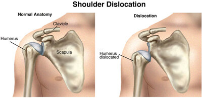
The shoulder is a ball-and-socket joint made up of 3 bones: the upper-arm bone (humerus), the shoulder blade (scapula), and the collar bone (clavicle). The ball at the top of the upper-arm bone is called the head of the humerus. The socket on the shoulder blade is called the glenoid fossa. A tendon is a fibrous bundle that attaches a muscle to a bone. The muscles and tendons of the rotator cuff hold the ball into the socket of the shoulder. The biceps muscle has 2 tendons that attach it to the shoulder and travel the length of the upper arm and insert just below the elbow. The biceps muscle is responsible for bending (flexing) the elbow and rotating the forearm. One of the tendons is called the "long head" of the biceps muscle; it attaches to the upper-arm bone. The second area of attachment is called the "short head" of the biceps; it attaches the muscle to a bony bump on the shoulder blade called the coracoid process.

Most commonly, the biceps tendon will tear at the long head of the biceps at the upper-arm bone, leaving the second attachment at the shoulder blade intact. The arm can still be used after this type of rupture, yet weakness will be present in the shoulder and upper arm. A tear can either be partial, when part of the tendon remains intact and only a portion is torn away from the bone, or complete, where the entire tendon is torn away from the bone.

How Does It Feel?

After sustaining a biceps tendon rupture, you may experience:

  • Sharp pain in the upper arm or elbow

  • Hearing a "pop" or snap at the shoulder or elbow

  • Bruising and swelling in the upper arm to elbow

  • Weakness in the arm when bending the elbow, rotating the forearm, or lifting the arm overhead

  • Tenderness in the shoulder or elbow

  • Muscle spasms in the shoulder and arm

  • A bulge or deformity in the lower part of the upper arm (a "Popeye arm")

How Is It Diagnosed?

In most cases, a thorough history and physical examination of the involved arm can diagnose a biceps tendon rupture. Your physical therapist will ask you several questions regarding your medical history, your regular daily tasks at home and at work, and your recreational or sports activities. Your physical therapist will ask how the injury happened and where you are having pain and/or weakness.

Your physical therapist will examine your entire upper arm for bruising or swelling, and gently touch it to determine if there is any tenderness over the biceps region at the shoulder, upper arm, or elbow. Your physical therapist also will examine the amount of motion and strength present on the involved side in the shoulder, forearm, and elbow, compared to the noninvolved side. Functional testing may also be performed to determine what daily tasks are difficult for you to perform (eg, lifting an object, reaching overhead, reaching behind the body, or rotating the forearm to open a door).


How Can a Physical Therapist Help?

A biceps tendon rupture often is treated without surgery. Your physical therapist will design an individualized treatment program to help heal your injury in the safest and most efficient way possible. Treatment may include:

Rest. You will be instructed in ways that allows the limb to rest to promote healing.

Icing. Your physical therapist will show you how to apply ice to the affected area to manage pain and swelling.

Range-of-Motion Activities. Your physical therapist will teach you gentle mobility exercises for the shoulder, elbow, and forearm, so your arm does not get stiff during the healing process.

Strengthening Exercises. As the pain and swelling ease, gentle strengthening exercises with resistant bands or light weights will be added.

Functional Activities. You will learn exercises to help you return to the activities you performed before the injury.

Education. Your physical therapist will teach you how to protect your joints from further injury. You will learn how to properly lift objects once the arm is healed, and how to avoid lifting objects that are simply too heavy.

Can This Injury or Condition Be Prevented?

To prevent a biceps tendon rupture, individuals should:

  • Maintain proper strength in the shoulder, elbow, and forearm.

  • Avoid repetitive overhead lifting and general overuse of the shoulder, such as performing forceful pushing or pulling activities, or lifting objects that are simply too heavy. Lifting more than 150 pounds can be dangerous for older adults.

  • Use special care when performing activities, such as lowering a heavy item to the ground.

  • Avoid smoking; it introduces carbon monoxide into the body and leaves less oxygen for the muscles to grow and heal.

  • Avoid steroid use, as it weakens muscles and tendons.

What Kind of Physical Therapist Do I Need?

All physical therapists are prepared through education and experience to treat biceps tendon ruptures. However, you may want to consider:

  • A physical therapist who is experienced in treating people with shoulder and elbow conditions or injuries. Some physical therapists have a practice with an orthopedic or sports medicine focus.

  • A physical therapist who is a board-certified clinical specialist or who completed a residency or fellowship in orthopedic physical therapy. This therapist has advanced knowledge, experience, and skills that may apply to your condition.

You can find physical therapists who have these and other credentials by using Find a PT, the online tool built by the American Physical Therapy Association to help you search for physical therapists with specific clinical expertise in your geographic area.

General tips when you're looking for a physical therapist (or any other health care provider):

  • Get recommendations from family, friends, or other health care providers.

  • When you contact a physical therapy clinic for an appointment, ask about the physical therapists' experience in helping people with a biceps tendon rupture.

  • Be prepared to describe your symptoms in as much detail as possible, and say what makes your symptoms worse

Athlete Assessments and Injury Prevention

At Pro Dynamic PT we’ve seen many sports injuries over the years and would like to help prevent them. Many individuals are unaware of weaknesses or techniques that place them at a higher risk for injury. Our assessments will seek those out and provide information that we’ll use to customize a program going forward to minimize injury risk. Our therapists are all athletes and understand what it takes to be successful in sport. This is open to adults as well.

3 Steps For Returning To Physical Activity After COVID-19

The following tips are designed to help people return to fitness after a typical case of COVID-19. Around 10% of people infected with COVID-19 will have problems that linger for months after the infection is gone. These individuals are called long-haulers, and the condition is known as "long COVID" or PASC, which stands for Post-Acute Sequelae of SARS-CoV-2. If you have symptoms of long COVID, contact your primary care doctor before starting an exercise program. Long COVID may include other health complications that require labs, tests, or imaging, before being referred to a physical therapist for an evaluation and treatment specific to your condition. Note: Exercise may not be appropriate for everyone living with long COVID.

A mild to moderate bout with COVID-19 can leave you feeling weak, with a loss of balance and coordination, a lack of endurance, and sometimes problems with memory. Physical activity can help you recover.

Exercise may be the last thing on your mind, but it is key to regaining your fitness. Regular physical activity benefits your physical, mental, and social health. It's important for COVID-19 survivors (after their initial recovery) to get moving. Physical activity helps to improve:

  • Strength.

  • Endurance.

  • Breathing capacity.

According to an article in BMJ, it's important to return to exercise after at least seven days free of COVID-19 symptoms, and to begin with at least two weeks of minimal exertion.

Listen to your body (and your doctor or physical therapist) for when it's safe to return to exercise. Then, take things slowly and follow this advice for returning to physical activity after a typical case of COVID-19.

1. Just Move, Even a Little

Your body has been through a lot. Take things slowly. For some, a trip from the bed or couch to the bathroom may be as much as you can handle in the early days. A flight of stairs may make you want to plop on the nearest easy chair. Get up and move as many times throughout the day as you can, even if it's just to stand from sitting several times in a row. Stretch for the sky with both arms and take several deep breaths each time you rise. Doing this light movement several times a day will help you start to build back strength. 

2. Take a Walk

If a little movement is not too challenging, try taking a brief walk. Begin at first by walking down the hall several times or around your house or apartment building. If that feels good, try a five-, 10-, or 15-minute walk around your neighborhood.

At this stage in your recovery, your intensity should be very light to light. At a light intensity, you should be able to easily carry on a conversation. If your intensity causes you to gasp for breath, you are pushing yourself too hard. The CDC provides a helpful description of Borg's Rating of Perceived Exertion to help you measure your intensity.

If you're a regular fitness fanatic and light intensity sounds too easy, be careful not to overdo it. It is important to allow your body time to get back to doing activities at your pre-COVID-19 pace. Gradually increase the intensity and length of your walks. With each day and each week, you'll be preparing your body to return to the full demands of a vigorous workout.

3. Ready To Run

If you tolerate walking, you may be ready to begin jogging, swimming, biking, or other activities. First, start your chosen activity at a slow pace for 10 minutes. Then, increase your pace for one minute before returning to the slower pace for another five to 10 minutes. Then repeat. When you're able to do these intervals for 30 minutes or more, you're ready to progress. Safely ease back into physical activity by slowly increasing the amount of intense exercise each day or week.

At this phase of recovery you may be ready for a higher intensity level. Aim for moderate intensity in which the exercise is somewhat hard, but not too hard. You should be breathing faster and deeper, but still be able to speak a full sentence and not be gasping for breath.

Everyone, regardless of age, condition, or ability should try to get the amount of daily physical activity recommended by the Department of Health and Human Services.

If you struggle with lingering side effects from COVID-19 and have trouble doing even minor physical activity, contact your doctor or a physical therapist experienced in treating COVID long-haulers. They can work with you on pacing, conserving energy, and addressing breathing pattern disorders to help you reach your goals.

Physical therapists are movement experts. They improve quality of life through hands-on care, patient education, and prescribed movement.

Physical Therapy Guide to Turf Toe

Turf toe is the common name for a sprain of the metatarsophalangeal, or MTP, joint. The MTP joint is located where the big toe meets the foot. This injury occurs when the big toe is forced back toward the top of the foot past its normal range of motion. It is more common in athletes, especially those who play football and similar sports. It can occur when an athlete pushes off to sprint or is tackled from behind. The front of the foot gets fixed and jammed into the ground, forcing the big toe to bend too far backward. In most cases, a turf toe injury does not require surgery. Physical therapy is effective for managing turf toe.


What Is Turf Toe?

There are two joints in the big toe. These joints allow the toe to flex downward and extend upward. The big toe plays a major role in the ability to walk and run. When the foot touches the ground and prepares to take another step, the big toe is the last joint through which the foot pushes off the ground to move the body forward. The primary joint involved in this motion is the MTP joint. This joint is where the metatarsal (the first long, straight bone of the foot) attaches to the phalange (the first shorter bone of the toe).

If the big toe is forced into a very unnatural position, the MTP joint can be injured, along with any surrounding structures such as:

  • Ligaments.

  • Muscle tendons.

  • Small bones that sit under the big toe, called the sesamoid bones.


All of these structures help to maintain the integrity and function of the MTP joint. When described together, they are called the plantar complex. Sometimes, one of the soft-tissue structures is simply stretched when the toe is bent back toward the top of the foot. However, a turf toe injury may result in one of the following:

  • Subluxation (where one bone of the joint slips out of place but comes back to its normal position).

  • Dislocation (where the two bones of the joint are completely separated).


How Does It Feel?

The most common symptoms of a turf toe injury are:

  • Localized pain at the first MTP joint.

  • Feeling a "pop" at or around the MTP joint at the time of the injury.

  • Swelling.

  • Bruising.

  • Tenderness to touch.

  • Cramping in the arch of the foot.

  • Pain with weight-bearing, especially if trying to rise up onto the toes.

  • A dislocation, in more severe injuries.


How Is It Diagnosed?

Health care providers classify turf toe injuries into one of three grades to describe the severity of the injury and guide treatment:

  • Grade 1. Stretching of the plantar complex.

  • Grade 2. Partial tearing of the plantar complex.

  • Grade 3. Complete tearing of the plantar complex.


Diagnosing a turf toe injury starts with an interview to learn about how your injury occurred and your symptoms. Your physical therapist also will perform a gentle physical examination to:

  • Assess the toe’s movement and muscle function

  • Note any swelling or tenderness in the area.

  • Analyze your gait pattern (how you walk, if you can).

  • Determine if you should see an orthopedic doctor for imaging (X-ray, MRI), splinting, or for casting your foot to restrict movement. A doctor may recommend surgery in severe cases.


How Can a Physical Therapist Help?

Immediately after a turf toe injury, the following approaches can help ease pain and prevent further injury. You can easily remember these using the acronym “PEACE”:

  • Protect: Limit movement and use pain as a guide to avoid causing discomfort.

  • Elevate: Put your feet up (above heart level if possible).

  • Avoid anti-inflammatories: Inflammation is the first stage of the body’s natural healing process. You don’t want to disrupt or delay your recovery.

  • Compress: Pressure on the toe/foot (such as when using a compression sock) may help limit swelling. Too much compression may restrict needed blood flow. Your physical therapist will choose the right amount of compression to treat your specific injury.

  • Educate: Your physical therapist will educate you about the injury and instruct you on an active approach to recovery and your options for treatment. They also can determine when it is safe for you to return to activity.


After a few days of “PEACE,” physical therapists recommend the following steps. (You can remember them using the acronym “LOVE”):

  • Loading: Your body needs a certain amount of stress to stimulate repair and recovery. Your physical therapist will work with you to determine how much weight (such as standing or walking) you can put on your toe, and decide when the time is right to do so.

  • Optimism: Stay positive, even though you’re injured.

  • Vascularization (improving blood flow): Cardiovascular (aerobic) exercise that does not put too much stress on your injured toe joint will help you:

    • Reduce pain.

    • Improve blood flow to the injured area.

    • Stay motivated through your recovery.

  • Exercise: Use pain as your guide for a gradual return to normal activity. Your physical therapist will design a treatment plan with specific and targeted exercises for your condition.


Most turf toe injuries do not require surgery. They can be managed by working with your physical therapist. Your treatment plan will depend on the severity of your injury and your goals. In all cases, the main goal of treatment is to restore your ability to return to normal activity.

The following are typical treatment options, depending on the grade of your injury:

  • Grade 1. Taping or inserts may be used to restrict painful motion at first. In many cases, an athlete may return to sports within a few weeks. Often, your physical therapist will have you do strength and weight-bearing exercises almost immediately.

  • Grade 2. A brace or walking boot may be prescribed for several weeks to restrict movement and allow rest. Your physical therapist will then start you on a structured exercise program and a gradual return to activity.

  • Grade 3. Surgery may be needed for a grade 3 injury. Your health care team will determine whether you need surgery based on the severity of the damage and its impact on your function. Surgery is more likely if there is:

    • Fracture of a bone.

    • Damage to the cartilage (the tissue that lines the bones of the joints).

    • Complete tearing of the tendon.

    • Excessive movement of the joint that causes ongoing instability (subluxation or dislocation).


With any grade of injury, your physical therapist will work with you to design a treatment program specific to your condition and goals. Your treatment plan may include:

Range-of-motion exercises. It is important to regain the full range of motion of your big toe and foot. If your injury required use of a brace or boot to restrict movement during healing, your toe and foot joints may be stiff. Your physical therapist will teach you gentle stretching and movement exercises, including guided toe exercises, to help restore normal movement.

Muscle strengthening. It is common to lose strength in the muscles of your foot, ankle, and leg after a turf toe injury. This is due to the change in activity and any bracing or boot used to restrict movement during healing. Your physical therapist will determine which muscles are weak and teach you specific exercises to strengthen them. Exercises may include:

  • Balance activities.

  • Climbing stairs.

  • Using resistance bands.


Manual therapy. Many physical therapists use manual (hands-on) therapy to gently move and treat muscles and joints to improve their function. These techniques can target areas that are difficult to treat on your own. Manual therapy can be especially effective to restore movement in joints that become stiff after being immobilized. Your physical therapist may gently move the joints involving your injury for you. This might feel like your foot is being gently “wiggled.”

Patient education. Your physical therapist will educate you to help ensure that your recovery goes smoothly. They will identify any activities you should avoid or limit at certain stages in your recovery. They also can help you understand how long it may take until you can return to full activity.


Can This Injury or Condition Be Prevented?

Certain external factors may increase the risk of a turf toe injury. These factors can include:

  • Competing on artificial turf surfaces.

  • Wearing shoes with highly flexible soles.


It is important to ensure that your footwear properly supports your foot and is the right type for the surface on which you play your sport. Also, performing regular flexibility and strengthening activities for the foot and ankle may improve your body's ability to withstand athletic activities. Your physical therapist can teach you these exercises and how often to do them.


What Kind of Physical Therapist Do I Need?

All physical therapists are prepared through education and clinical experience to treat a variety of conditions or injuries. You may want to consider:

  • A physical therapist who is experienced in treating people with orthopedic or musculoskeletal (muscle, bone, and joint) injuries.

  • A physical therapist who is a board-certified specialist or who has completed a residency in orthopedic and/or sports physical therapy. This physical therapist will have advanced knowledge, experience, and skills that apply to athletes and turf toe injuries.


You can find physical therapists who have these and other credentials by using Find a PT, the online tool built by the American Physical Therapy Association. This tool will help you search for physical therapists with specific clinical expertise in your geographic area.

Here are some general tips for when you are looking for a physical therapist (or any other health care provider):

  • Get recommendations from family, friends, or other health care providers.

  • When you contact a physical therapy clinic for an appointment, ask about the physical therapist’s experience in helping people with turf toe injury.

  • During your first visit with the physical therapist, be prepared to describe your symptoms in as much detail as possible, and report activities that make your symptoms worse and better.

Further Reading

The American Physical Therapy Association believes that consumers should have access to information to help them make informed health care decisions and prepare them for their visit with a health care provider.

The following resources offer some of the best scientific evidence related to physical therapy treatment for turf toe. They report recent research and give an overview of the standards of practice both in the United States and internationally. They link to a PubMed* abstract, which also may offer free access to the full text or to other resources. You can read them or print out a copy to bring with you to your health care provider.

Dubois B, Esculier JF. Soft-tissue injuries simply need PEACE and LOVE. Br J Sports Med. 2020;54(2):72–73. Article Summary in PubMed.

Fraser TW, Doty JF. Turf toe: review of the literature and surgical technique. Ann Jt. 2019;12(4). doi: 10.21037/aoj/.2019.05.03

Najefi AA, Jeyaseelan L, Welck M. Turf toe: a clinical update. EFORT Open Rev. 2018;3:501–506. Article Summary in PubMed .

George E, Harris AH, Dragoo JL, Hunt KJ. Incidence and risk factors for turf toe injuries in intercollegiate football: data from the national collegiate athletic association injury surveillance system. Foot Ankle Int. 2014;35(2):108–115. Article Summary in PubMed .

Anandan N, Williams PR, Dalavaye SK. Turf toe injury. Emerg Med J. 2013;30(9):776–777. Article Summary in PubMed .

McCormick JJ, Anderson RB. Turf toe: anatomy, diagnosis, and treatment. Sports Health. 2010;2(6):487–494. Article Summary in PubMed .

* PubMed is a free online resource developed by the National Center for Biotechnology Information. PubMed contains millions of citations to biomedical literature, including citations in the National Library of Medicine’s MEDLINE database.


Revised in 2021 by Megan James, PT DPT, and reviewed by James E. Zackazewski, board-certified clinical specialist in sports physical therapy, on behalf of the American Academy of Sports Physical Therapy. Authored in 2014 by Laura Stanley, PT, DPT, board-certified clinical specialist in sports physical therapy.


Physical Therapists Help You Overcome Barriers to Physical Activity

Enjoying Physical Activity_GettyImages-200464866-001.jpg

According to the Department of Health and Human Services' Facts & Statistics on physical activity, more than 80% of American adults do not get enough physical activity despite the proven benefits, such as a reduced risk of some cancers and chronic diseases, as well as improved bone health, cognitive function, weight control, and quality of life. As a result, half of adults — 117 million people — have one or more chronic diseases. The good news is that regular physical activity can help prevent and improve many chronic conditions.

Barriers to movement and physical activity can be small or large, real or perceived. Whatever barriers may be preventing you from enjoying the many important health benefits of physical activity, physical therapists will partner with you to create a safe and effective program to get you moving.

Physical therapists are movement experts who improve quality of life through hands-on care, patient education, and prescribed movement. Using the latest evidence, physical therapists design physical activity plans for people of all ages and abilities specific to your needs, challenges, and goals.

Physical therapists and physical therapist assistants work together and collaborate with other members of your health care team to maximize your movement and empower you to be an active participant in your care.

You can contact a physical therapist directly for an evaluation.

11 Barriers to Physical Activity, and How to Overcome Them

1. It's too late to start, I'm too old, or I've been physically inactive for a long time.

It's never too late to get moving. According to a recent JAMA Network Open study, adding physical activity at any age has benefits. In addition to an increased life span, adding the recommended amount of physical activity for your age and ability to your daily routine can help you manage stress, improve memory and brain function, avoid chronic disease, and much more.

2. It hurts when I …

Movement is crucial to a person's health, quality of life, and independence. For some people, pain makes movement a challenge. Pain is one of the most common reasons people seek health care. A physical therapist can help you move better and safely manage your pain.

3. I don't have time.

Some physical activity is better than nothing. Try to fit in a few short bursts of physical activity a few times a day for a total of 30 minutes. Make sure that the activity increases your heart rate.

Parents should make physical activity part of their family's daily routine to establish a lifelong commitment to health for their children. Play an outdoor game like hopscotch or tag with the kids (playing is for adults too!). If you're a caretaker, maintaining your health is vital to being there for those you love. Determine when it makes the most sense to fit small amounts of physical activity into your daily routine. If possible, include movement as part of the care you provide your loved ones. They need to move, too, and you'll both benefit.

4. I don't have access to a gym or equipment.

You don't need a gym membership or fancy equipment to enjoy the benefits of physical activity. You can get plenty in and around your home. Dancing, jogging, walking, climbing stairs, and gardening are all examples of physical activity that you can do without any equipment. To improve your balance, flexibility, and strength, try doing body-weight exercises at least two days a week. Use household objects, e.g., cans, milk jugs, to strengthen muscles.

Try one of these physical therapist- and physical therapist assistant-led home exercise videos.

5. I don't like to exercise.

Competitive sports and hour-long fitness classes are not for everyone. Physical activity doesn't have to involve things you don't like doing to be effective. Discover the types of activities that you enjoy and make them part of your daily physical activity routine.

6. I can't get motivated, it's too big of a hurdle, or I don't know where to begin.

Physical activity releases endorphins, and the feeling of well-being you get after a good workout will become its own reward. To help you get started, offer yourself a small reward each time you are physically active until it becomes a habit. Perhaps looking forward to a special reward will help you reach the recommended physical activity guidelines for your age and ability. Resolve not to watch your favorite TV show unless you have met your daily movement goal. Break down long-term goals into small goals and work on achieving them one at a time. Keep a journal of how you feel after you've been physically active and each time you reach a goal.

Set yourself up for success with these tips:

  • Make it convenient (walking shoes, hand weights, or resistance bands within easy reach of your desk or where you spend the most time).

  • Schedule time for a daily physical activity break and set a calendar reminder.

  • Track your steps daily. Increase your step count goal each week.

7. I have a chronic disease, condition, or disability.

Movement is essential for everyone. Whether you use a wheelchair or other assistive device to get around or have mobility challenges due to a chronic condition or a prior injury, there are activities that you can do to challenge your muscles and lungs and improve your health and quality of life. Physical activity can even improve some chronic conditions and prevent others.

8. I'm afraid of hurting myself.

The right activity for you depends on your age, ability, and goals. A physical therapist can help you identify a safe and effective physical activity plan for your age and ability that addresses your fears and helps you reach your goals.

9. I feel out of breath when I move/walk/exercise even a little bit.

It is normal to feel a bit winded when doing physical activities in which you exert yourself more than usual.

If you worry for any reason that physical activity will be unsafe, contact a physical therapist before you begin. After an evaluation, a physical therapist can work with you to find the right duration and type of physical activity to improve your stamina and overall health.

10. I'm tired all the time; I have no energy to exercise.

Research shows that exercise boosts energy levels. Physical activity helps deliver oxygen and nutrients to our tissues and vascular system, and other body functions work more efficiently. Physical activity also improves brain function and mental health, lowers anxiety, promotes better sleep, and aids in weight management. All of these enhance our energy and lead to feelings of well-being.

11. I work out all the time but can't reach my goal.

Finding the right plan for you is essential to your success. If you are having trouble meeting strength and conditioning goals, despite your best effort, a physical therapist can work with you to identify any issues and design a program to maximize your movement and enhance performance.

Physical Therapy Guide to Acromioclavicular Joint Injuries

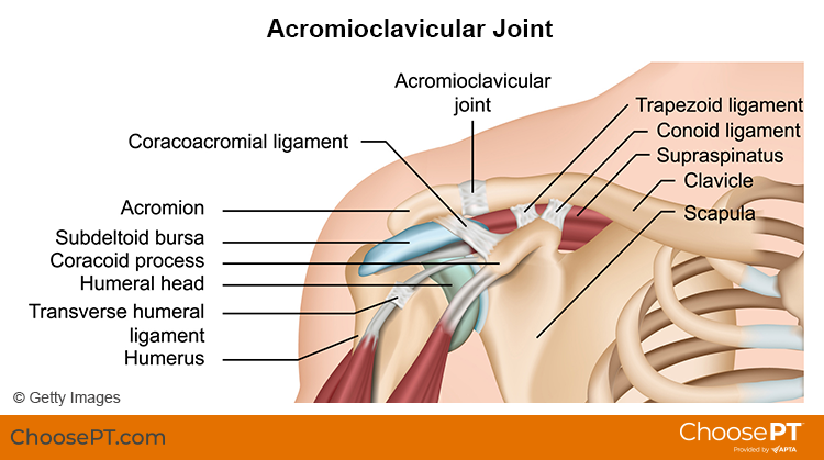
The acromioclavicular, or AC, joint is part of the shoulder girdle (the collar bone and shoulder blade that support the shoulder joint). An AC joint injury describes an injury to the top of the shoulder. It occurs where the front of the shoulder blade (acromion) attaches to the collarbone (clavicle). Most often, trauma, such as a fall directly on the outside of the shoulder, causes an AC joint injury. Overuse (repeated lifting of heavy weights or objects overhead with poor mechanics) also can result in an AC joint injury.

AC joint injuries are most common in people younger than age 35. Males sustain five times more traumatic AC joint injuries than females. Younger athletes who take part in activities like football, biking, skiing, and hockey have the highest risk for this injury.

Physical therapists can identify and effectively treat AC joint injuries, often avoiding the need for surgery.


What Are Acromicioclavicular (AC) Joint Injuries?

Acromioclavicular Joint Injury_ChoosePT_frame.png

There are four ligaments that hold the two bones of the AC joint together. When an AC joint injury occurs, these ligaments are stressed. This stress results in some degree of joint separation. There are two types of injuries of the AC joint: traumatic and overuse injuries.

Traumatic AC joint injury. This type of injury occurs when the joint is disrupted. The ligaments that hold the two bones of the joint together get stretched too far. This is called a shoulder separation. It is different from a shoulder dislocation, which involves the ball-and-socket joint.

Traumatic AC joint injuries are most common in people who fall and land on the outside of the shoulder or hand. Examples include a:

  • Football player who is tackled.

  • Cyclist who crashes.

  • Worker who falls off a ladder.

Traumatic AC joint injuries can range from a mild to severe grade. Grading is based on the amount of joint separation involved. Mild cases can be treated by a physical therapist. More severe cases may require surgery followed by physical therapy.

Overuse AC joint injury. This type of injury occurs over time due to repeated and too much stress on the joint. Cartilage at the end of the acromion and clavicle bones protects the joint from daily wear and tear. Over time, the demand placed on this cartilage may be more than it can endure. The result is an overuse injury that can lead to major wearing of the cartilage and arthritis. Overuse AC joint injury is most common in people who do repeated tasks. Examples include:

  • Heavy weightlifting (bench and military presses).

  • Jobs that require physical work with the arms stretched over the head.

How Does It Feel?

With an AC joint injury, you may experience:

  • General shoulder pain and swelling.

  • Swelling and tenderness over the AC joint.

  • Loss of shoulder strength.

  • A visible bump above the shoulder.

  • Pain when lying on the involved side.

  • Loss of shoulder motion.

  • A “popping” sound or feeling that your shoulder “catches” with movement.

  • Discomfort with daily activities that stress the AC joint. Examples include lifting objects overhead, reaching across your body, or carrying heavy objects at your side.

How Is It Diagnosed?

Physical therapists can diagnose an AC joint injury through a shoulder exam. Your physical therapist will conduct a full evaluation to find out the degree of your injury and identify all the factors that may contribute to it.

They will begin by interviewing you to learn about your health history. They may be helped by forms you fill out before your first session. The interview will become more specific to the condition of your shoulder. Your physical therapist may ask you questions such as:

  • How did your injury occur?

  • How have you taken care of the condition, such as seeing other health care providers? Have you had imaging (X-ray, MRI) or other tests and received their results?

  • What are your current symptoms? How have they changed your typical day and activities?

  • Do you have pain, and if so, what is the location and intensity of your pain? Does the pain vary during the day?

  • Do you have trouble doing any activities? What activities are you unable to do?

This information allows the physical therapist to better understand what you are going through. It also helps determine the course of your physical exam.

The physical exam will vary depending on your interview. Most often it will begin with observing the region of your symptoms and any movements or positions that cause pain. Your physical therapist also may examine other areas of your body that may have changed due to problems with your shoulder function. They may:

  • Watch you move your arm and shoulder overhead, and while doing other reaching tasks.

  • Assess the mobility and strength of your shoulder.

  • Check other regions of the body as needed. This will help to determine if other areas also require treatment to improve your condition.

  • Gently, but skillfully, feel around your shoulder and the AC joint to find exactly where it is most painful.

Your physical therapist will discuss their findings with you. They will work with you to develop a program for your specific needs and goals and to help you heal.

In some cases, your physical therapist may refer you for diagnostic imaging. Ultrasound, X-ray, or MRI can help to confirm the diagnosis and find out how severe the injury is.

How Can a Physical Therapist Help?

Your physical therapist will design a treatment plan to help you safely return to your desired activities. Your treatment plan may include:

  • Patient education. Your physical therapist will educate you about your AC joint and shoulder injury. They will work with you to identify any external factors causing your pain, including the amount and type of exercises and activities you do. Your physical therapist will recommend improvements to your activities.

  • Pain management. Your physical therapist will address your pain. This may include applying ice to the affected area and other methods. They also may recommend changing some activities that cause pain. Physical therapists are experts in prescribing pain-management methods. These can reduce or eliminate the need for medicines, including opioids.

  • Range-of-motion exercise. The mobility of the AC joint and shoulder may be limited, causing increased stress on the shoulder. Your physical therapist may teach you self-stretching methods. These can decrease tension and help restore normal motion of your injured joints.

  • Manual therapy. Your physical therapist may apply hands-on treatments to gently move your muscles and joints. These techniques help to improve movement. Your physical therapist also may use manual therapy to guide your shoulder area into a less stressful movement pattern.

  • Muscle strength. Muscle weaknesses or imbalances can contribute to problems of the AC joint and shoulder. They also can cause continued symptoms. Based on the how serious your injury is, your physical therapist will design a safe resistance program to aid your recovery. Exercises may include using resistance machines in the clinic and doing exercises to strengthen your core (midsection). You may begin doing exercises while lying on a table or at home on the bed or floor. You then may advance to exercises done in a standing position. Your physical therapist will choose what exercises are right for you based on your diagnosis, age, and condition. They will determine when it is safe for you to exercise on your own at home or in a gym.

  • Functional training. Once your pain, strength, and motion improve, functional training can help you safely resume more demanding activities. To minimize the stress to the AC joint and shoulder it is important to teach your body safe, controlled movements. Your physical therapist will create a series of activities to help you learn how to use and move your body correctly and safely. These may include retraining your movements and positioning when throwing, swinging a racket, lifting objects overhead, or doing other daily activities.

Can This Injury or Condition Be Prevented?

Accidents happen and it can be difficult to prevent many traumatic AC joint injuries. However, much can be done to prevent the string of events that lead to overuse injuries. Physical therapists can help reduce overuse injuries by:

  • Teaching you how to properly lift objects overhead at work.

  • Demonstrating good form for overhead resistance training or sports activities.

  • Helping you maintain general shoulder strength and motion to safely perform tasks.

Consult a physical therapist as soon as possible if you have persistent constant or worsening symptoms.

What Kind of Physical Therapist Do I Need?

All physical therapists are prepared through education and clinical experience to treat a variety of conditions or injuries. You may want to consider:

  • A physical therapist who is experienced in treating people with orthopedic or musculoskeletal (muscle, bone, and joint injuries).

  • A physical therapist who is a board-certified specialist or who has completed a residency in orthopedic or sports physical therapy. This physical therapist will have advanced knowledge, experience, and skills that apply to those who are physically active.

You can find physical therapists who have these and other credentials by using Find a PT, the online tool built by the American Physical Therapy Association. This tool can help you search for physical therapists with specific clinical expertise in your area.

General tips when you're looking for a physical therapist (or any other health care provider):

  • Get recommendations from family, friends, or other health care providers.

  • When you contact a physical therapy clinic for an appointment, ask about the physical therapists' experience in helping patients with shoulder pain.

  • During your first visit with the physical therapist, be prepared to describe your symptoms in as much detail as possible, and report activities and movements that make your symptoms worse.

Further Reading

The American Physical Therapy Association believes that consumers should have access to information to help them make informed health care decisions and prepare them for their visit with a health care provider.

The following resources offer some of the best scientific evidence related to physical therapy treatment for AC joint injuries. They report recent research and give an overview of the standards of practice both in the United States and internationally. They link to a PubMed* abstract that also may offer free access to the full text, or to other resources. You can read them or print out a copy to bring with you to your health care provider.

Frank RM, Cotter EJ, Leroux TS, Romeo AA. Acromioclavicular joint injuries: evidence-based treatment. J Am Acad Orthop Surg. 2019;27(17):e775–e788. Article Summary in PubMed.

Li X, Ma R, Bedi A, Dines DM, Altchek DW, Dines JS. Management of acromioclavicular joint injuries. J Bone Joint Surg Am. 2014;96(1):73–84. Article Summary in PubMed .

Harris KD, Deyle GD, Gill NW, Howes RR. Manual physical therapy for injection-confirmed nonacute acromioclavicular joint pain. J Orthop Sports Phys Ther. 2012;42(2):66–80. Article Summary in PubMed.

Pallis M, Cameron KL, Svoboda SJ, Owens BD. Epidemiology of acromioclavicular joint injury in young athletes. Am J Sports Med. 2012;40(9):2072–2077. Article Summary in PubMed .

*PubMed is a free online resource developed by the National Center for Biotechnology Information. PubMed contains millions of citations to biomedical literature, including citations in the National Library of Medicine's MEDLINE database.


Reviewed and revised in 2021 by Erin Hayden, PT, DPT, board-certified clinical specialist in orthopedic physical therapy, and Stephen Reischl, PT, DPT, board-certified clinical specialist in orthopedic physical therapy, on behalf of the Academy of Orthopaedic Physical Therapy. Authored in 2014 by Allison Mumbleau, PT, DPT, board-certified clinical specialist in sports physical therapy.


Physical Therapy Guide to Compartment Syndrome

What is Compartment Syndrome?

Our limbs (arms and legs) are divided into compartments that contain different muscles, nerves, and blood vessels. Each compartment is separated by fascia, a thick sheet-like tissue that does not stretch.

Our bodies are able to handle small changes in the pressure levels within these compartments. For example, our tissues may swell slightly after a hard workout or a mild injury. However, when there is excessive swelling within a compartment due to a severe acute injury or chronic overuse, pressure builds within that compartment as the fascia does not expand to accommodate the increased volume. In rare circumstances, this condition can be more than our bodies can handle, and the blood supply to the area is restricted. If the condition persists, the muscle and nerve tissue can be harmed. It is essential to relieve the pressure immediately to avoid permanent damage.

Compartment syndrome is typically classified into 2 categories—acute or chronic—based on its cause and symptoms.

Unknown.jpeg

Acute Compartment Syndrome

Acute compartment syndrome (ACS) is a medical emergency. It can develop as early as several hours following a severe injury. If left untreated for even a few hours, irreversible tissue damage can occur. ACS most often develops in the lower leg and forearm.

ACS is typically caused by a serious injury, such as:

  • A direct hit or blow to the limb (athletics, a significant fall)

  • Crush injuries (motor vehicle accident, work-site injury)

  • Highly restrictive bandages

How Does It Feel?

The most common signs and symptoms of ACS include:

  • Severe pain in the involved limb that may be out of proportion to the typical response to a certain injury

  • Changes in sensation (tingling, burning, numbness)

  • A sense that the limb is tight or full (from the swelling and increase in pressure)

  • Discoloration of the limb

  • Severe pain with stretching of the involved muscle

  • Severe pain when the involved area is touched

  • Significant pain or an inability to bear weight throughout the involved limb

How Is It Diagnosed?

It is critical that ACS is identified and treated immediately. Following a severe injury, if an individual is showing signs of ACS, the individual should be taken to the emergency room right away for evaluation by a physician. The physician will be able to objectively measure the levels of pressure in the involved compartment. If necessary, surgery will be performed to alleviate pressure in the compartment using a procedure called a fasciotomy. During surgery, an incision is made through the skin and fascia to drain the swelling and relieve the pressure within the compartment. A patient undergoing a fasciotomy will have to spend a period of time in the hospital to ensure that the pressure normalizes and the wound heals properly. Following a fasciotomy, physical therapy is necessary to restore the motion, strength, and function of the limb.

Chronic Compartment Syndrome

Chronic compartment syndrome (CCS) is often referred to as “exertional” compartment syndrome, and is typically caused by exercise that involves repetitive movements, such as walking, running, biking, or jumping. Usually, excessive exercise causes the tissues of the leg to be overworked without time to recover. The development of CCS may be influenced by external factors, such as poor body control during movement, poor footwear, uneven or too-firm training surfaces, or too much training. There have also been cases where excessive steroid use has been linked to CCS.

How Does It Feel?

The symptoms for CCS may be similar to that of ACS, but less severe and not a result of an acute traumatic injury. These may include:

  • Pain and cramping in the involved limb that usually worsens with activity and subsides with rest

  • Mild swelling

  • Pain with stretching

  • Numbness or tingling in the limb

  • Weakness

How Is It Diagnosed?

Because the symptoms of CCS are similar to many other conditions, it is important that a physician or physical therapist rules out other possible diagnoses, such as tendinitis, stress fractures, shin splints, or other inflammatory conditions. The examination may include the use of diagnostic imaging, such as an ultrasound, x-ray, or MRI to assess the tissues in the painful area.

If CCS is suspected, an individual will likely be referred to a physician for a specific test called the "compartment pressure measurement." This test is only used in cases where CCS is strongly suspected. It is performed in a medical office. During the test, the pressure in the involved compartment is measured before, during, and after exercise. The goal of the test is to reproduce symptoms as they occur during real-life activities. If CCS is diagnosed, your medical team will devise a plan to best treat your specific condition. For more mild cases of CCS, you will likely be referred directly to physical therapy. In more severe cases, individuals are likely to be referred to a surgeon to discuss the option of a fasciotomy.


How Can a Physical Therapist Help?

If you are diagnosed with compartment syndrome, your physical therapist will play an important role in the treatment of the condition, whether it requires surgery or not. Your physical therapist will work with you to design an individualized treatment program based on your condition and your personal goals. Your physical therapist may recommend:

Range-of-Motion Exercises. Restrictions in the motion of your knee, foot, or ankle may be causing increased strain in the muscles housed within the compartments of your lower leg. Stretching techniques can be used to help restore motion in these joints to minimize undue muscle tension.

Muscle Strengthening. Hip and core weakness can influence how your lower body moves, and can cause imbalanced forces through the lower-leg muscle groups that may contribute to compartment syndrome. Building core strength (in the muscles of the abdomen, low back, and pelvis) is important; a strong midsection allows greater stability through the body as the arms and legs perform different motions. For athletes engaged in endurance sports, it is important to have a strong core to stabilize the hip and knee joints during repetitive leg motions. Your physical therapist will be able to determine which muscles are weak, and provide specific exercises to target these areas.

Manual Therapy. Many physical therapists are trained in manual (hands-on) therapy, using their hands to move and manipulate muscles and joints to improve motion and strength. These techniques can target areas that are difficult to treat on your own.

Modalities. Your physical therapist may use modalities ( e.g., ultrasound, iontophoresis, moist heat, cold therapy) as a part of your rehabilitation program. These tools can help improve tissue mobility and flexibility, and enhance recovery. Your physical therapist will discuss the purpose of each modality with you.

Education. Your treatment will include education about how to safely return to your previous activities, particularly if your condition required a fasciotomy. Your physical therapist may recommend:

  • Wearing more appropriate footwear

  • Choosing more appropriate surfaces and terrain for exercise

  • Pacing your activities

  • Avoiding certain activities altogether

  • Mastering strategies for recovery and maintenance of good health (e.g., allowing your muscles and joints proper rest time)

  • Modifying your workplace to lower risk of injury


How Can a Physical Therapist Help Before & After Surgery?

In the event that your case of compartment syndrome requires surgery (either due to an acute injury or chronic condition), postoperative physical therapy will be essential to a successful recovery. Your physical therapist will be in close communication with your surgeon regarding the nature of your procedure, expected timelines for healing, and your progress during rehabilitation. As a health care team, your providers will develop a plan to ensure your body has adequate time to heal, while incorporating strategies to restore your motion, mobility, strength, and function.


Real Life Experiences

Caleb is a 14-year-old baseball player. One hot summer day, he and his best friend Bobby decided to get in some batting practice at the ballpark down the street. Unfortunately, the batting cages were being replaced, so they decided to practice on the actual field. Caleb offered to pitch first, as he knew Bobby needed more work on his batting to get ready for fall tryouts.

A few hits into the second bucket of balls, Bobby nailed a pitch right back at Caleb. The baseball hit him very hard in the side of his calf. He fell to the ground and was in a great deal of pain. He tried to get up, but had a hard time putting weight on his injured leg. Bobby felt so bad, he carried Caleb home on his back. That afternoon, Caleb started to feel better and was able to limp around the house. However, his leg still hurt a lot, and after dinner, he noticed his lower leg was extremely swollen, tender to touch, and warm. Caleb said that his toes were tingling, and he was having a more difficult time walking because his leg felt heavy and weak. He showed his dad, who immediately recognized that this was no ordinary bruise and took Caleb to the emergency room.

Upon examination by the emergency room medical team, Caleb was diagnosed with acute compartment syndrome. His injury required a fasciotomy to release the compartment and allow the swelling to dissipate so the pressure would decrease. He had surgery that night, and spent several days recuperating in the hospital. Bobby brought him ice cream every day.

One week after he left the hospital, Caleb was referred to physical therapy. His lower leg had lost a lot of muscle mass, his skin was very tight and tender around his incision, and he was still nervous about bearing his full weight on the injured leg. Caleb knew he would miss his fall baseball season, but was hoping to try out for JV basketball that winter. After a comprehensive evaluation, his physical therapist developed a rehabilitation plan based on Caleb's goals, and drew up a timeline for reaching them.

For the next several months, Caleb and his physical therapist worked on restoring motion at his knee and ankle. She gently stretched the muscles of his lower leg, and progressively began incorporating strengthening exercises into Caleb's routine. She also designed a home-exercise program that Caleb followed diligently.

Once he was able to walk normally without pain, Caleb and his physical therapist started working on more advanced strengthening exercises, building up to running, jumping, and "cutting" activities. Toward the end of his rehabilitation, they performed basketball-specific drills. His physical therapist was in constant communication with his surgeon, parents, and coaches to make sure everyone was on the same page regarding his recovery.

Three months later, Caleb attended basketball tryouts and made the JV squad as the starting point guard! Luckily, Bobby made the team, too. Caleb and Bobby were thrilled to be back playing sports together—although Caleb often reminded Bobby that he owed him ice cream for the rest of his life.


What Kind of Physical Therapist Do I Need?

All physical therapists are prepared through education and clinical experience to treat a variety of conditions or injuries. You may want to consider:

  • A physical therapist who is experienced in treating people with orthopedic or musculoskeletal injuries.

  • A physical therapist who is a board-certified specialist or who has completed a residency in orthopedic or sports physical therapy, as they will have advanced knowledge, experience, and skills that apply to an athletic population.

You can find physical therapists that have these and other credentials by using Find a PT, the online tool built by the American Physical Therapy Association to help you search for physical therapists with specific clinical expertise in your geographic area.

General tips when you're looking for a physical therapist (or any other health care provider):

  • Get recommendations from family and friends or from other health care providers.

  • When you contact a physical therapy clinic for an appointment, ask about the physical therapists' experience in helping people with compartment syndrome.

  • During your first visit with the physical therapist, be prepared to describe your symptoms in as much detail as possible, and report activities that make your symptoms worse.


Further Reading

The American Physical Therapy Association (APTA) believes that consumers should have access to information that could help them make health care decisions and also prepare them for their visit with their health care provider.

The following articles provide some of the best scientific evidence related to physical therapy treatment of compartment syndrome. The articles report recent research and give an overview of the standards of practice both in the United States and internationally. The article titles are linked either to a PubMed* abstract of the article or to free full text, so that you can read it or print out a copy to bring with you to your health care provider.

Irion V, Magnussen RA, Miller TL, Kaeding CC. Return to activity following fasciotomy for chronic exertional compartment syndrome. Eur J Orthop Surg Traumatol. 2014 March 25. [E-pub ahead of print.] Article Summary in PubMed.

Davis DE, Raikin S, Garras DN, et al. Characteristics of patients with chronic exertional compartment syndrome. Foot Ankle Int. 2013;34(10):1349–1354. Article Summary in PubMed.

Gill CS, Halstead ME, Matava MJ. Chronic exertional compartment syndrome of the leg in athletes: evaluation and management. Phys Sportsmed. 2010;38(2): 126–132. Article Summary in PubMed.

McCaffrey DD, Clarke J, Bunn J, McCormack MJ. Acute compartment syndrome of the anterior thigh in the absence of fracture secondary to sporting trauma. J Trauma. 2009;66(4):1238–1242. Article Summary in PubMed.


* PubMed is a free online resource developed by the National Center for Biotechnology Information (NCBI). PubMed contains millions of citations to biomedical literature, including citations in the National Library of Medicine’s MEDLINE database.

Authored by Laura Stanley, PT, DPT, board-certified clinical specialist in sports physical therapy. Reviewed by the editorial board.


Physical Therapy Guide to Anterior Cruciate Ligament (ACL) Tear

ACLAttachment_Small.jpg

An anterior cruciate ligament (ACL) tear is an injury to the knee commonly affecting athletes, such as soccer players, basketball players, skiers, and gymnasts. Nonathletes can also experience an ACL tear due to injury or accident. Approximately 200,000 ACL injuries are diagnosed in the United States each year. It is estimated that there are 95,000 ruptures of the ACL and 100,000 ACL reconstructions performed per year in the United States. Approximately 70% of ACL tears in sports are the result of noncontact injuries, and 30% are the result of direct contact (player-to-player, player-to-object). Women are more likely than men to experience an ACL tear. Physical therapists are trained to help individuals with ACL tears reduce pain and swelling, regain strength and movement, and return to desired activities.

What is an ACL Tear?

The ACL is one of the major bands of tissue (ligaments) connecting the thigh bone (femur) to the shin bone (tibia) at the knee joint. It can tear if you:

  • Twist your knee while keeping your foot planted on the ground.

  • Stop suddenly while running.

  • Suddenly shift your weight from one leg to the other.

  • Jump and land on an extended (straightened) knee.

  • Stretch the knee farther than its usual range of movement.

  • Experience a direct hit to the knee.

ACL Attachment: See More Detail

How Does It Feel?

When you tear the ACL, you may feel a sharp, intense pain or hear a loud "pop" or snap. You might not be able to walk on the injured leg because you can’t support your weight through your knee joint. Usually, the knee will swell immediately (within minutes to a few hours), and you might feel that your knee "gives way" when you walk or put weight on it.

How Is It Diagnosed?

Immediately following an injury, you may be examined by a physical therapist, athletic trainer, or orthopedic surgeon. If you see your physical therapist first, your therapist will conduct a thorough evaluation that includes reviewing your health history. Your physical therapist will ask:

  • What you were doing when the injury occurred.

  • If you felt pain or heard a "pop" when the injury occurred.

  • If you experienced swelling around the knee in the first 2 to 3 hours following the injury.

  • If you felt your knee buckle or give out when you tried to get up from a chair, walk up or down stairs, or change direction while walking.

Your physical therapist may perform gentle "hands-on" tests to determine the likelihood that you have an ACL tear, and may use additional tests to assess possible damage to other parts of your knee.

An orthopedic surgeon may order further tests, including magnetic resonance imaging (MRI), to confirm the diagnosis and rule out other possible damage to the knee.

Surgery

Most people who sustain an ACL tear will undergo surgery to repair the tear; however, some people may avoid surgery by modifying their physical activity to relieve stress on the knee. A select group can actually return to vigorous physical activity following rehabilitation without having surgery.

Your physical therapist, together with your surgeon, can help you determine if nonoperative treatment (rehabilitation without surgery) is a reasonable option for you. If you elect to have surgery, your physical therapist will help you prepare both for surgery and to recover your strength and movement following surgery.


How Can a Physical Therapist Help?

Once an ACL tear has been diagnosed, you will work with your surgeon and physical therapist to decide if you should have surgery, or if you can recover without surgery. If you don’t have surgery, your physical therapist will work with you to restore your muscle strength, agility, and balance, so you can return to your regular activities. Your physical therapist may teach you ways to modify your physical activity in order to put less stress on your knee. If you decide to have surgery your physical therapist can help you before and after the procedure.

Treatment Without Surgery

Current research has identified a specific group of patients (called "copers") who have the potential for healing without surgery following an ACL tear. These patients have injured only the ACL, and have experienced no episodes of the knee "giving out" following the initial injury. If you fall into this category, based on the specific tests your physical therapist will conduct, your therapist will design an individualized physical therapy treatment program for you. It may include treatments such as gentle electrical stimulation applied to the quadriceps muscle, muscle strengthening, and balance training.

Treatment Before Surgery

If your orthopedic surgeon determines that surgery is necessary, your physical therapist can work with you before and after your surgery. Some surgeons refer their patients to a physical therapist for a short course of rehabilitation before surgery. Your physical therapist will help you decrease your swelling, increase the range of movement of your knee, and strengthen your thigh muscles (quadriceps).

Treatment After Surgery

Your orthopedic surgeon will provide postsurgery instructions to your physical therapist, who will design an individualized treatment program based on your specific needs and goals. Your treatment program may include:

Bearing weight. Following surgery, you will use crutches to walk. The amount of weight you are allowed to put on your leg and how long you use the crutches will depend on the type of reconstructive surgery you have received. Your physical therapist will design a treatment program to meet your needs and gently guide you toward full weight bearing.

Icing and compression. Immediately following surgery, your physical therapist will control your swelling with a cold application, such as an ice sleeve, that fits around your knee and compresses it.

Bracing. Some surgeons will give you a brace to limit your knee movement (range of motion) following surgery. Your physical therapist will fit you with the brace and teach you how to use it safely. Some athletes will be fitted for braces as they recover and begin to return to their sports activities.

Movement exercises. During your first week following surgery, your physical therapist will help you begin to regain motion in the knee area, and teach you gentle exercises you can do at home. The focus will be on regaining full movement of your knee. The early exercises help with increasing blood flow, which also helps reduce swelling.

Electrical stimulation. Your physical therapist may use electrical stimulation to help restore your thigh muscle strength, and help you achieve those last few degrees of knee motion.

Strengthening exercises. In the first 4 weeks after surgery, your physical therapist will help you increase your ability to put weight on your knee, using a combination of weight-bearing and non-weight-bearing exercises. The exercises will focus on your thigh muscles (quadriceps and hamstrings) and might be limited to a specific range of motion to protect the new ACL. During subsequent weeks, your physical therapist may increase the intensity of your exercises and add balance exercises to your program.

Balance exercises. Your physical therapist will guide you through exercises on varied surfaces to help restore your balance. Initially, the exercises will help you gently shift your weight on to the surgery leg. These activities will progress to standing on the surgery leg, while on firm and unsteady surfaces to challenge your balance.

Return to sport or activities. As athletes regain strength and balance, they may begin running, jumping, hopping, and other exercises specific to their individual sport. This phase varies greatly from person-to-person. Physical therapists design return-to-sport treatment programs to fit individual needs and goals.

Can This Injury or Condition Be Prevented?

Much of the research on ACL tears has been conducted with female collegiate athletes, because women are 4 to 6 times more likely to experience the injury. Preventive physical therapy programs have proven to lower ACL injury rates by 41% for female soccer players. Researchers have made the following recommendations for a preventive exercise program:

  • The program should be designed to improve balance, strength, and sports performance. Strengthening your core (abdominal) muscles is key to preventing injury, in addition to strengthening your thigh and leg muscles.

  • Exercises should be performed 2 or 3 times per week and should include sport-specific exercises.

  • The program should last no fewer than 6 weeks.

Although most exercise studies have been conducted with female athletes, the findings may benefit male athletes as well.

Real Life Experiences

Anita is a 20-year-old student at a local university, and a star basketball player. Her team is off to a great start this year; the buzz around campus is that this could be a dream team!

But tonight, when Anita goes up for a rebound and lands off-balance, she hears a "pop" in her left knee and feels a sharp pain. When she tries to walk, she realizes that she can't put weight on her left leg. She's led back to the training room, where the school physical therapist conducts an evaluation. The test results indicate injury, and the physical therapist notices an increase in swelling around the knee just 30 minutes after the incident. She suspects an ACL tear, and refers Anita to an orthopedic surgeon. The next day, the surgeon confirms the diagnosis of an ACL tear, and tells Anita that her injury requires surgery.

After a short course of treatment by her new local physical therapist, including pain and swelling management, manual (hands-on) therapy, and knee range-of-motion and strengthening exercises, Anita has surgery the following month. Her surgeon schedules her to receive physical therapy 3 days after her surgery. She is advised to ice and elevate the knee several times per day.

Three days after surgery, Anita returns to her local physical therapist to begin her rehabilitation. He shows her how to use her crutches properly to gently begin to put weight on the operative knee. He guides her to contract/tighten the quadriceps muscle, and gently performs manual (hands-on) stretches for her to straighten the knee.

Over the next few weeks, Anita is able to gradually stop using her crutches, and begins to put her full weight on her left leg. She can also fully straighten her knee and tighten her quadriceps muscle without help from her physical therapist. She learns exercises she can safely perform at home.

After 5 weeks, Anita is able to walk normally, fully extending her knee with no pain or feelings of instability. During the next 2 months, she and her physical therapist work on her strength and balance. She finds the hardest exercises are the balance exercises, which require her to balance on a piece of foam or a rocker board while throwing a ball.

About 4 months after surgery, Anita's physical therapist designs a gentle jogging program for her. At 5 months, he allows her to begin a running program. He also adds exercises during Anita's physical therapy sessions that mimic basketball activities such as rebounding or taking a jump shot. During these activities, Anita’s physical therapist teaches her proper landing techniques to lessen the chance of reinjuring her knee when she returns to play.

After 8 months, Anita is allowed to practice with her team. They are thrilled and excited to see their star player is back. Last year was a good year for the team, but it ended in the first round of the playoffs.

Anita and her team begin a new year of full competition 11 months after her surgery. With Anita back in top form, they make the playoffs, blast through to the finals – and bring home the trophy!

This story was based on a real-life case. Your case may be different. Your physical therapist will tailor a treatment program to your specific case.

What Kind of Physical Therapist Do I Need?

Although all physical therapists are prepared through education and experience to treat a variety of conditions or injuries, you may want to consider:

  • A physical therapist who is experienced in treating people with orthopedic (musculoskeletal) problems.

  • A physical therapist who is a board-certified clinical specialist or who has completed a residency or fellowship in orthopedic physical therapy and has advanced knowledge, experience, and skills that may apply to your condition.

You can find physical therapists with these and other credentials by using Find a PT, the online tool built by the American Physical Therapy Association to help you search for physical therapists with specific clinical expertise in your geographic area.

General tips when you're looking for a physical therapist:

  • Get recommendations from family and friends or from other health care providers.

  • When you contact a physical therapy clinic for an appointment, ask about the physical therapist's experience in helping people with ACL tears.

During your first visit with the physical therapist, be prepared to describe your symptoms in as much detail as possible, and say what makes your symptoms worse.

Further Reading

The American Physical Therapy Association (APTA) believes that consumers should have access to information that could help them make health care decisions and also prepare them for their visit with their health care provider.

The following articles provide some of the best scientific evidence related to physical therapy treatment of ACL tears. The articles report recent research and give an overview of the standards of practice for treatment both in the United States and internationally. The article titles are listed by year and are linked either to a PubMed* abstract of the article or to free access of the full article, so that you can read it or print out a copy to bring with you to your health care provider.

Nyland J, Mattocks A, Kibbe S, Kalloub A, Greene JW, Caborn DN. Anterior cruciate ligament reconstruction, rehabilitation, and return to play: 2015 update. Open Access J Sports Med. 2016;7:21–32. Free Article.

Anderson MJ, Browning WM III, Urband CE, Kluczynski MA, Bisson LJ. A systematic summary of the systematic reviews on the topic of the anterior cruciate ligament. Orthop J Sports Med. 2016;4:2325967116634074. Free Article.

Anterior cruciate ligament injury. Medscape website. Accessed June 16, 2016.

Logerstedt DS, Snyder-Mackler L, Ritter RC, Axe MJ, Godges JJ; Orthopaedic Section of the American Physical Therapy Association. Knee stability and movement coordination impairments: knee ligament sprain. J Orthop Sports Phys Ther. 2010;40:A1–A37. Free Article.

Eitzen I, Moksnes H, Snyder-Mackler L, Risberg MA. A progressive 5-week exercise therapy program leads to significant improvement in knee function early after anterior cruciate ligament injury. J Orthop Sports Phys Ther. 2010;40:705-721. Free Article.

Nyland J, Brand E, Fisher B. Update on rehabilitation following ACL reconstruction. Open Access J Sports Med. 2010;1:151–166. Free Article.

Risberg MA, Holm I. The long-term effect of 2 postoperative rehabilitation programs after anterior cruciate ligament reconstruction: a randomized controlled clinical trial with 2 years of follow-up. Am J Sports Med. 2009;37:1958–1966. Free Article.

Gilchrist J, Mandelbaum BR, Melancon H, et al. A randomized controlled trial to prevent noncontact anterior cruciate ligament injury in female collegiate soccer players. Am J Sports Med. 2008;36:1476–1483. Article Summary on PubMed.

Hurd WJ, Axe MJ, Snyder-Mackler L. A 10-year prospective trial of a patient management algorithm and screening examination for highly active individuals with anterior cruciate ligament injury: Part 1, outcomes. Am J Sports Med. 2008;36:40-47. Free Article.

Benjaminse A, Gokeler A, van der Schans CP. Clinical diagnosis of an anterior cruciate ligament rupture: a meta-analysis. J Orthop Sports Phys Ther. 2006;36:267–288. Article Summary on PubMed.

Hewett TE, Ford KR, Myer GD. Anterior cruciate ligament injuries in female athletes: part 2, a meta-analysis of neuromuscular interventions aimed at injury prevention. Am J Sports Med. 2006;34:490–498. Article Summary on PubMed.

Beynnon BD, Johnson RJ, Abate JA, Fleming BC, Nichol CE. Treatment of anterior cruciate ligament injuries, part 2. Am J Sports Med. 2005;33:1751–1767. Article Summary on PubMed.

Fitzgerald GK, Piva SR, Irrgang JJ. A modified neuromuscular electrical stimulation protocol for quadriceps strength training following anterior cruciate ligament reconstruction. J Orthop Sports Phys Ther. 2003;33:492–501. Article Summary on PubMed.

*PubMed is a free online resource developed by the National Center for Biotechnology Information (NCBI). PubMed contains millions of citations to biomedical literature, including citations in the National Library of Medicine’s MEDLINE database.

Authored by Christopher Bise, PT, DPT, MS. Revised by Julie Mulcahy, PT. Reviewed by the editorial board.

Physical Therapy Guide to Turf Toe

Turf toe injury is an injury to the main joint of the big toe. The formal medical name for the condition is metatarsophalangeal (MTP) joint sprain. This injury occurs when the big toe is forced into extreme positions of hyperextension (where the toe moves back toward the top of the foot past its normal range of motion). It occurs primarily in athletic environments, particularly in football, such as when an athlete pushes off to sprint or is tackled with the front of the foot fixed and jammed into the ground, causing the toe to get stuck or caught in a hyperextended position. In most circumstances, a turf toe injury does not require surgery and can be treated effectively by a physical therapist.

What is Turf Toe?

There are 2 joints in the big toe. These joints allow the toe to move in an upward motion and bend in a downward motion. The big toe plays a significant role in our ability to walk and run; when the foot touches the ground and prepares to take another step, the big toe is the last joint through which the foot pushes off to move the body forward. The primary joint that this motion occurs through is the metatarsophalangeal joint, where the metatarsal, the first long, straight bone of the foot, attaches to the phalange, the first shorter bone of the toe.


If the big toe is forced into an extremely unnatural position, the MTP joint and surrounding structures may be injured. These structures may include ligaments, muscle tendons, or the small bones that sit under the big toe, called the sesamoid bones. All of these structures play a role in maintaining the integrity and function of the MTP joint; they are often grouped together and termed the plantar complex. Sometimes, 1 of the soft-tissue structures is simply stretched when the toe is bent back toward the top of the foot. However, a turf toe injury may involve a subluxation (where 1 bone of the joint slips out of place, but comes back to its normal position) or a dislocation (where the 2 bones of the joint are completely separated).

How Does It Feel?

The most common symptoms associated with a turf toe injury are:

  • Localized pain at the MTP joint

  • Feeling a "pop" at or around the MTP joint at the time of the injury

  • Swelling

  • Bruising

  • Tenderness to touch

  • Cramping in the arch of the foot

  • In more severe injuries, a disfiguring of the MTP joint (as in a dislocation)

How Is It Diagnosed?

Turf toe injuries are typically classified into grades 1 to 3 to describe the severity of the injury and to guide treatment:

  • Grade 1: stretching of the plantar complex

  • Grade 2: partial tearing of the plantar complex

  • Grade 3: complete tearing of the plantar complex

Diagnosis of turf toe injury starts with an interview to learn the mechanism of injury and your symptoms. Your physical therapist will perform a gentle clinical examination to assess the toe's movement and muscle function as well as to note any swelling or tenderness in the area. Your physical therapist may ask you if you are able to walk on your foot and, if so, will analyze your gait pattern. If your therapist suspects a fracture of 1 of the bones or a tearing of the muscle-tendon unit, your physical therapist may refer you to an orthopedic physician who specializes in foot and ankle injuries for diagnostic imaging (i.e., x-ray, MRI).

How Can a Physical Therapist Help?

Immediately following a turf toe injury, the RICE protocol is recommended: Rest, Ice, Compression, and Elevation. The goal of the RICE protocol is to decrease pain and swelling and protect the joint from further injury until it can be more thoroughly assessed. Most turf toe injuries do not require surgery and are treated with physical therapy. The treatment depends on the severity of the injury.

  • Grade 1. To treat a Grade 1 injury, your physical therapist may use narrow athletic tape to immobilize your big toe with your second toe to restrict painful motion. Your physical therapist may also place a firm insert in your shoe to limit motion and promote healing. In many cases, an athlete may be able to return to sport soon after a Grade 1 injury.

  • Grade 2. Treating Grade 2 injuries may require immobilizing the foot in a brace or walking boot, and allowing several weeks of rest.

  • Grade 3. Treatment of Grade 3 injuries is dependent on the severity of the damage to the structures of the foot. Surgery may be required if there is a fracture of a bone, damage to the cartilage (the tissue that lines the bones of the joints), a complete tearing of the tendon, or excessive movement of the joint that causes repetitive instability (subluxation or dislocation).

In each case, your physical therapist will work with you to design an individualized treatment program specific to the exact nature of your condition and your goals. Treatment may include:

Range of Motion Exercises. It is important to regain a full range of motion of your big toe. Your motion may be limited after a turf toe injury, particularly one that requires immobilization in a brace or boot. Your physical therapist will teach you gentle stretching exercises to help regain motion.

Muscle Strengthening. It is common to lose strength in the muscles of your leg, particularly around your foot and ankle after a turf toe injury due to the limited weight-bearing and activity that is required to allow the injury to heal. Your physical therapist will determine which muscles are weak and teach you specific exercises to treat them, such as strengthening with resistance bands, balance activities, and functional activities, like stair climbing.

Manual Therapy. Many physical therapists use manual (hands-on) therapy to gently move and manipulate muscles and joints to improve their motion and strength. These techniques can target areas that are difficult to treat on your own. Manual therapy can be especially effective for joints that become stiff following immobilization; with turf toe injury, your physical therapist will use different techniques to mobilize your big toe as well as the other joints of your foot and ankle that may have become stiff during your recovery.

Patient Education. Your physical therapist will educate you on the dos and don’ts following turf toe injury to ensure that your recovery is a smooth one. Your physical therapist will work with you to develop an individualized rehabilitation program, including expected timelines and goals to give you a roadmap for your return to full activity.

Can This Injury or Condition Be Prevented?

There are certain external factors that may increase the risk of turf toe injury, such as competing on artificial turf surfaces and wearing shoes with highly flexible soles. Care can be made to ensure that your footwear is supportive and appropriate for the surface on which the sport is being played. Additionally, performing preventative flexibility and strengthening activities for the foot and ankle may improve your body's ability to withstand the stresses placed on the body during athletic activities.

Real Life Experiences

Chris is the starting running back on his high-school football team, the Jets. His team had a great summer of training and is ready to compete for the state championship after a heartbreaking playoff loss last year. Several teams are looking strong; the Jets are prepping for a challenging journey to the playoffs.

In the third game of the season, the Jets are facing the Knights for their first road contest of the year. Over the summer the Knights renovated their stadium, including the installation of artificial turf. Chris is excited to play under the lights in this new venue.

On a play in the third quarter, Chris finds a narrow hole and just before breaking loose for the end zone, is tackled from behind and goes down. His coach tells him later that it looked like his foot got stuck in the artificial turf. When Chris tries to get up and walk it off, he can't; he is pulled off the field for the next play. He complains of sharp pain under his big toe and notices some swelling. His coach keeps him out of the rest of the game. Chris applies ice to his foot and elevates his leg when he gets home that night. When his foot pain fails to improve over the weekend, his father takes him to see his physical therapist.

Chris's physical therapist asks him about the injury. He says his toe got pushed back when he was tackled and he felt a small pop with stabbing pain as he fell forward. She performs an examination, assessing his range of motion and strength, and gently pressing around his foot to find where it hurts. She suspects a turf toe injury. Because Chris is unable to bear his full weight on his foot and has limited motion, she refers him to the orthopedic physician next door for an x-ray to examine the bones of his foot.

Fortunately, the x-ray results show there is no fracture; his physical therapist and the physician agree Chris has a grade 2 MTP sprain—a turf toe injury. Chris is fitted for a walking boot; his physical therapist tells him he will not be able to play football for 4 to 6 weeks.

Chris attends physical therapy twice a week. Together, he and his physical therapist develop a plan to get him back on the field by the end of the season. They first work on gentle movement and strengthening exercises, and Chris performs low-impact cardio activities, like aqua jogging and stationary biking.

As he continues to heal, his physical therapist teaches him more advanced exercises, like squatting and lunging. Chris works hard on his exercise program in and out of the clinic, and is able to return to practice after 5 weeks, using a firm shoe insert to protect his toe.

With the help of his physical therapist and coach, Chris is able to return to game-day action in the district playoffs. He leads the team with carries, and helps the Jets win the district championship in a comeback effort on their home field!

What Kind of Physical Therapist Do I Need?

All physical therapists are prepared through education and clinical experience to treat a variety of conditions or injuries. You may want to consider:

  • A physical therapist who is experienced in treating people with orthopedic or musculoskeletal injuries.

  • A physical therapist who is a board-certified specialist or who has completed a residency in orthopedic or sports physical therapy, as they will have advanced knowledge, experience, and skills that apply to an athletic population.

You can find physical therapists that have these and other credentials by using Find a PT, the online tool built by the American Physical Therapy Association to help you search for physical therapists with specific clinical expertise in your geographic area.

General tips when you're looking for a physical therapist (or any other health care provider):

  • Get recommendations from family and friends or from other health care providers.

  • When you contact a physical therapy clinic for an appointment, ask about the physical therapists' experience in helping people with turf toe injury.

  • During your first visit with the physical therapist, be prepared to describe your symptoms in as much detail as possible, and report activities that make your symptoms worse.

Further Reading

The American Physical Therapy Association (APTA) believes that consumers should have access to information that could help them make health care decisions, and also prepare them for their visit with their health care provider.

The following articles provide some of the best scientific evidence related to physical therapy treatment of turf toe injury. The articles report recent research and give an overview of the standards of practice both in the United States and internationally. The article titles are linked either to a PubMed* abstract of the article or to free full text, so that you can read it or print out a copy to bring with you to your health care provider.

George E, Harris AH, Dragoo JL, Hunt KJ. Incidence and risk factors for turf toe injuries in intercollegiate football: data from the national collegiate athletic association injury surveillance system. Foot Ankle Int. 2014;35(2):108–115. Article Summary in PubMed.

Anandan N, Williams PR, Dalavaye SK. Turf toe injury. Emerg Med J. 2013;30(9):776–777. Article Summary in PubMed.

McCormick JJ, Anderson RB. Turf toe: anatomy, diagnosis, and treatment. Sports Health. 2010; 2(6):487–494. Free Article.

* PubMed is a free online resource developed by the National Center for Biotechnology Information (NCBI).  PubMed contains millions of citations to biomedical literature, including citations in the National Library of Medicine’s MEDLINE database.

Authored by Laura Stanley, PT, DPT, board-certified clinical specialist in sports physical therapy. Reviewed by the editorial board.

Impact of sedentarism due to the COVID-19 home confinement on neuromuscular, cardiovascular and metabolic health

Impact of sedentarism due to the COVID-19 home confinement on neuromuscular, cardiovascular and metabolic health: Physiological and pathophysiological implications and recommendations for physical and nutritional countermeasures

Marco Naricia, Giuseppe De Vitoa, Martino Franchib, Antonio Paolic, Tatiana Moroc, GiuseppeMarcolinc, Bruno Grassid, Giovanni Baldassarred, Lucrezia Zuccarellid, Gianni Bioloe, Filippo Giorgio di Girolamoe, Nicola Fiottie, Flemming Delaf,g, Paul Greenhaffh, and ConstantinosMaganarisi

a Department of Biomedical Sciences, CIR-MYO Myology Center, Neuromuscular Physiology Laboratory, University of Padova, Padua, Italy

b Department of Biomedical Sciences, Neuromuscular Physiology Laboratory, University of Padova, Padua, Italy

c Department of Biomedical Sciences, Nutrition and Exercise Physiology Laboratory, University of Padova, Padua, Italy

d Department of Medicine, University of Udine, Udine, Italy

e Department of Internal Medicine, University of Trieste, Ospedale di Cattinara, Trieste, Italy

f Xlab, Department of Biomedical Sciences, University of Copenhagen, Copenhagen, Denmark

g Department of Geriatrics, Bispebjerg-Frederiksberg University Hospital, Copenhagen, Denmark

hMRC Versus Arthritis Centre for Musculoskeletal Ageing Research, Centre for Sport, Exercise and Osteoarthritis Research Versus Arthritis, National Institute for Health Research Nottingham Biomedical Research Centre, School of Life Sciences, The Medical School, University of Nottingham, Queen's Medical Centre, Nottingham, UK

i School of Sport and Exercise Sciences, Liverpool John Moores University, Liverpool, UK

ABSTRACT

The COVID-19 pandemic is an unprecedented health crisis as entire populations have been asked to self-isolate and live in home-confinement for several weeks to months, which in itself represents a physiological challenge with significant health risks. This paper describes the impact of sedentarism on the human body at the level of the muscular, cardiovascular, metabolic, endocrine and nervous systems and is based on evidence from several models of inactivity, including bed rest, unilateral limb suspension, and step-reduction. Data form these studies show that muscle wasting occurs rapidly, being detectable within two days of inactivity. This loss of muscle mass is associated with fibre denervation, neuromuscular junction damage and upregulation of protein breakdown, but is mostly explained by the suppression of muscle protein synthesis. Inactivity also affects glucose homeostasis as just few days of step reduction or bed rest, reduce insulin sensitivity, principally in muscle. Additionally, aerobic capacity is impaired at all levels of the O2 cascade, from the cardiovascular system, including peripheral circulation, to skeletal muscle oxidative function. Positive energy balance during physical inactivity is associated with fat deposition, associated with systemic inflammation and activation of antioxidant defences, exacerbating muscle loss. Importantly, these deleterious effects of inactivity can be diminished by routine exercise practice, but the exercise dose–response relationship is currently unknown. Nevertheless, low to medium-intensity high volume resistive exercise, easily implementable in home-settings, will have positive effects, particularly if combined with a 15–25% reduction in daily energy intake. This combined regimen seems ideal for preserving neuromuscular, metabolic and cardiovascular health.

Highlights•This paper describes the impact of sedentarism, caused by the COVID-19 home confinement on the neuromuscular, cardiovascular, metabolic and endocrine systems.•Just few days of sedentary lifestyle are sufficient to induce muscle loss, neuromuscular junction damage and fibre denervation, insulin resistance, decreased aerobic capacity, fat deposition and low-grade systemic inflammation.•Regular low/medium intensity high volume exercise, together with a 15-25% reduction in caloric intake are recommended for preserving neuromuscular, cardiovascular, metabolic and endocrine health.

KEYWORDS

COVID-19, sedentarism, neuromuscular system, cardiovascular system, glucose homeostasis, body composition, nutrition, exercise

CONTACT Correspondence: Marco Narici. E-mail: marco.narici@unipd.it

© 2020 European College of Sport Science

Introduction

The COVID-19 pandemic is posing a very serious challenge to our societies as entire populations have been asked to restrict their social interactions and in many countries even to self-isolate and live in home-confinement for several weeks to months. This period of restricted movement affects all citizens regardless of age, sex and ethnicity. It forces people, even the youngest and fittest, to become suddenly inactive and adopt sedentary behaviours.

This short position-point paper aims to explain the impact of sedentarism on the human body at the level of the muscular, cardiovascular, metabolic, endocrine and nervous systems and is based on knowledge derived from several models of inactivity, including bed rest, unilateral limb suspension, and step-reduction. Evidence is provided on the degree and speed of muscle atrophy we can expect when undergoing a period of complete inactivity caused by bed rest. Notably, muscle atrophy is a very fast phenomenon detectable after just two days of inactivity. The novel and concerning findings of muscle denervation and damage to the neuromuscular junction associated with inactivity are also discussed. The mechanisms of disuse muscle atrophy are also examined in terms of muscle protein metabolism and cellular signalling, highlighting the different temporal contributions of changes in muscle protein synthesis and degradation and how these processes differ between young and older populations and can impact on muscle mass restoration during recovery. Additionally the concept of anabolic resistance, in the context of inactivity and ageing, and its role in impairing the anabolic response to feeding and exercise is considered. This paper also critically addresses the impact of bed rest and of step-reduction on glucose metabolism and on the pivotal role of skeletal muscle in inactivity-induced insulin resistance. Evidence is provided that inactivity leads to a specific reduction in muscle insulin sensitivity without affecting that of the liver. The noteworthy observations that just few days of step-reduction can induce insulin resistance and that changes in insulin sensitivity precede muscle atrophy and changes in body composition are also brought to the reader’s attention. Bed-rest and step reduction also have a major impact on aerobic capacity, yielding remarkably similar losses in VO2max within two weeks of inactivity (bed rest)/reduced activity (∼7%). It is also noteworthy that the impairment of VO2max after this period of inactivity is twice as large in older (aged 60 years) compared to younger individuals. A decrease in VO2max is associated with an increased mortality rate. Fundamentally, the available data shows that few days/weeks of inactivity impair the O2 pathway at all levels, from the cardiovascular system, including peripheral circulation, to the oxidative function of skeletal muscles. This paper also examines the relevance of nutritional intake versus energy expenditure on lean muscle loss, body fat and systemic inflammation. In particular, the observations that excess fat deposition during physical inactivity is associated with greater muscle loss and greater activation of systemic inflammation and antioxidant defences are highlighted. The contribution of these mechanisms to long-term changes in body composition and to the development of cardiometabolic risk in healthy sedentary persons are also explained. The importance of reducing caloric intake to match the energy expenditure is emphasised in this paper, and recommendations are given for maintaining a normal number of meals/day per day, without snacking and with a long overnight fast. The role of fasting on inflammation and on the immune response are also addressed.

Finally, this paper provides recommendations for lifestyle, exercise and nutritional interventions to prevent loss of muscle mass, aerobic capacity, insulin sensitivity and of neuromuscular integrity during long periods of home-confinement, and also to increase muscle mass restoration following prolonged periods of inactivity or immobilisation.

Impact of inactivity on the neuromuscular system and the protective action of exercise: don’t stop the music, your muscles are still listening!

The negative consequences of inactivity on the muscular system have long been recognised since the early 20’s by Cuthbertson (1929) who suggested that prolonged rest in healthy subjects leads to a loss of nitrogen, phosphorous and calcium due to non-use of muscles and bones. Forty years later, Saltin et al. (1968), a pioneer in human applied physiology, showed that in response to 20-day bed confinement, young healthy individuals lose on average 28% of maximum oxygen uptake (VO2max) and 11% of heart volume.

It is now firmly established that inactivity, induced by bed rest, limb casting, limb suspension or by simple sedentarism, causes a rapid loss of muscle mass, particularly of the antigravity muscles that are constantly used for sustaining an upright posture, to perform movement and for maintaining balance. The resulting loss of muscle function affects both muscle strength and power and it is noteworthy that the loss of muscle function typically exceeds that of muscle size, indicating that muscle with disuse becomes intrinsically weaker. Atrophy and loss of contractile force and force per unit cross-sectional area are also found at single fibre level, together with a gradual shift in myosin isoforms towards the fast type. Recent evidence shows that inactivity also causes damage to the neuromuscular junction and muscle denervation (Narici et al., 2020), which suggest that muscle atrophy not only arises from the reduction in mechanical loading but also from neurodegenerative processes. The significant deterioration of the muscular system caused by inactivity emphasises the fundamental importance of exercise for preserving muscle mass and neuromuscular function when unexpected conditions, such as the latest COVID-19 outbreak, cause a drastic restriction of daily movement compared to habitual life.

The impact of sedentarism on muscle mass

A recent survey performed on the impact of sedentarism on 6733 people aged 18–98 years showed a clear association between low physical activity or age, and fat-free mass and body fat, normalised to body height (Kyle, Morabia, Schutz, & Pichard, 2004). Essentially, the study demonstrated that physical activity was successful for maintaining fat-free mass, prevented excess body fat and resulted in lower rates of obesity. Also, when comparing muscle mass and muscle power of sedentary people aged 20–80 years to those of a population of age-matched master power athletes, it is clear that maintaining a high physical activity level preserves muscle mass and power throughout the lifespan (Grassi, Cerretelli, Narici, & Marconi, 1991). This benefit translates into a gain of 20–25 years in terms of biological age when muscle mass and performance of master athletes and sedentary peers and of master weightlifters and active older peers are compared (Grassi et al., 1991; Pearson et al., 2002). Similarly, lifelong trained individuals show 30% greater muscle strength compared to age-matched sedentary people (Aagaard, Magnusson, Larsson, Kjaer, & Krustrup, 2007). Remarkably, the benefits conferred by an active lifestyle protect not only against the loss of muscle mass and strength but also seem to protect against the progressive muscle denervation that accompanies the ageing process and is exacerbated by inactivity. In fact, when comparing muscle biopsies of older sedentary people with those of seniors with a long history of high-level recreational sport activities, significantly fewer denervated fibres are found in the life-long athletes (Mosole et al., 2014).

Lessons from prolonged bed-rest and unloading studies in man

Preservation of muscle mass requires a constant supply of mechanical stimuli that stimulate directly, or indirectly protein synthesis. When we stop loading our muscles, these essential stimuli required for muscle anabolism are removed (see Sect. Physical inactivity and the regulation of muscle mass) and the balance between protein synthesis and protein degradation tips towards degradation. Within a few days, objective signs of muscle atrophy can be found. Indeed, significant quadriceps atrophy is found after just 2 days of leg immobilisation (1.7%) (Kilroe, Fulford, Jackman, Van Loon, & Wall, 2020), 3 days of dry-immersion (2%) (Demangel et al., 2017) or 5 days bed rest (2%) (Mulder et al., 2015), associated with an even greater loss of muscle strength (8–9%) (de Boer, Maganaris, Seynnes, Rennie, & Narici, 2007; Demangel et al., 2017; Mulder et al., 2015). Over the following days and weeks, quadriceps atrophy progresses at an inexorable pace, 6% ca. after 10 days (Narici et al., 2020), 10% after 29 days (Alkner & Tesch, 2004a), 13% after 60 days (Mulder et al., 2015), reaching 18% after 90 days (Alkner & Tesch, 2004b). This rate of muscle atrophy follows an exponential time course, predicting a ∼10% loss of muscle mass in 30 days and ∼15% in 60 days. Similar results are found in other disuse paradigms, such as in unilateral lower limb suspension (ULLS). The lack of use of one lower limb for 3 weeks results in 5% muscle loss after 10 days and 10% after 21 days of ULLS (de Boer, Maganaris, et al., 2007).

Hence it is clear that complete inactivity of the entire body, or segments of it, will lead to an unavoidable and predictable muscle loss.

Inactivity also compromises muscle innervation and nerve-muscle cross-talk

Up to recent times, it was assumed that muscle loss caused by inactivity was simply due to the lack of mechanical loading of muscles. However, there is now increasing evidence that chronic inactivity, caused by bed rest for example, triggers muscle fibre denervation and damage to the neuromuscular junction (NMJ). In humans, the presence of muscle denervation may be demonstrated by measuring neural cell adhesion molecule (NCAM)-positive muscle fibres. NCAM is a glycoprotein normally expressed during embryonic development but absent in adult muscle; hence, its presence in adult muscle is indicative of an ongoing denervation/reinnervation process, as seen in paralysis or in other neurodegenerative disease conditions (Dickson et al., 1987). Indeed, an increase in NCAM positive muscle fibres has been found in three separate bed rest studies lasting 3, 10 and 15 days, respectively (Arentson-Lantz, English, Paddon-Jones, & Fry, 2016; Demangel et al., 2017; Narici et al., 2020). Also, inactivity leads to damage to the NMJ. A decreased expression of Homer protein, a component of the NMJ involved in translating of neuromuscular synaptic input to the calcineurin-NFAT signalling cascade in skeletal muscle fibres, has been found after 60-day bed rest (Salanova et al., 2011). Similarly, increased levels of c-terminal Agrin fragment, a serum marker of NMJ damage (Hettwer et al., 2013), have been recently found after 10 days of bed rest (Narici et al., 2020). Collectively, these findings provide evidence that chronic inactivity triggers neurodegenerative processes inducing muscle denervation and NMJ damage. The speed with which these changes occur emphasise even more the essentiality of exercise as not only muscle, but also innervation and muscle-nerve cross-talk, are compromised by periods of chronic inactivity.

Exercise for neuromuscular health

The evidence that exercise is of vital importance for preserving the integrity and function of the neuromuscular system is incontrovertible. Numerous studies have shown that when resistive exercise, in various forms, is applied during bed rest periods, the loss of muscle mass is significantly mitigated or fully prevented (Alkner & Tesch, 2014a, 2014b; Belavý, Miokovic, Armbrecht, Rittweger, & Felsenberg, 2009; Kawakami et al., 2001). Likewise, the comparison of neuromuscular decline in sedentary versus active seniors, confirms the essential role of exercise for the prevention of neuromuscular system impairment with inactivity. When dealing with inactivity, or reduced activity, the essential goal of any exercise countermeasure programme should be to preserve normal physiological function. In this respect, we should provide our muscular system with loading activities (intensity and duration) similar to those encountered during habitual, unrestricted, ambulatory activities. In so doing we would also “keep in tune” motoneurons and motor end-plates, ensuring uncompromised nerve-muscle cross-talk. As motoneurons are particularly rich in mitochondria, regular physical activity, particularly if aerobic in nature, seems essential for preventing mitochondrial dysfunction and oxidative damage to the motoneuron and the NMJ. Also, exercise is known to maintain neurotrophin release, whose action plays an essential role in maintaining neuromuscular system integrity (Nishimune, Stanford, & Mori, 2014).

Thus to achieve protection of the neuromuscular system, exercise should involve both high intensity resistive exercises for preserving muscle mass as well as aerobic exercise for preserving neuromuscular system integrity and mitochondrial function (see Sect. Physical inactivity and the cardiorespiratory system). Performing high-intensity resistive exercises typically requires the use of weights and specialised machines, such as those found in gyms. However, experimental evidence shows that exercising with slow contractions at a relatively low intensity, about ∼50% of 1 RM (3 s concentric and 3 s eccentric contraction with no rest in between), produces the same gains in muscle size as training at ∼80% of the 1RM (1 s concentric, 1 s eccentric, 1 s rest) (Tanimoto & Ishii, 2006). Performing such lower intensity contractions is possible in home-settings without any specialised equipment or machines, e.g., by bodyweight exercises and resistance elastic bands. It thus seems likely that preserving muscle mass can be achieved at home, without access to classical weight training or sophisticated equipment. It is also noteworthy that training with low loads high volume contractions (30% 1RM, 24 repetitions), has been found to lead to a greater increase in protein synthesis than training with high-load, low volume (90% 1RM, 5 repetitions) contractions (Burd et al., 2010). Hence low to medium-intensity high volume resistive exercise seem particularly effective for preserving, or most likely developing, muscle mass. This seems particularly relevant for the present home-confinement period, in which training with high loads is not feasible and does not seem anyway to produce a greater anabolic response.

As for the aerobic exercise, any workouts involving repeated exercises with large muscle groups such as rope-skipping, jogging in place, burpees, mountain climbers, seem suitable. These exercises could take the form of a circuit training where aerobic exercises are alternated with resistive ones trying to complete a fixed set of repetitions in rapid succession. The intensity and the volume could be manipulated by increasing either the number of repetitions/circuits completed or the speed of execution. This form of training can have many advantages such as reduced monotony, improvements in both aerobic capacity and muscle strength, and ultimately overall health (Muñoz-Martínez, Rubio-Arias, Ramos-Campo, & Alcaraz, 2017).

An extremely effective workout, particularly suited for a young and fit population, is full body high intensity interval training (HIIT). Home-based HIIT workouts do not require any equipment and provide rapid improvements in terms of muscle power, cardiorespiratory fitness and glucose metabolism (Blackwell et al., 2017; Karlsen, Aamot, Haykowsky, & Rognmo, 2017).

Hence, when facing period of restricted activity due to home confinement as in the present COVID-19 pandemic, the main recommendation for preserving neuromuscular health is to exercise daily with slow, low/medium-intensity high volume contractions and to perform aerobic exercise workouts involving large muscle groups. Remember that exercise is music for your muscles, don’t stop playing as they are still listening!

Physical inactivity and the regulation of muscle mass: you keep on moving

A number of factors are reported to increase risk for poor metabolic health and functional decline, including mental disorders, physical disabilities, physical inactivity and sedentary time (time spent sitting). Of these, physical inactivity and time spent sitting appear to be the most prevalent risk factors (de Rezende, Rey-López, Matsudo, & do Carmo Luiz, 2014; Matthews et al., 2012; Wilmot et al., 2012), but unfortunately most individuals are currently unaware of the potential insidious health risks associated with not moving. Time spent sitting has been linked with increased risk of all-cause mortality (Katzmarzyk, Church, Craig, & Bouchard, 2009), cause specific mortality (Katzmarzyk et al., 2009; Wilmot et al., 2012), cardiovascular disease (Stamatakis, Hamer, & Dunstan, 2011) and poor metabolic health (Ford et al., 2010; Hu, Li, Colditz, Willett, & Manson, 2003). A large scale (3720 men and 1412 women) 16-year follow-up study, in which a total of 450 deaths was recorded, however reported no clear associations between any of 5 different indicators of sitting time with mortality risk, and pointed to physical inactivity per se as the central driver of mortality risk (Pulsford, Stamatakis, Britton, Brunner, & Hillsdon, 2015). It is therefore of genuine concern that physical inactivity and sedentary behaviours are likely to be common place during the current coronavirus (COVID-19) pandemic. Moreover, it is vital to raise awareness of the associated health risks. This section will focus on the impact of inactivity on the regulation of muscle mass and what we understand about maintaining muscle mass during and after such physiological insult. Please be aware that inactivity is indeed a physiological insult, and its effects manifest very quickly.

Immobilisation studies

The maintenance of muscle mass is dependent on the balance between rates of muscle protein synthesis and muscle protein breakdown, where a chronic imbalance results in either the loss or gain of muscle mass. Much insight regarding the regulation of muscle mass in humans during inactivity has been gleaned from bed-rest or single limb immobilisation (casting) studies. From such studies it is generally agreed that immobilisation induced suppression of muscle protein synthesis is the primary driver of muscle mass loss in humans. For example, de Boer, Selby, et al., (2007) detected a 50% decline in the rate of post-absorptive myofibrillar protein synthesis measured over several hours following 10 days of limb suspension in healthy, young volunteers when compared to baseline. The authors concluded that the immobilisation induced suppression of muscle protein synthesis was of sufficient magnitude to fully account for the loss of muscle cross sectional area recorded, i.e. the contribution from muscle protein breakdown to total muscle mass loss during immobilisation was small (de Boer, Selby, et al., 2007). It is important to recognise, however, this does not preclude a role for muscle protein breakdown during immobilisation in humans. Indeed, increased amounts of markers of muscle protein breakdown, such as ubiquitin protein conjugates (Abadi et al., 2009) and increased 3-methylhistidine release (Tesch, von Walden, Gustafsson, Linnehan, & Trappe, 2008), have been observed in the first few days of muscle disuse in volunteers pointing to an early and possibly transient contribution of muscle protein breakdown to muscle mass loss. Of further health importance, despite a clear appreciation of the importance of muscle mass to longevity with ageing (Srikanthan & Karlamangla, 2014), some authors have reported three-fold greater muscle mass loss during immobilisation in older compared to young people (Paddon-Jones et al., 2006), whilst others report the diametric opposite (Suetta et al., 2009). In short, we do not yet fully understand the interaction between muscle ageing processes and immobilisation induced muscle mass loss.

Reduced step count studies

In the limited number of studies where semi-quantitative approaches have been used to control physical activity levels, reduced levels of physical activity (from 10,500–1300 steps/day for 2 weeks) induced muscle insulin resistance and the loss of lean leg mass in young males (Krogh-Madsen et al., 2010). Further evidence reports that 2 weeks of reduced physical activity (from >3500 to <1500 steps/day) in healthy older people (>65 years and normally the most inactive proportion of the population) induced a small but measurable increase in whole-body insulin resistance and blunted post-prandial rates of muscle protein synthesis (Breen et al., 2013). Rather alarmingly, severe reductions in daily step counts to far below the recommendation of remaining >5000 steps per day to avoid sedentarism, such as that seen in hospitalised older women (>65 years, n = 239) with an acute medical illness where ambulatory activity was found to be on average 740 steps/day (interquartile range 89–1014 steps/day) (Fisher et al., 2011), can initiate a downward spiral resulting in severe deconditioning and long-lasting functional deficits (Hirsch, Sommers, Olsen, Mullen, & Winograd, 1990). Importantly, what is astonishing is that the time-course of inactivity induced metabolic dysfunction appears to be far quicker than the positive impact of increasing physical activity levels. For example, a 2 week transition period from an ambulatory lifestyle (without structured exercise training) to inactivity, induces insulin resistance, increases central adiposity and reduces muscle mass in healthy, young volunteers (Thyfault & Krogh-Madsen, 2011), whilst restoration of metabolic function and muscle volume, particularly following marked inactivity such as immobilisation, can take longer than might be expected, especially in older people. For example, 4 weeks of supervised strength training involving three sessions each week did not restore muscle volume following only 2 weeks of immobilisation in older males (Suetta et al., 2009). This is clearly of significant concern in the current circumstances of social distancing and isolation that is likely to continue for several months, and moreover where metabolic and physiological fitness appear to be associated with disease susceptibility.

Cellular and molecular mechanisms controlling inactivity induced muscle mass loss

Research has highlighted protein translation initiation, where the ribosomal structure is formed and the associated mRNA transcript becomes bound in response to increased dietary protein intake and/or muscle contraction, as a key point of regulation of muscle protein synthesis. The Akt/mTOR/p70S6K signalling cascade has been assigned a central role in this nutrient and/or contraction mediated activation of protein translation initiation, and is founded on elegant experiments demonstrating high frequency electrical stimulation of rodent muscle occurs in parallel with increased phosphorylation of these signalling proteins (Atherton et al., 2005) and muscle specific over-expression of Akt in transgenic mice results in muscle hypertrophy (Bodine et al., 2001). However, accumulating evidence suggests that the Akt/mTOR/p70S6K signalling cascade has no obvious role in the regulation of the decline in muscle protein synthesis seen during immobilisation, given neither the phosphorylation state nor content of Akt, p70S6K, 4E-BP1 or eIF4E were altered in the post-absorptive state following 10 or 21 days of limb suspension (de Boer, Selby, et al., 2007). Furthermore, although immobilisation blunted the increase in muscle protein synthesis in response to increased amino acid availability (so called anabolic blunting) in healthy volunteers when compared to a non-immobilised contralateral limb (even under conditions of high amino acid provision), this anabolic blunting occurred in the face of similar changes in the phosphorylation state of the Akt/mTOR/p70S6K signalling pathway in both limbs (Glover et al., 2008). Collectively these findings from volunteer studies highlight that Akt/mTOR/p70S6K signalling pathway is unlikely to be regulating the deficits in post-absorptive or post-prandial muscle protein synthesis observed during immobilisation in humans. On balance, it would seem the precise mechanisms responsible for the decline in muscle mass observed during immobilisation in humans remain to be elucidated.

You can’t always get what you want: but if you try sometimes you might just find you get what you need

On a positive note, interventional research trials have indicated that intermittent walking breaks during prolonged periods of sitting can improve indices of metabolic health (Dunstan et al., 2012; Healy et al., 2008), and that reducing sedentary behaviour has measurable positive effect on cardio-metabolic health that can be differentiated from exercise training (Macfarlane, Taylor, & Cuddihy, 2006). From the perspective of the maintenance of muscle mass, we do not yet know the precise relationship between exercise dose (daily frequency and intensity) and muscle mass retention during prolonged periods of immobilisation or inactivity. However, it is known that resistance exercise will be an effective intervention. For example, it has been shown that undertaking resistance exercise during 60 days bed rest maintained, and increased, the cross-sectional area of the soleus and vastus lateralis leg muscles, respectively (Trappe, Creer, Slivka, Minchev, & Trappe, 2007). It also prevented decrements in type I and IIa fibre diameters, maintained the proportion of hybrid fibres (Trappe et al., 2007), and prevented increases in markers of muscle protein breakdown (Salanova, Schiffl, Püttmann, Schoser, & Blottner, 2008). Such findings highlight the effectiveness of resistance exercise countermeasures to prevent muscle atrophy. Furthermore, observations of greater calf muscle cross sectional area compared to baseline in subjects 3, 6 and 12 months after 90 days bedrest (Rittweger & Felsenberg, 2009) highlights the enormous plasticity of the muscle to exercise intervention following prolonged immobilisation, at least in young people. Indeed, most of the exercise induced restoration of calf muscle volume occurred in the first phase of recovery in this study (Rittweger & Felsenberg, 2009), pointing to growth rates not being directly proportional to the magnitude of the exercise stimulus, i.e. muscle is more sensitised to grow in the early period following immobilisation induced atrophy (although it is not clear why). These studies highlight the effectiveness of muscle contraction as a countermeasure to prevent muscle loss during immobilisation and inactivity in young volunteers, and also to increase muscle mass restoration following prolonged periods of inactivity or immobilisation (but maybe less so in older people; Suetta et al., 2009). Importantly, the molecular mechanisms by which exercise exerts such positive effect(s) remain unknown, but such insight would greatly help our understanding of how to maintain muscle mass and metabolic health in any future public health crisis requiring social distancing and isolation.

Physical inactivity and glucose homeostasis

In the present coronavirus disease (COVID-19) pandemic, millions of people world-wide are being confined to little social activity and stay-at-home restrictions. This means that for almost every individual the level of daily physical activity will be reduced considerably and very quickly. We have well-documented information on the importance of being physically active to maintain health, and therefore the present situation of markedly reduced physical activity to levels well below the daily recommendation of 7500–10,000 steps per day will exacerbate health problems arising from physical inactivity (Blair, 2009; Booth, Roberts, Thyfault, Rugsegger, & Toedebusch, 2017). Indeed, unfavorable indicators of body composition and cardiometabolic risk have been consistently associated with taking <5000 steps/day. Importantly, negative health effects can be seen relatively quickly (3–14 days) when the transition is marked, e.g. from >10,000 to less than 5000 or as low as 1500 daily step counts (Tudor-Locke, Craig, Thyfault, & Spence, 2013), as will be happening around the world in the current pandemic.

This section will cover the consequences of inactivity on glucose homeostasis and provide advice on simple measures to offset the negative effects of physical inactivity. The first study demonstrating the deleterious effect of physical inactivity on glucose tolerance was published 75 years ago in patients confined to bed for various length of time, such as patients with hip or femoral fractures, multiple sclerosis, hemiplegia, coxa vara etc. (Blotner, 1945). It is now well established that sedentary activities such as desk work, TV viewing, sitting (Dunstan et al., 2005; Katzmarzyk et al., 2009; Van der Ploeg, Chey, Korda, Banks, & Bauman, 2012) are associated with increased all-cause mortality and increased morbidity (metabolic syndrome, cardiovascular disease). The association is summarised in a recent review that concluded: “Higher levels of total physical activity, at any intensity, and less time spent sedentary are associated with a substantially reduced risk for premature mortality, with evidence of a non-linear dose–response pattern in middle aged and older adults” (Ekelund et al., 2019).

Bed-rest studies

Structured intervention studies with advanced end-point measurements have been carried out in healthy volunteers confined to strict bed-rest. Such studies with 7–10 days bed-rest in healthy individuals (Mikines, Richter, Dela, & Galbo, 1991; Sonne et al., 2010; Stuart, Shangraw, Prince, Peters, & Wolfe, 1988) have shown that immobilisation leads to a 10–34% decrease whole body insulin sensitivity (measured by the hyperinsulinemic, glucose clamp technique). However, the decrease of insulin sensitivity measured by the arterio-venous balance technique in the forearm (Sonne et al., 2010; Stuart et al., 1988) or the leg (Mikines et al., 1991), was much greater, 47–75%. Metabolically speaking, the forearm and the leg consist of predominantly skeletal muscle, emphasising the pivotal role of skeletal muscle in inactivity induced insulin resistance, which appears to be attributable to the reduction in muscle contraction per se (Crossland, Skirrow, Puthucheary, Constantin-Teodosiu, & Greenhaff, 2019). The central role of skeletal muscle in inactivity induced insulin resistance is also highlighted by the fact that hepatic insulin sensitivity is not affected by short-term bed rest (Mikines et al., 1991; Stuart et al., 1988).

Therefore, the mechanisms behind the inactivity induced decrease in whole-body insulin sensitivity and impaired glucose tolerance (Alibegovic et al., 1988; Arciero, Smith, & Calles-Escandon, 1998; Dolkas & Greenleaf, 1977; Hamburg et al., 2007; Knudsen et al., 2012; Krogh-Madsen et al., 2010; Lipman, Schnure, Bradley, & Lecocq, 1970; Mikines et al., 1991; Myllynen, Koivisto, & Nikkila, 1987; Richter, Kiens, Mizuno, & Strange, 1989; Rogers, King, Hagberg, Ehsani, & Holloszy, 1990; Sonne et al., 2010; Sonne et al., 2011; Stuart et al., 1988; Vukovich et al., 1996) are coupled to changes within the skeletal muscle. The decrease in skeletal muscle insulin sensitivity with physical inactivity is not linked to changes in body composition (loss of muscle mass, increase in body fat percentage) (Knudsen et al., 2012) as the insulin resistance develops rapidly (within a few days) and long before muscle atrophy and increases in body fat (or ectopic fat deposition) sets in.

In contrast, there is a reduction in skeletal muscle metabolic capacity with inactivity. GLUT4 transporter protein content and glycogen synthase activation decreases (Bienso et al., 2012; Op’t, Urso, Richter, Greenhaff, & Hespel, 2001; Tabata et al., 1999; Vukovich et al., 1996) as well as mitochondrial DNA content, hexokinase II and sirtuin 1 protein content (Ringholm et al., 2011). Also insulin-induced Akt phosphorylation and 3-hydroxyacyl-CoA dehydrogenase (HAD) activity have been found to decrease in some (Bienso et al., 2012; Krogh-Madsen et al., 2010; Ringholm et al., 2011), but not all (Mikines et al., 1991; Mortensen et al., 2014) bed-rest studies. Muscle capillary density does not change with short-term bed-rest (∼7 days) (Mikines et al., 1991; Ringholm et al., 2011), but microvascular dysfunction will develop (Hamburg et al., 2007; Sonne et al., 2010; Sonne et al., 2011). Of note, after 7-day bed-rest the normal exercise induced response of AMP-activated protein kinase phosphorylation, peroxisome proliferator activated receptor- coactivator-1, and VEGF mRNA content in skeletal muscle is abolished (Ringholm et al., 2011), underlining the profound effect of physical inactivity on muscle metabolism.

On the gene expression level, it has been found that 9 days of bed-rest altered the expression of ∼4500 genes, with downregulation of 34 pathways, mostly associated with mitochondrial function (PPARγ coactivator-1α, NADH dehydrogenase 1 β-subcomplex 6) and insulin resistance (e.g. hexokinase II, ras-related associated with diabetes (RRAD)) (Alibegovic et al., 2010). Notably, these genes reversed to pre- bed-rest levels after 4 wks of re-training (Alibegovic et al., 2010).

Parallel to, and possibly linked with inactivity induced insulin resistance, is the elevated inflammatory burden which may occur with prolonged bed rest (Crossland et al., 2019; Kwon et al., 2015). However, not all studies have found that skeletal muscle inflammation plays a role in short-term bed rest induced insulin resistance (Friedrichsen et al., 2012).

Studies with reduced ambulatory activity

Strict bed-rest is a drastic model, which may not comply with the situation for most people affected by the COVID-19 restrictions, but just reducing daily physical activity has a negative impact on glucose homeostasis. There are many observational studies demonstrating this, but only a few interventional studies. By employing accelerometer controlled reductions in daily step counts from ∼10,000–12,000/day to ∼1000 steps/day, two studies showed that glycemic control and indices of insulin sensitivity markedly deteriorated already after 3 days of daily step reductions (Knudsen et al., 2012; Mikus et al., 2012) in young healthy men. After a total of 14 days of step count reduction, insulin sensitivity was decreased by 17–44% (Knudsen et al., 2012; Krogh-Madsen et al., 2010). Notably, hepatic insulin sensitivity did not change (Krogh-Madsen et al., 2010), again underscoring the important role of skeletal muscle. In older (∼69 years) pre-diabetic people, the same negative effect of reducing daily steps for 2 weeks is seen, but even worse, glycaemic control did not recover after additional 2 weeks with return to normal physical activity (McGlory et al., 2018).

Measures to offset the negative effects of physical inactivity

How low can you go? At the present time we do not know the exercise dose or frequency required to offer protection from inactivity. Exact thresholds for specific minimal physical activity is not possible to define accurately, but measurements of daily steps (measured by pedometers or accelerometers) have provided useful insight. Less than 5000 steps per day seems to be associated with unfavourable indicators of body composition, cardiometabolic risk, insulin sensitivity and glycemic control (Tudor-Locke et al., 2013). For this reason <5000 steps/day has been proposed as the threshold defining a sedentary lifestyle for adults (Tudor-Locke et al., 2013). Fundamentally, the aim must be to increase energy expenditure through muscular work, as light as it may be. Simple measures, such as alternating between sitting and standing for 30 min periods during desk-top work, will result in a small, but meaningfully and significant increase in energy expenditure (Gibbs, Kowalsky, Perdomo, Grier, & Jakicic, 2017).

Intervention studies involving interruption of sitting time with standing (Benatti et al., 2017) or light-intensity walking (Pulsford, Blackwell, Hillsdon, & Kos, 2017) have been carried out in healthy males, aged ∼30 and ∼40 years of age, respectively. One study found that breaking up prolonged sitting with non-ambulatory standing during 9 h, acutely reduced post-prandial glycemic response (Benatti et al., 2017), while another study found the opposite, namely that during 8.5 h interrupting sustained sitting with brief repeated bouts of light-intensity walking but not standing improved glycemic control (Pulsford et al., 2017). Similar experiments have been done over four days in patients with type 2 diabetes, in whom the importance for daily physical activity is even greater. The authors compared breaking up sitting ∼14 h/day with either structured ergometer exercise for ∼1 h or breaking up sitting every half hour with standing (in total 3 h) and light intensity walking (in total 2 h) (Duvivier et al., 2017). The “Sit Less” (interrupting sitting with standing/walking) was superior to structured exercise in terms of glycemic control (Duvivier et al., 2017); a conclusion that was also reached in healthy young individuals with a comparable intervention (Duvivier et al., 2013). Furthermore, the findings in patients with type 2 diabetes (Duvivier et al., 2017) is supported by an earlier study in patients with type 2 diabetes showing that 3 × 10 min exercise per day is preferable to 1 × 30 min per day (Eriksen, Dahl-Petersen, Haugaard, & Dela, 2007).

The cellular mechanisms linking physical inactivity and/or sedentary time to impaired metabolic health are not known in details. Only pieces of information are available, as described above. Unfortunately, most individuals are currently unaware/and or unconvinced of the potential insidious health risks associated with prolonged periods of inactivity and/or sitting. What is also remarkable is that the time-course of inactivity induced metabolic dysfunction appears to be far quicker than the positive impact of increasing physical activity levels. In the times of restrictions due to the COVID16 pandemic it is important to realise that a modest amount of moderate-intensity daily exercise (equivalent to 30 min per day) is necessary (Slentz, Houmard, & Kraus, 2007). Any addition to this minimal regimen will lead to improvements in many health measures. In the words of the Rolling Stones “You Gotta Move”!

Physical inactivity and the cardiorespiratory system: a matter of survival

A study carried out a few years ago by Prof. Bente Pedersen’s group (Krogh-Madsen et al., 2010) anticipated the condition to which hundreds of millions of people, around the world, are now exposed as a consequence of the home confinement in response to the COVID-19 pandemic: a drastic reduction in the level of physical activity. In that study (Krogh-Madsen et al., 2010) a group of healthy young males acutely reduced the number of steps per day, from a baseline of ∼10,000 to ∼1350, and maintained this lower level of activity for 2 weeks. To put this value into the right context: (i) a number of steps/day lower than ∼5000 identifies a “sedentary life-style” (Slentz et al., 2007); (ii) a threshold of ∼4500–6000 steps/day is considered the minimum necessary to avoid an increased cardio-metabolic risk (1); and (iii) ∼10,000 steps/day represents a reasonable target for healthy adults (Slentz et al., 2007). After the 2 weeks of reduced activity the subjects presented a ∼7% decrease in maximal O2uptake (, taken as an index of “cardiorespiratory fitness”), a ∼3% decrease in lean leg mass and a decreased insulin sensitivity (Krogh-Madsen et al., 2010). The parallel decreases of , leg lean mass and insulin sensitivity were considered clinically relevant, since all three factors independently increase mortality.

Decrease in  and impairment of O2 transport and utilisation mechanisms

Interestingly, the rate of decrease in described in the study by Krogh-Madsen et al. (2010), that is ∼7% over 2 weeks, corresponding to a rate of decrease of ∼0.5%/day, was remarkably similar to the average rate of decrease observed in bed rest studies (Ried-Larsen, Aarts, & Joyner, 2017). This rate of decrease is linear over bed rest durations from ∼4 h to 90 days (Ried-Larsen et al., 2017). If we assume, as a first approximation, that the rate of decrease of is linear also following a forced inactivity not associated with bed rest (such as the COVID-19 confinement), over a 2-month period the decrease would be a terrifying −30%! Realistically, during an inactivity such as that elicited by the COVID-19 pandemic, the decrease could be slightly less pronounced, considering that in the studies evaluated by Ried-Larsen et al. (2017) the bed rest was strict, and no countermeasures were provided. But, still, the decrease associated with a prolonged period of forced inactivity would likely be substantial.

The studies mentioned above, were exclusively (Krogh-Madsen et al., 2010), or almost exclusively (Ried-Larsen et al., 2017), conducted on young subjects. What could be the situation in the elderly? Inactivity studies in the elderly are very scarce, mainly for ethical reasons. Some insights could be derived from the limited number of bed rest studies carried out in elderly or middle-aged subjects. In the study by Pišot et al. (2016), for example, the percentage decrease in during a 2-wk bed rest was twice greater in 60-yr old subjects (−15%) vs. that observed in young controls. During a 2 wk-rehabilitation period following the bed rest, moreover, young subjects recovered the pre-bed rest baseline, whereas in the elderly the recovery was minor and incomplete (Pišot et al., 2016). Thus, it is reasonable to assume that also during a period of forced inactivity, not associated with bed rest, the decrease would be more pronounced in the elderly with respect to younger counterparts.

A direct dose–response relationship is observed between exercise “volume” (duration x intensity) and cardiorespiratory fitness. According to Joyner and Green (2009) ∼50% of the protective effects of physical activity are accounted for by a reduction of traditional cardiovascular risk factors, such as high blood pressure and blood lipids. Other protective effects presumably relate to decreased low-grade inflammation of visceral fat tissue and to decreased insulin resistance.

During exercise, sheer stress and other hemodynamic stimuli induce positive effects on the peripheral circulation, favouring vasodilation, proliferation of blood vessels and an anti-atherogenic phenotype. Inactivity inevitably goes in the opposite direction. According to Boyle et al. (2013) a reduction of physical activity to <5000 steps/day for only a few days impairs flow-mediated vasodilation. Preliminary data from our group suggest that 10 days of bed rest induces, in healthy young subjects, an impaired microvascular function, as shown by a blunted blood flow increase during passive leg movement of one leg (an index of nitric oxide [NO]-mediated vasodilation [Gifford & Richardson, 2017]) (Zuccarelli et al., 2020), and by a less pronounced reactive microvascular hyperaemia following a transient ischaemia, in association with signs of impaired NO metabolism (Porcelli et al., 2020).

In terms of mitochondrial respiration in skeletal muscle fibres, the studies dealing with the effects of short-term exposures to bed rest are somewhat controversial. Whereas Miotto et al. (2019) and Dirks et al. (2020) described an impaired mitochondrial function following bed rest periods of 3 and 7 days, respectively, other authors (Larsen et al., 2019; Salvadego et al., 2016; Zuccarelli et al., 2020) did not see impairments following 4 and 10 days of bed rest exposure. An impaired mitochondrial respiration was seen after 21 days of bed rest (Salvadego et al., 2018), confirming the impairment of skeletal muscle oxidative function described in that study by other methods (Salvadego et al., 2018). In a broader perspective, it could be concluded that a few days/weeks of inactivity impair the O2 pathway at all levels, from the cardiovascular system to the oxidative function of skeletal muscles.

“Cardiorespiratory fitness”: effects on health and mortality

is classically considered a variable evaluating the maximal performance of the cardiorespiratory system and skeletal muscles in the transport and in the utilisation of O2 for the purpose of oxidative phosphorylation. Besides being one of the main determinants of exercise tolerance, is considered an index of “cardiorespiratory fitness”. As such, (or a “proxy” of , such as or the number of METs, multiples of resting metabolic rate, that can be reached during exercise) is inversely related to mortality. According to Myers et al. (2002), both in normal subjects and in patients with cardiovascular diseases “exercise capacity (number of METs reached during exercise) is a more powerful predictor of mortality than other established risk factors for cardiovascular diseases”. According to the same authors, for every 1 MET drop in cardiorespiratory fitness mortality increases by 12% (Myers et al., 2002). In an hypothetical sedentary 70-yr subject with a of ∼25 ml kg−1 min−1, a forced inactivity of 4 weeks would likely translate into a ∼15% decrease in (see above), corresponding to a decrease of ∼3.75 ml kg−1 min−1, corresponding to ∼1 MET: this, in turn, would translate into a ∼12% increase in mortality! According to Blair, Kohl, Paffenbarger, Clark, and Gibbons (1989), when cardiorespiratory fitness decreases from 10 to 4 METs the death rate increases ∼4.5 times.

A reduced cardiorespiratory fitness negatively affects mortality also independently from its effects on cardiovascular diseases. According to Booth et al. (2017) for at least 35 chronic diseases/conditions, very relevant in terms of their impact on public health, physical activity has a role in the prevention or as a therapy (see also the review by Pedersen & Saltin, 2015) including: ischaemic heart disease, stroke, hypertension, deep vein thrombosis, chronic heart failure, endothelial dysfunction, peripheral artery disease, type 2 diabetes, metabolic syndrome, osteoporosis, osteoarthritis, falls, balance problems, rheumatoid arthritis, chronic pain, non-alcoholic fatty liver disease, colon cancer, diverticulitis, constipation, breast cancer, ovarian cancer, polycistic ovaric syndrome, gestational diabetes, preeclampsia, cognitive dysfunction, anxiety, depression, sarcopenia, and several others.

Is there a “minimum amount” of exercise to recommend?

What is the minimum amount of exercise needed to prevent the impairment of cardiovascular fitness and prevent, or at least attenuate, the negative health consequences of enforced “lockdown”? Whereas the Physical Activity Guidelines for Americans normally recommend 150–300 min per week of moderate-intensity aerobic physical activity, and 2 sessions per week of muscle strength training, the minimum amount of exercise to recommend in an emergency situation, such as home confinement during the present COVID-19 pandemic, is not clear. Very little is known about this topic, and good quality research is badly needed. According to the 2008 version of the U.S. Physical Activity Guidelines “some physical activity is better than none”. According to Slentz et al. (2007) “a prudent approach would be to recommend that all adults aim for 30 min of moderate-intensity activity each day, and then let body mass changes be the surrogate measure for determining if this amount of activity is adequate”. As mentioned above, a threshold of ∼4500–6000 steps/day has been identified as the minimum required to avoid an increased cardio-metabolic risk (Adams et al., 2019). The vagueness of these recommendations, together with the extraordinary burden of physical inactivity put on hundreds of millions of people by the COVID-19 pandemic, stress the need for more research on the topic.

Awareness of energy intake in physical inactivity to maintain energy balance and prevent metabolic alterations: everything will be all right (Italian motto during the COVID-19 epidemics)

The European population is aging with an increasingly higher percentage of people above 60 years. The absence of vaccines to deal with the sudden COVID-19 pandemic leaves home restriction as the only “therapeutic option”. This countermeasure mainly is going to benefit older people as they seem to be the most affected by the virus. However, domestic restriction has a physio-pathological, psychological and metabolic impact on people.

Physical exercise is a critical element to maintain humans in good health. Humans developed and evolved during ages through continuous physical activity and a human body reaches an optimal physical and mental state when physical activity is balanced with energy intake. While Paleolithic hunter-gatherers (as well as humans living nowadays in a Paleolithic state) are reported to walk up to 16 km per day, civilisation limited walking as a necessity (O'Keefe, Vogel, Lavie, & Cordain, 2010). The effects of reduced mobility have been balanced through history by leisure activities (e.g. sports) but are going to be detrimental in these days, when limitations in walking and outdoor activities are mandatory. This might lead to negative changes in mental and physical status associated with physical inactivity. It is required to define and quantify these alterations in order to counteract their negative effects. In order to maintain body composition and efficiency, a precise matching between exercise-associated energy expenditure and energy intake with nutrition is required.

Negative energy balance

Physical inactivity, bed rest and sedentary lifestyle are associated with decreased activity-associated energy expenditure. Nonetheless, energy intake may not be reduced in parallel with expenditure due to inefficient appetite regulation or to maladaptive behaviour (Panahi & Tremblay, 2018). Indeed, experimental works demonstrate a complex scenario. Experimental bed rest in healthy volunteers as well as long term space-flight are suitable models to investigate physiologic and psychological adaptation to confinement and inactivity. Sixty days of strict bed rest (an experimental approach to study the effects of physical inactivity) in lean healthy women did not change gut hormones or fat mass but reduced muscle mass and, surprisingly, the desire to consume food. In another arm of the study, exercise-induced energy expenditure in bed rest did not induce hunger and directly promoted a negative energy balance (Bergouignan et al., 2010).

The combination of low energy intake and physical inactivity, typically observed in bedridden sick patients, may lead to protein-energy malnutrition, skeletal muscle and fat mass loss, increased complications and, possibly, poor clinical outcome (Ritz & Elia, 1999). Poor energy intake is often observed in astronauts during space missions in microgravity. Astronauts may exhibit alterations in body composition and efficiency commonly observed in bedridden patients (Ritz & Elia, 1999; Wade et al., 2002; Wilson & Morley, 2003). In addition to decreased energy intake, physical inactivity is characterised by anabolic resistance, i.e. a decreased ability to utilise dietary amino acids for synthesis of body proteins. Anabolic resistance to dietary amino acids in association with muscle unloading leads to protein catabolism (Biolo et al., 2004; Ferrando, Lane, Stuart, Davis-Street, & Wolfe, 1996; Stein, Leskiw, Schluter, Donaldson, & Larina, 1999; Stevenson, Giresi, Koncarevic, & Kandarian, 2003) and, ultimately, to muscle dysfunction and atrophy (di Prampero & Narici, 2003; Jackman & Kandarian, 2004). Major triggers of anorexia and decreased food intake in bedridden patients, sedentary healthy humans and astronauts are cytokines and systemic inflammation, disruption of circadian rhythms, alteration in gastrointestinal functions and alterations in neuroendocrine mediators (Da Silva et al., 2002; Stein et al., 1999). Evidence indicates that anorexia in astronauts during long-term space flight can lead to 20–30% decrease in food intake as compared to pre- and/or post-flight conditions (Da Silva et al., 2002; Stein et al., 1999; Wade et al., 2002). By this mechanism, the body weight of an astronaut can decrease by about 0.5 kg for each week spent in space (Wade et al., 2002).

Positive energy balance

In contrast to bedridden sick patients and astronauts, sedentary behaviour in healthy humans may not be associated with decreased appetite. In sedentary healthy humans, humoral and psychological mechanisms of appetite regulation may be altered. Appetite and food intake may not be matched by the decrease in energy requirement associated with inactivity. In this condition, mental work or leisure activities carried out while sedentary may increase the appetite and desire to eat, possibly linked to changes in hormones, neuromediators and gluco-metabolic pattern. Thus, the problem of appetite in sedentariness may not only be attributed to a lack of movement, but also to the stimulation provided by replacing activities (Panahi & Tremblay, 2018). When physical exercise is restricted on condition of sedentary behaviour, energy intake largely depends on psychological mechanisms. Regardless of physiological or psychological mechanisms, positive energy balance in physical inactivity greatly influences metabolic regulation, body composition, muscle efficiency and cardiometabolic risk profile. The combination of positive energy balance with inactivity leads to insulin resistance (Blanc et al., 1998; Stuart et al., 1988), fat accumulation preferentially in the visceral compartments (Olsen, Krogh-Madsen, Thomsen, Booth, & Pedersen, 2008), and lean body mass catabolism (Barbe et al., 1999; Blanc, Normand, Pachiaudi, Duvareille, & Gharib, 2000; Ferrando et al., 1996; Gretebeck, Schoeller, Gibson, & Lane, 1995; Krebs, Schneider, Evans, Kuo, & LeBlanc, 1990; Lovejoy et al., 1999; Scheld et al., 2001; Shackelford et al., 2004; Stein et al., 1999). Excess of food intake, inactivity and fat accumulation trigger a low-grade inflammatory response and enhance oxidative stress (Schaffler, Muller-Ladner, Scholmerich, & Buchler, 2006; Van Guilder, Hoetzer, Greiner, Stauffer, & Desouza, 2006). Inflammation and redox stress lower muscle protein synthesis and accelerate proteolysis (Powers, Kavazis, & McClung, 2007; Schaap, Pluijm, Deeg, & Visser, 2006). It was demonstrated that, in animal muscle, overfeeding lowered protein fractional synthesis rate (Glick, McNurlan, & Garlick, 1982). While, in conditions of activated systemic inflammation and redox imbalance, the rate of utilisation of the tripeptide glutathione, the major cellular defender against oxidative stress, is accelerated (Lu, 1999; Richards, Roberts, Dunstan, McGregor, & Butt, 1998).

Another important component to take in account is ghrelin. This is a circulating hormone produced by enteroendocrine cells especially in the stomach. It is often called a “hunger hormone” because it increases food intake. Blood levels of ghrelin are highest before meals and return to lower levels after feeding. Ghrelin response is altered during overfeeding and may contribute to muscle catabolism (Nagaya et al., 2005; Robertson, Henderson, Vist, & Rumsey, 1998).

We tested the hypothesis that during inactivity (bed rest), positive energy balance leading to fat deposition would accelerate inactivity-induced loss of lean mass and activation of systemic inflammation, free radical production and antioxidant defenses (Biolo et al., 2007). We demonstrated that, during 35 days of bed rest in healthy young volunteers at different levels of energy intake, fat gain was associated with the greatest loss of skeletal muscle mass. Moreover, we also found that a positive energy balance during experimental inactivity, greatly activated the glutathione system (Lu, 1999), providing both local and systemic antioxidant protection (Richards et al., 1998). In contrast, maintenance of near-neutral balance (no significant change in body fat) during bed rest was associated with lower muscle loss and no alteration in systemic inflammation, redox balance and glutathione synthesis. Evidence indicates that proinflammatory mediators up-regulates glutathione synthesis and oxidative stress (Lu, 1999). Plasma C-reactive protein and myeloperoxidase are suitable markers for detecting activation of systemic inflammation (Podrez, Abu-Soud, & Hazen, 2000). After 5 weeks of bed rest at positive energy balance, C-reactive protein levels were higher (p = .04) than in subjects with neutral balance (Biolo et al., 2007). The effects of inactivity and overfeeding on systemic inflammation and redox balance can contribute to muscle mass catabolism during bed rest at positive energy balance (Glick et al., 1982; Powers et al., 2007; Schaap et al., 2006).

We also investigated changes of TNF related apoptosis induction ligand (TRAIL) following bed rest at different levels of energy intake. We showed a strict relationship between TRAIL and levels of energy intake during sedentariness. TRAIL was significantly higher in overfed subjects as compared to those following an eucaloric diet. Energy restriction significantly decreased circulating TRAIL. (Biolo, Secchiero, De Giorgi, Tisato, & Zauli, 2012).

Long-term physical inactivity affected also lipid metabolism (Mazzucco, Agostini, Mangogna, Cattin, & Biolo, 2010). Inactivity, in fact, led to insulin resistance and dyslipidemia, namely an increased levels of triglycerides associated with decreased HDL concentration. CETP is a plasma protein transferring cholesteryl esters and triglycerides from HDL to VLDL and LDL. We have demonstrated that its availability significantly increased after bed rest (Mazzucco et al., 201) explaining how inactivity decreased the ratio between HDL and non-HDL cholesterol. We suggest, therefore, that changes in CETP availability contributes to inactivity-mediated alterations of plasma lipid pattern.

In media stat virtus

Physical inactivity is frequently associated with spontaneous reduction in caloric intake especially in stress conditions such as acute or chronic diseases or long-term space flight. Loss of muscle mass in persons with very low physical activity is faster when energy intake is not adequate and this alteration may rapidly lead to severe malnutrition. This catabolic response may be further amplified by stress mediators, such as cortisol and cytokines. Other potential causes for this weight loss may involve variations in circadian rhythms and busy work schedules.

In contrast to sick or stressed people, reduction of physical activity in healthy humans may lead to excess nutrient intake. It has been shown that, excess fat deposition during physical inactivity is associated with greater muscle loss and greater activation of systemic inflammation and antioxidant defenses. These mechanisms potentially contribute to long-term changes in body composition and to development of cardiometabolic risk in healthy sedentary persons.

Media and science communicators often represent energy balance as the mathematical difference between energy expenditure and energy intake. Nonetheless, food intake and energy expenditure are not independent variables and may influence each other to complicate the physiological scenario and therapeutic strategies. Psychology and personal behaviour further complicate such relationship between food intake and energy expenditure. Increasing the awareness of physiological and psychological mechanisms of overfeeding will contribute to the maintenance of energy balance and metabolic health in conditions of reduced physical activity.

Physical inactivity during COVID-19: nutritional strategies to counteract its effects on metabolism and body composition

The perfect storm

Humans’ evolutionary history suggests that our ancestors were forced to be physically active in order to survive (hunters-gatherers). Only in the last few centuries physical activity has became a leisure/hobby and, until recently, only for the rich and noble. In fact, the treadmill was invented in England 200 years ago as a prison rehabilitation device (Shayt, 1989) but was banned as a cruel and inhumane practice at the beginning of the 1900s (BMJ, 1885). Hunters-gatherers were forced to walk and run during daily activity and also, during non-ambulatory rest, they performed many movements that increase muscle activity unlike the typical sedentary posture of industrialised populations (Raichlen et al., 2020). This fact may explain, in part, the paradoxical negative effect of physical inactivity (PI) on health, considering the evolutionary pressure to save energy. The other side of the coin is, obviously, diet. Even though the diet-centric paradigm has been demonstrated to be partially, flawed (Archer, Lavie, & Hill, 2018), energy intake, dietary nutrients composition, and distribution influence health outcomes and body fat. In this regard it has been demonstrated that physical activity (PA) is important not only for its effects on energy expenditure but also for its influence on energy intake (Shook et al., 2015; Stubbs et al., 2004). That being said, it follows that the relationship between PA and metabolic control is more complex that a simple increase or decrease of energy expenditure; PA and its influence on metabolic flux (liver and muscle glycogen, adipose tissue liposynthesis and lipolysis) may be considered, quite rightly, the major determinant of energy control (energy intake and energy expenditure) and of metabolic control. It follows that PI and sedentary behaviour have a clear negative effect on health. The two terms “physical inactivity” and “sedentary behaviour” have been recently defined (Tremblay et al., 2017) as “an insufficient physical activity level to meet present physical activity recommendations (i.e. for adults (≥ 18 years): not achieving 150 min of moderate-to-vigorous-intensity physical activity per week or 75 min of vigorous-intensity physical activity per week or an equivalent combination of moderate- and vigorous-intensity activity)” and “any waking behaviour characterized by an energy expenditure ≤1.5 metabolic equivalents (METs), while in a sitting, reclining or lying posture”, respectively. It is clear that the recent, COVID-19-related strict limitation to mobility in many countries and the prohibition of moving from home unless for reasons related to work, real necessity or health care, have drastically reduced the citizens’ possibility to walk, run and to exercise in gyms, swimming pools, etc. We define this situation as being more related to PI than to sedentary behaviour, even though the forced lockdown may exacerbate previous bad sedentary habits (i.e. increasing the time spent lying, sitting, etc). Another dangerous factor is the increase of the number of hours devoted to television watching: high levels of moderate intensity PA (60–75 min per day) eliminates the increased risk of death associated with great sitting time but only blunts the increased risk associated with high TV-viewing time (Ekelund et al., 2016). During this period of home isolation, a good indicator of PI is the step-reduction. Many studies have investigated the effects of step-reduction on health parameters, demonstrating that even a short-term reduction in PA has a negative effect on skeletal muscle protein and carbohydrate metabolism. These changes may lead to muscle anabolic resistance, muscle and adipose tissue insulin resistance, and liver triglyceride accumulation with consequent hepatic insulin resistance. The final result is dyslipidaemia, a decrease of muscle mass and strength and, in general, an overall decline in function. Thus, the obligation to stay at home, the high number of tv “on-demand” channels, the increase in spare time, boredom and hunger represent the “perfect storm” for a dramatic future increase of metabolic diseases.

Exercise and nutritional countermeasures to physical inactivity and its consequences

Obviously, the best countermeasure to PI is PA, i.e. trying to exercise even when confined at home; but it is also of paramount importance to modulate the diet to fit the new physical activity context.

In general, many studies provide strong bases for amino acids/protein supplementation in older adults (Volpi et al., 2013) whilst the existence of anabolic resistance related to age is not well defined (Moro et al., 2018). Anyhow, in older healthy adults and healthy adults the ability of amino acids/protein supplementation to improve muscle mass/function is related to the amount and the kind of exercise performed (Churchward-Venne, Holwerda, Phillips, & van Loon, 2016; Morton et al., 2018; Morton, McGlory, & Phillips, 2015). Resistance training, that can be done without the so-called free weights (barbells, dumbbells, kettlebells) but also with bodyweight exercises (Suchomel, Nimphius, Bellon, & Stone, 2018), is the best choice to maintain or increase muscle mass and function. Subjects requested to stay at home during this time of social distancing and isolation should modify their diet according to the reduced activity-induced energy expenditure (up to 35–40%) (EVIDATION, 2020), reducing the total energy intake by about 15–20% (the average activity-induced energy expenditure in general population is about one-third of total energy expenditure) (Westerterp, 2003). It is important to refrain from multiple snacks during the day (nibbling) because, if not well controlled, this behaviour risks an increase in daily energy intake. In addition, in this period, social distancing, isolation and concerns about COVID-19 may increase depression, anxiety and boredom (Wang et al., 2020), factors which are related to an increase of energy intake (Braden, Musher-Eizenman, Watford, & Emley, 2018); thus, it would be helpful to maintain 2–3 meals per day, with a long overnight fast. Kahleova and colleagues (Kahleova, Lloren J, Mashchak, Hill, & Fraser G, 2017) investigated more than 50 thousand adult members of Seventh-day Adventist churches in the United States and Canada. The results showed that eating 1 or 2 meals daily was associated with better health outcomes compared with 3 meals daily. The Seventh-day Adventist is a unique population in which the consumption of alcohol, tobacco, and pork is prohibited and the majority of members adhere to a lacto-ovovegetarian diet (Beeson, Mills, Phillips, Andress, & Fraser, 1989; Phillips, Lemon, Beeson, & Kuzma, 1978). This religious group has a low meal frequency and also a regular meal timing that, together, may positively influence their health (Paoli, Tinsley, Bianco, & Moro, 2019). Seventh-day Adventists have an early dinner and a prolonged fast until breakfast. The long period of fasting may have beneficial effects on inflammation (Paoli et al., 2019; Vasconcelos et al., 2014) and immune system response (Faris et al., 2012; Han et al., 2018; Mindikoglu et al., 2020). We demonstrated that in healthy subjects (Moro et al., 2016) a normal energy time-restricted eating protocol (i.e. a window of 16 h of fasting and a window of 8 h of eating) may reduce many markers of inflammation such as tumour necrosis factor alpha, interleukin 6, and interleukin 1 beta and, at the same time, may increase the anti-inflammatory cytokine adiponectin. Also the energy distribution during the day is important: Jakubowicz, Barnea, Wainstein, and Froy (2013) demonstrated that diets with the same energy but differing in the distribution of calories during the day (high calorie in the morning vs. high calorie in the evening) may have influences on body weight, insulin resistance indices, and subjective appetite feeling in overweight/obese women. These suggest that is preferable for health to consume more calories earlier in the day (breakfast).

We therefore posit that during “stay at home” period, the following dietary practices may be beneficial:•a reduced meal frequency, regular meals and a long fasting period between dinner and breakfast (i.e. more than 12 h);•a reduced energy intake (from 15 up to 20–25%) compared to usual;•consumption of fresh vegetables (if possible), good quality protein sources (fish, poultry, lean meat);•at least 1.3 grams of good quality protein per kilogram of body weight (for an average subjects of 70 Kg it means 91 grams of protein, divided equally between meals);•moderate consumptions of seed and nuts and monounsaturated fat e.g. olive oil, due to the high energy content of fats;•avoid refined foods;•reduce the intake of high glycaemic index, glycaemic load and/or high insulinemic foods;•consume more energy during breakfast (about 40%), less during lunch (30%) and dinner (30%).

In these strange times that reflect some life habits of the mediaeval period, it may be useful to follow this twelfth-century recommendation: “Eat like a king in the morning, a prince at noon, and a peasant at dinner”

Take-home messages

Neuromuscular system

Sedentarism causes a very rapid loss of muscle mass, detectable after just two days from the onset of inactivity; after 10 days the loss of muscle mass is ∼ 6% and after 30 days ∼10%

Inactivity also leads to degenerative changes of the neuromuscular system: signs of damage to the neuromuscular junction are found after just 10 days and signs of denervation can be observed after just 3-days of inactivity

Daily exercise is essential for counteracting the effects of inactivity: low to medium-intensity, high volume resistive exercise seems ideal for preventing neuromuscular degeneration, maximising protein synthesis and combating muscle atrophy

Neuromuscular integrity is closely linked to mitochondrial function, hence a combination of aerobic as well as low-intensity, high-volume strength training are likely to afford protection against neurodegenerative changes and muscle atrophy

Muscle protein metabolism

Physical inactivity and time spent sitting increase risk of poor metabolic health, functional decline and all-cause mortality

Suppression of muscle protein synthesis is the primary driver of muscle mass loss during immobilisation or step count reduction in young, healthy people, and is evident within days

The precise cellular and molecular mechanisms responsible for the decline in muscle mass observed during immobilisation in humans remain to be elucidated

We do not yet fully understand the interaction between ageing processes and inactivity induced muscle mass loss

The precise relationship between exercise dose (daily frequency and intensity) and muscle mass retention during prolonged periods of immobilisation or inactivity is not yet clear, but muscle contraction is a very effective countermeasure to dampen muscle mass loss during inactivity in young volunteers, although maybe less so in older people

It will take several months to restore muscle mass loss completely following prolonged periods of inactivity or immobilisation in the absence of structured rehabilitation exercise

Glucose homeostasis

Skeletal muscle has a pivotal role in inactivity-induced insulin resistance

Inactivity leads to a specific reduction in muscle insulin sensitivity without affecting that of the liver

Just few days of step-reduction can induce insulin resistance

Changes in insulin sensitivity precede muscle atrophy and changes in body composition

Start monitoring your physical activity (smart-phone, wearables)

Strive to achieve >5000 steps per day

Any form of energy expenditure is of help to avoid the deleterious effects of sedentarism;

If possible, go outside in the nature (walking, jogging, running)

The advices are important for all, but particularly important for people at risk of diabetes (family history of diabetes) and cardio-vascular disease (elevated blood pressure, overweight/obese, elevated cholesterol, smokers)

Cardiorespiratory system

A reduced level of physical activity is inevitably associated with a reduced “cardiorespiratory fitness”, as estimated by the maximal O2 uptake (VO2max) or by other variables

Various steps along the O2 pathway are impaired by inactivity, from central and peripheral cardiovascular function to skeletal muscle oxidative metabolism

During profound inactivity the rate of loss of VO2max (about −0.5%/day) is similar to that described in bed rest studies. An accelerated decrease may occur in middle-aged and elderly subjects

A lower or decreased VO2max is associated with an increased mortality

The minimum amount of aerobic exercise needed to counteract the VO2max decrease due to inactivity is not clear. More research is needed. A reasonable estimate could be 4500–6000 steps/day

Energy balance, inflammation, lean and fat body mass

Overfeeding and excess fat deposition in healthy sedentary persons is associated with greater muscle loss and activation of systemic inflammation, leading to development of cardio-metabolic risk

Increasing the awareness of physiological and psychological mechanisms of overfeeding will contribute to the maintenance of energy balance and metabolic health of sedentary persons

Bed rest or inactivity in patients with diseases or in subjects in stressed conditions (e.g. strict home confinements or extreme environments) may be associated with decreased energy intake, rapidly leading to muscle wasting. Nutritional support and/or anabolic countermeasures may be required

Nutritional intake, metabolism and body composition

Subjects requested to stay at home during this time of social distancing and isolation reduce their daily activity-induced energy expenditure up to 35–40%;

The obligation to stay at home, the high number of tv “on-demand” channels, the increase of spare time, boredom and hunger represent the “perfect storm” for a dramatic future increase of metabolic diseases;

Practical suggestions

Reduce the daily energy intake (from 15 up to 20–25%) compared to usual;

Consume more fresh vegetables (if possible), good quality protein sources (fish, poultry, lean meat);

Consume at least 1.3 grams of good quality protein per kilogram of body weight.

Consume (moderately due to the high energy content) seeds. nuts and monounsaturated fats e.g. olive oil;

Avoid refined foods

Reduce the intake of high glycaemic index, glycaemic load and/or high insulinemic foods;

A reduced meal frequency, regular meals and a long fasting period between dinner and breakfast (i.e. more than 12 hours) may have some beneficial effects on metabolism and some health outcomes

Consume more energy during breakfast (about 40% of daily total), less during lunch (30% of daily total) and dinner (30% of daily total)

Disclosure statement

No potential conflict of interest was reported by the author(s).

Funding

This work was funded by ASI, MARS-PRE Project, n. DC-VUM-2017-006.

References

Aagaard, P., Magnusson, P. S., Larsson, B., Kjaer, M., & Krustrup, P. (2007). Mechanical muscle function, morphology, and fiber type in lifelong trained elderly. Medicine & Science in Sports & Exercise, 39(11), 1989–1996. Crossref. PubMed.

Abadi, A., Glover, E. I., Isfort, R. J., Raha, S., Safdar, A., Yasuda, N., … Tarnopolsky, M. (2009). Limb immobilization induces a coordinate down-regulation of mitochondrial and other metabolic pathways in men and women. PLoS One, 4(8), e6518. Crossref. PubMed.

Adams, B., Fidler, K., Demoes, N., Aguiar, E. J., Ducharme, S. W., McCullough, A. K., … Thomas, D. (2019). Cardiometabolic thresholds for peak 30-min cadence and steps/day. PLoS One, 14(8), e0219933. Crossref. PubMed.

Alibegovic, A. C., Hojbjerre, L., Sonne, M. P., Van, H. G., Stallknecht, B., Dela, F., & Vaag, A. (2009). Impact of 9 days of bed rest on hepatic and peripheral insulin action, insulin secretion, and whole-body lipolysis in healthy young male offspring of patients with type 2 diabetes. Diabetes, 58, 2749–2756. Crossref. PubMed.

Alibegovic, A. C., Sonne, M. P., Hojbjerre, L., Bork-Jensen, J., Jacobsen, S., Nilsson, E., … Vaag, A. (2010). Insulin resistance induced by physical inactivity is associated with multiple transcriptional changes in skeletal muscle in young men. American Journal of Physiology-Endocrinology and Metabolism, 299, E752–E763. Crossref. PubMed.

Alkner, B. A., & Tesch, P. A. (2004a). Efficacy of a gravity-independent resistance exercise device as a countermeasure to muscle atrophy during 29-day bed rest. Acta Physiologica Scandinavica, 181(3), 345–357.Crossref. PubMed.

Alkner, B. A., & Tesch, P. A. (2004b). Knee extensor and plantar flexor muscle size and function following 90 days of bed rest with or without resistance exercise. European Journal of Applied Physiology, 93(3), 294–305. Crossref. PubMed.

Archer, E., Lavie, C. J., & Hill, J. O. (2018). The contributions of ‘diet’, ‘genes’, and physical activity to the etiology of obesity: Contrary evidence and consilience. Progress in Cardiovascular Diseases, 61(2), 89–102. Crossref. PubMed.

Arciero, P. J., Smith, D. L., & Calles-Escandon, J. (1998). Effects of short-term inactivity on glucose tolerance, energy expenditure, and blood flow in trained subjects. Journal of Applied Physiology, 84, 1365–1373. Crossref. PubMed.

Arentson-Lantz, E. J., English, K. L., Paddon-Jones, D., & Fry, C. S. (2016). Fourteen days of bed rest induces a decline in satellite cell content and robust atrophy of skeletal muscle fibers in middle-aged adults. Journal of Applied Physiology, 120(8), 965–975. Crossref. PubMed.

Atherton, P. J., Babraj, J., Smith, K., Singh, J., Rennie, M. J., & Wackerhage, H. (2005). Selective activation of AMPK-PGC-1alpha or PKB-TSC2-mTOR signaling can explain specific adaptive responses to endurance or resistance training-like electrical muscle stimulation. The FASEB Journal, 19, 786–788. Crossref. PubMed.

Barbe, P., Galitzky, J., Thalamas, C., Langin, D., Lafontan, M., Senard, J. M., & Berlan, M. (1999). Increase in epinephrine-induced responsiveness during microgravity simulated by head-down bed rest in humans. Journal of Applied Physiology, 87(5), 1614–1620. Crossref. PubMed.

Beeson, W. L., Mills, P. K., Phillips, R. L., Andress, M., & Fraser, G. E. (1989). Chronic disease among seventh-day adventists, a low-risk group. Rationale, methodology, and description of the population. Cancer, 64(3), 570–581.Crossref. PubMed.

Belavý, D. L., Miokovic, T., Armbrecht, G., Rittweger, J., & Felsenberg, D. (2009). Resistive vibration exercise reduces lower limb muscle atrophy during 56-day bed-rest. Journal of Musculoskeletal & Neuronal Interactions, 9(4), 225–235. PubMed.

Benatti, F. B., Larsen, S. A., Kofoed, K., Nielsen, S. T., Harder-Lauridsen, N. M., Lyngbaek, M. P., … Ried-Larsen, M. (2017). Intermittent standing but not a moderate exercise bout reduces postprandial glycemia. Medicine & Science in Sports & Exercise, 49, 2305–2314. Crossref. PubMed.

Bergouignan, A., Momken, I., Schoeller, D. A., Normand, S., Zahariev, A., Lescure, B., … Blanc, S. (2010). Regulation of energy balance during long-term physical inactivity induced by bed rest with and without exercise training. The Journal of Clinical Endocrinology & Metabolism, 95(3), 1045–1053. Crossref. PubMed.

Bienso, R. S., Ringholm, S., Kiilerich, K., Aachmann-Andersen, N. J., Krogh-Madsen, R., Guerra, B., … Wojtaszewski, J. F. (2012). GLUT4 and glycogen synthase are key players in bed rest-induced insulin resistance. Diabetes, 61, 1090–1099. Crossref. PubMed.

Biolo, G., Agostini, F., Simunic, B., Sturma, M., Torelli, L., Preiser, J. C., … Narici, M. V. (2008). Positive energy balance is associated with accelerated muscle atrophy and increased erythrocyte glutathione turnover during 5 wk of bed rest. The American Journal of Clinical Nutrition, 88(4), 950–958. Crossref. PubMed.

Biolo, G., Ciocchi, B., Lebenstedt, M., Barazzoni, R., Zanetti, M., Platen, P., … Guarnieri, G. (2004). Short-term bed rest impairs amino acid-induced protein anabolism in humans. The Journal of Physiology, 558(2), 381–388.Crossref. PubMed.

Biolo, G., Secchiero, P., De Giorgi, S., Tisato, V., & Zauli, G. (2012). The energy balance positively regulates the levels of circulating TNF-related apoptosis inducing ligand in humans. Clinical Nutrition, 31(6), 1018–1021.Crossref. PubMed.

Blackwell, J., Atherton, P. J., Smith, K., Doleman, B., Williams, J. P., Lund, J. N., & Phillips, B. E. (2017). The efficacy of unsupervised home-based exercise regimens in comparison to supervised laboratory-based exercise training upon cardio-respiratory health facets. Physiological Reports, 5(17), e13390. Crossref. PubMed.

Blair, S. N. (2009). Physical inactivity: The biggest public health problem of the 21st century. British Journal of Sports Medicine, 43, 1–2. Crossref. PubMed.

Blair, S. N., Kohl, H. W., Paffenbarger, R. S., Clark, D. G., & Gibbons, L. W. (1989). Physical fitness and all-cause mortality. A prospective study of healthy men and women. JAMA, 262, 2395–2401. Crossref. PubMed.

Blanc, S., Normand, S., Pachiaudi, C., Duvareille, M., & Gharib, C. (2000). Leptin responses to physical inactivity induced by simulated weightlessness. American Journal of Physiology-Regulatory, Integrative and Comparative Physiology, 279, R891–R898. Crossref. PubMed.

Blanc, S., Normand, S., Ritz, P., Pachiaudi, C., Vico, L., Gharib, C., & Gauquelin-Koch, G. (1998). Energy and water metabolism, body composition, and hormonal changes induced by 42 days of enforced inactivity and simulated weightlessness. Journal of Clinical Endocrinology and Metabolism, 83(12), 4289–4297. PubMed.

Blotner, H. (1945). Effect of prolonged physical inactivity on tolerance of sugar. Archives of Internal Medicine, 75, 39–44. Crossref.

Bodine, S. C., Stitt, T. N., Gonzalez, M., Kline, W. O., Stover, G. L., Bauerlein, R., … Yancopoulos, G. D. (2001). Akt/mTOR pathway is a crucial regulator of skeletal muscle hypertrophy and can prevent muscle atrophy in vivo. Nature Cell Biology, 3(11), 1014–1019. Crossref. PubMed.

Booth, F. W., Roberts, C. K., Thyfault, J. P., Rugsegger, G. N., & Toedebusch, R. G. (2017). Role of inactivity in chronic diseases: Evolutionary insight and pathophysiological mechanisms. Physiological Reviews, 97, 1351–1402. Crossref. PubMed.

Boyle, L. J., Credeur, D. P., Jenkins, N. T., Padilla, J., Leidy, H. J., Thyfault, J. P., & Fadel, P. J. (2013). Impact of reduced daily physical activity on conduit artery flow-mediated dilation and circulating endothelial microparticles. Journal of Applied Physiology, 115, 1519–1525. Crossref. PubMed.

Braden, A., Musher-Eizenman, D., Watford, T., & Emley, E. (2018). Eating when depressed, anxious, bored, or happy: Are emotional eating types associated with unique psychological and physical health correlates? Appetite, 125, 410–417. Crossref. PubMed.

Breen, L., Stokes, K. A., Churchward-Venne, T. A., Moore, D. R., Baker, S. K., Smith, K., … Phillips, S. M. (2013). Two weeks of reduced activity decreases leg lean mass and induces “anabolic resistance” of myofibrillar protein synthesis in healthy elderly. The Journal of Clinical Endocrinology & Metabolism, 98(6), 2604–2612. Crossref. PubMed.

British Medical Journal. (1885). Death on the treadmill. BMJ, 1(1265), 667–668.

Burd, N. A., West, D. W., Staples, A. W., Atherton, P. J., Baker, J. M., Moore, D. R., … Phillips, S. M. (2010). Low-load high volume resistance exercise stimulates muscle protein synthesis more than high-load low volume resistance exercise in young men. PLoS One, 5(8), e12033. Crossref. PubMed.

Churchward-Venne, T. A., Holwerda, A. M., Phillips, S. M., & van Loon, L. J. (2016). What is the optimal amount of protein to support post-exercise skeletal muscle reconditioning in the older adult? Sports Medicine, 46(9), 1205–1212. Crossref. PubMed.

Crossland, H., Skirrow, S., Puthucheary, Z. A., Constantin-Teodosiu, D., & Greenhaff, P. L. (2019). The impact of immobilisation and inflammation on the regulation of muscle mass and insulin resistance: Different routes to similar end-points. The Journal of Physiology, 597, 1259–1270. Crossref. PubMed.

Cuthbertson, D. P. (1929). The influence of prolonged muscular rest on metabolism. Biochemical Journal, 23(6), 1328–1345. Crossref. PubMed.

Da Silva, M. S., Zimmerman, P. M., Meguid, M. M., Nandi, J., Ohinata, K., Xu, Y., … Inui, A. (2002). Anorexia in space and possible etiologies: An overview. Nutrition, 18(10), 805–813. Crossref. PubMed.

de Boer, M. D., Maganaris, C. N., Seynnes, O. R., Rennie, M. J., & Narici, M. V. (2007). Time course of muscular, neural and tendinous adaptations to 23 day unilateral lower-limb suspension in young men. The Journal of Physiology, 583(3), 1079–1091. Crossref. PubMed.

de Boer, M. D., Selby, A., Atherton, P., Smith, K., Seynnes, O. R., Maganaris, C. N., … Rennie, M. J. (2007). The temporal responses of protein synthesis, gene expression and cell signalling in human quadriceps muscle and patellar tendon to disuse. The Journal of Physiology, 585, 241–251. Crossref. PubMed.

Demangel, R., Treffel, L., Py, G., Brioche, T., Pagano, A. F., Bareille, M. P., … Millet, C. (2017). Early structural and functional signature of 3-day human skeletal muscle disuse using the dry immersion model. The Journal of Physiology, 595(13), 4301–4315. Crossref. PubMed.

de Rezende, L. F., Rey-López, J. P., Matsudo, V. K., & do Carmo Luiz, O. (2014). Sedentary behaviour and health outcomes among older adults: A systematic review. BMC Public Health, 14, 333. Crossref. PubMed.

Dickson, G., Gower, H. J., Barton, C. H., Prentice, H. M., Elsom, V. L., Moore, S. E., … Walsh, F. S. (1987). Human muscle neural cell adhesion molecule (N-CAM): Identification of a muscle-specific sequence in the extracellular domain. Cell, 50(7), 1119–1130. Crossref. PubMed.

di Prampero, P. E., & Narici, M. V. (2003). Muscles in microgravity: From fibres to human motion. Journal of Biomechanics, 36, 403–412. Crossref. PubMed.

Dirks, M. L., Miotto, P. M., Goossens, G. H., Senden, J. M., Petrick, H. L., van Kranenburg, J., … Holloway, G. P. (2020). Short-term bed rest-induced insulin resistance cannot be explained by increased mitochondrial H2O2emission. The Journal of Physiology, 598, 123–137. Crossref. PubMed.

Dolkas, C. B., & Greenleaf, J. E. (1977). Insulin and glucose responses during bed rest with isotonic and isometric exercise. Journal of Applied Physiology, 43, 1033–1038. Crossref. PubMed.

Dunstan, D. W., Kingwell, B. A., Larsen, R., Healy, G. N., Cerin, E., Hamilton, M. T., … Owen, N. (2012). Breaking up prolonged sitting reduces postprandial glucose and insulin responses. Diabetes Care, 35(5), 976–983.Crossref. PubMed.

Dunstan, D. W., Salmon, J., Owen, N., Armstrong, T., Zimmet, P. Z., Welborn, T. A., … Shaw, J. E. (2005). Associations of TV viewing and physical activity with the metabolic syndrome in Australian adults. Diabetologia, 48, 2254–2261. Crossref. PubMed.

Duvivier, B. M., Schaper, N. C., Bremers, M. A., Van, C. G., Menheere, P. P., Kars, M., & Savelberg, H. H. (2013). Minimal intensity physical activity (standing and walking) of longer duration improves insulin action and plasma lipids more than shorter periods of moderate to vigorous exercise (cycling) in sedentary subjects when energy expenditure is comparable. PLoS One, 8, e55542. Crossref. PubMed.

Duvivier, B. M., Schaper, N. C., Hesselink, M. K., Van, K. L., Stienen, N., Winkens, B., … Savelberg, H. H. (2017). Breaking sitting with light activities vs structured exercise: A randomised crossover study demonstrating benefits for glycaemic control and insulin sensitivity in type 2 diabetes. Diabetologia, 60, 490–498. Crossref. PubMed.

Ekelund, U., Steene-Johannessen, J., Brown, W. J., Fagerland, M. W., Owen, N., Powell, K. E., & Bauman, A. (2016). Does physical activity attenuate, or even eliminate, the detrimental association of sitting time with mortality? A harmonised meta-analysis of data from more than 1 million men and women. The Lancet, 388(10051), 1302–1310. Crossref. PubMed.

Ekelund, U., Tarp, J., Steene-Johannessen, J., Hansen, B. H., Jefferis, B., Fagerland, M. W., … Lee, I. M. (2019). Dose-response associations between accelerometry measured physical activity and sedentary time and all cause mortality: Systematic review and harmonised meta-analysis. BMJ, 366, l4570. Crossref. PubMed.

Eriksen, L., Dahl-Petersen, I., Haugaard, S. B., & Dela, F. (2007). Comparison of the effect of multiple short-duration with single long-duration exercise sessions on glucose homeostasis in type 2 diabetes mellitus. Diabetologia, 50, 2245–2253. Crossref. PubMed.

EVIDATION. (2020, March 27). COVID-19 pulse: Delivering weekly insights on the pandemic from a 150,000+ person connected cohort. Retrieved from https://evidation.com/news/covid-19-pulse-first-data-evidation/

Faris, M. A., Kacimi, S., Al-Kurd, R. A., Fararjeh, M. A., Bustanji, Y. K., Mohammad, M. K., & Salem, M. L. (2012). Intermittent fasting during Ramadan attenuates proinflammatory cytokines and immune cells in healthy subjects. Nutrition Research, 32(12), 947–955. Crossref. PubMed.

Ferrando, A. A., Lane, H. W., Stuart, C. A., Davis-Street, J., & Wolfe, R. R. (1996). Prolonged bed rest decreases skeletal muscle and whole body protein synthesis. American Journal of Physiology, 270, E627–E633. PubMed.

Fisher, S. R., Goodwin, J. S., Protas, E. J., Kuo, Y. F., Graham, J. E., Ottenbacher, K. J., & Ostir, G. V. (2011). Ambulatory activity of older adults hospitalized with acute medical illness. Journal of the American Geriatrics Society, 59(1), 91–95. Crossref. PubMed.

Ford, E. S., Schulze, M. B., Kröger, J., Pischon, T., Bergmann, M. M., & Boeing, H. (2010). Television watching and incident diabetes: Findings from the European prospective investigation into cancer and nutrition-Potsdam study. Journal of Diabetes, 2(1), 23–27. Crossref. PubMed.

Friedrichsen, M., Ribel-Madsen, R., Mortensen, B., Hansen, C. N., Alibegovic, A. C., Hojbjerre, L., … Vaag, A. (2012). Muscle inflammatory signaling in response to 9 days of physical inactivity in young men with low compared to normal birth weight. European Journal of Endocrinology, 167, 828–838. Crossref.

Gibbs, B. B., Kowalsky, R. J., Perdomo, S. J., Grier, M., & Jakicic, J. M. (2017). Energy expenditure of deskwork when sitting, standing or alternating positions. Occupational Medicine, 67, 121–127. Crossref. PubMed.

Gifford, J. R., & Richardson, R. S. (2017). CORP: Ultrasound assessment of vascular function with the passive leg movement technique. Journal of Applied Physiology, 123, 1708–1720. Crossref. PubMed.

Glick, Z., McNurlan, M. A., & Garlick, P. J. (1982). Protein synthesis rate in liver and muscle of rats following four days of overfeeding. The Journal of Nutrition, 112, 391–397. Crossref. PubMed.

Glover, E. I., Phillips, S. M., Oates, B. R., Tang, J. E., Tarnopolsky, M. A., Selby, A., … Rennie, M. J. (2008). Immobilization induces anabolic resistance in human myofibrillar protein synthesis with low and high dose amino acid infusion. The Journal of Physiology, 586(24), 6049–6061. Crossref. PubMed.

Grassi, B., Cerretelli, P., Narici, M. V., & Marconi, C. (1991). Peak anaerobic power in master athletes. European Journal of Applied Physiology and Occupational Physiology, 62(6), 394–399. Crossref. PubMed.

Gretebeck, R. J., Schoeller, D. A., Gibson, E. K., & Lane, H. W. (1995). Energy expenditure during antiorthostatic bed rest (simulated microgravity). Journal of Applied Physiology, 78, 2207–2211. Crossref. PubMed.

Hamburg, N. M., McMackin, C. J., Huang, A. L., Shenouda, S. M., Widlansky, M. E., Schulz, E., … Vita, J. A. (2007). Physical inactivity rapidly induces insulin resistance and microvascular dysfunction in healthy volunteers. Arteriosclerosis, Thrombosis, and Vascular Biology, 27, 2650–2656. Crossref. PubMed.

Han, K., Nguyen, A., Traba, J., Yao, X., Kaler, M., Huffstutler, R. D., … Sack, M. N. (2018). A pilot study to investigate the immune-modulatory effects of fasting in steroid-naive mild asthmatics. The Journal of Immunology, 201(5), 1382–1388. Crossref. PubMed.

Healy, G. N., Dunstan, D. W., Salmon, J., Cerin, E., Shaw, J. E., Zimmet, P. Z., & Owen, N. (2008). Breaks in sedentary time: Beneficial associations with metabolic risk. Diabetes Care, 31(4), 661–666. Crossref. PubMed.

Hettwer, S., Dahinden, P., Kucsera, S., Farina, C., Ahmed, S., Fariello, R., … Vrijbloed, J. W. (2013). Elevated levels of a C-terminal agrin fragment identifies a new subset of sarcopenia patients. Experimental Gerontology, 48(1), 69–75. Crossref. PubMed.

Hirsch, C. H., Sommers, L., Olsen, A., Mullen, L., & Winograd, C. H. (1990). The natural history of functional morbidity in hospitalized older patients. Journal of the American Geriatrics Society, 38(12), 1296–1303.Crossref. PubMed.

Hu, F. B., Li, T. Y., Colditz, G. A., Willett, W. C., & Manson, J. E. (2003). Television watching and other sedentary behaviors in relation to risk of obesity and type 2 diabetes mellitus in women. JAMA, 289(14), 1785–1791.Crossref. PubMed.

Jackman, R. W., & Kandarian, S. C. (2004). The molecular basis of skeletal muscle atrophy. American Journal of Physiology-Cell Physiology, 287, C834–C843. Crossref. PubMed.

Jakubowicz, D., Barnea, M., Wainstein, J., & Froy, O. (2013). High caloric intake at breakfast vs. dinner differentially influences weight loss of overweight and obese women. Obesity, 21(12), 2504–2512. Crossref. PubMed.

Joyner, M. J., & Green, D. J. (2009). Exercise protects the cardiovascular system: Effects beyond traditional risk factors. The Journal of Physiology, 587, 5551–5558. Crossref. PubMed.

Kahleova, H., Lloren J, I., Mashchak, A., Hill, M., & Fraser G, E. (2017). Meal frequency and timing are associated with changes in body mass index in adventist health study 2. Journal of Nutrition, 147(9), 1722–1728. PubMed.

Karlsen, T., Aamot, I. L., Haykowsky, M., & Rognmo, Ø. (2017). High intensity interval training for maximizing health outcomes. Progress in Cardiovascular Diseases, 60(1), 67–77. Crossref. PubMed.

Katzmarzyk, P. T., Church, T. S., Craig, C. L., & Bouchard, C. (2009). Sitting time and mortality from all causes, cardiovascular disease, and cancer. Medicine & Science in Sports & Exercise, 41, 998–1005. Crossref. PubMed.

Kawakami, Y., Akima, H., Kubo, K., Muraoka, Y., Hasegawa, H., Kouzaki, M., … Fukunaga, T. (2001). Changes in muscle size, architecture, and neural activation after 20 days of bed rest with and without resistance exercise. European Journal of Applied Physiology, 84(1–2), 7–12. Crossref. PubMed.

Kilroe, S. P., Fulford, J., Jackman, S. R., Van Loon, L. J. C., & Wall, B. T. (2020). Temporal muscle-specific disuse atrophy during one week of leg immobilization. Medicine & Science in Sports & Exercise, 52(4), 944–954.Crossref. PubMed.

Knudsen, S. H., Hansen, L. S., Pedersen, M., Dejgaard, T., Hansen, J., Hall, G. V., … Krogh-Madsen, R. (2012). Changes in insulin sensitivity precede changes in body composition during 14 days of step reduction combined with overfeeding in healthy young men. Journal of Applied Physiology, 113, 7–15. Crossref. PubMed.

Krebs, J. M., Schneider, V. S., Evans, H., Kuo, M. C., & LeBlanc, A. D. (1990). Energy absorption, lean body mass, and total body fat changes during 5 weeks of continuous bed rest. Aviation, Space and Environmental Medicine, 61, 314–318. PubMed.

Krogh-Madsen, R., Thyfault, J. P., Broholm, C., Mortensen, O. H., Olsen, R. H., Mounier, R., … Pedersen, B. K. (2010). A 2-wk reduction of ambulatory activity attenuates peripheral insulin sensitivity. Journal of Applied Physiology, 108, 1034–1040. Crossref. PubMed.

Kwon, O. S., Tanner, R. E., Barrows, K. M., Runtsch, M., Symons, J. D., Jalili, T., … Drummond, M. J. (2015). Myd88 regulates physical inactivity-induced skeletal muscle inflammation, ceramide biosynthesis signaling, and glucose intolerance. American Journal of Physiology-Endocrinology and Metabolism, 309, E11–E21. Crossref. PubMed.

Kyle, U. G., Morabia, A., Schutz, Y., & Pichard, C. (2004). Sedentarism affects body fat mass index and fat-free mass index in adults aged 18 to 98 years. Nutrition, 20(3), 255–260. Crossref. PubMed.

Larsen, S., Lundby, A.-K. M., Dandanell, S., Oberholzer, L., Keiser, S., Andersen, A. B., … Lundby, C. (2018). Four days of bed rest increases intrinsic mitochondrial respiratory capacity in young healthy males. Physiological Reports, 6(18), e13793. Crossref. PubMed.

Lipman, R. L., Schnure, J. J., Bradley, E. M., & Lecocq, F. R. (1970). Impairment of peripheral glucose utilization in normal subjects by prolonged bed rest. Journal of Laboratory and Clinical Medicine, 76, 221–230. PubMed.

Lovejoy, J. C., Smith, S. R., Zachwieja, J. J., Bray, G. A., Windhauser, M. M., Wickersham, P. J., … de la Bretonne, J. A. (1999). Low-dose T(3) improves the bed rest model of simulated weightlessness in men and women. American Journal of Physiology, 277(2), E370–E379. PubMed.

Lu, S. C. (1999). Regulation of hepatic glutathione synthesis: Current concepts and controversies. The FASEB Journal, 13, 1169–1183. Crossref. PubMed.

Macfarlane, D. J., Taylor, L. H., & Cuddihy, T. F. (2006). Very short intermittent vs continuous bouts of activity in sedentary adults. Preventive Medicine, 43(4), 332–336. Crossref. PubMed.

Matthews, C. E., George, S. M., Moore, S. C., Bowles, H. R., Blair, A., Park, Y., … Schatzkin, A. (2012). Amount of time spent in sedentary behaviors and cause-specific mortality in US adults. The American Journal of Clinical Nutrition, 95(2), 437–445. Crossref. PubMed.

Mazzucco, S., Agostini, F., Mangogna, A., Cattin, L., & Biolo, G. (2010). Prolonged inactivity up-regulates cholesteryl ester transfer protein independently of body fat changes in humans. The Journal of Clinical Endocrinology & Metabolism, 95(5), 2508–2512. Crossref. PubMed.

McGlory, C., von Allmen, M. T., Stokes, T., Morton, R. W., Hector, A. J., Lago, B. A., … Phillips, S. M. (2018). Failed recovery of glycemic control and myofibrillar protein synthesis with 2 wk of physical inactivity in overweight, prediabetic older adults. The Journals of Gerontology: Series A, 73, 1070–1077. Crossref. PubMed.

Mikines, K. J., Richter, E. A., Dela, F., & Galbo, H. (1991). Seven days of bed rest decrease insulin action on glucose uptake in leg and whole body. Journal of Applied Physiology, 70(3), 1245–1254. Crossref. PubMed.

Mikus, C. R., Oberlin, D. J., Libla, J. L., Taylor, A. M., Booth, F. W., & Thyfault, J. P. (2012). Lowering physical activity impairs glycemic control in healthy volunteers. Medicine & Science in Sports & Exercise, 44, 225–231.Crossref. PubMed.

Mindikoglu, A. L., Abdulsada, M. M., Jain, A., Choi, J. M., Jalal, P. K., Devaraj, S., … Jung, S. Y. (2020). Intermittent fasting from dawn to sunset for 30 consecutive days is associated with anticancer proteomic signature and upregulates key regulatory proteins of glucose and lipid metabolism, circadian clock, DNA repair, cytoskeleton remodeling, immune system and cognitive function in healthy subjects. Journal of Proteomics, 217, 103645.Crossref. PubMed.

Miotto, P. M., McGlory, C., Bahniwal, R., Kamal, M., Phillips, S. M., & Holloway, G. P. (2019). Supplementation with dietary omega-3 mitigates immobilization-induced reductions in skeletal muscle mitochondrial respiration in young women. The FASEB Journal, 33, 8232–8240. Crossref. PubMed.

Moro, T., Brightwell, C. R., Deer, R. R., Graber, T. G., Galvan, E., Fry, C. S., … Rasmussen, B. B. (2018). Muscle protein anabolic resistance to essential amino acids does not occur in healthy older adults before or after resistance exercise training. The Journal of Nutrition, 148(6), 900–909. Crossref. PubMed.

Moro, T., Tinsley, G., Bianco, A., Marcolin, G., Pacelli, Q. F., Battaglia, G., … Paoli, A. (2016). Effects of eight weeks of time-restricted feeding (16/8) on basal metabolism, maximal strength, body composition, inflammation, and cardiovascular risk factors in resistance-trained males. Journal of Translational Medicine, 14(1), 290. Crossref. PubMed.

Mortensen, B., Friedrichsen, M., Andersen, N. R., Alibegovic, A. C., Hojbjerre, L., Sonne, M. P., … Vaag, A. (2014). Physical inactivity affects skeletal muscle insulin signaling in a birth weight-dependent manner. Journal of Diabetes and Its Complications, 28, 71–78. Crossref. PubMed.

Morton, R. W., McGlory, C., & Phillips, S. M. (2015). Nutritional interventions to augment resistance training-induced skeletal muscle hypertrophy. Frontiers in Physiology, 6, 245. Crossref. PubMed.

Morton, R. W., Murphy, K. T., McKellar, S. R., Schoenfeld, B. J., Henselmans, M., Helms, E., … Phillips, S. M. (2018). A systematic review, meta-analysis and meta-regression of the effect of protein supplementation on resistance training-induced gains in muscle mass and strength in healthy adults. British Journal of Sports Medicine, 52(6), 376–384. Crossref. PubMed.

Mosole, S., Carraro, U., Kern, H., Loefler, S., Fruhmann, H., Vogelauer, M., … Zampieri, S. (2014). Long-term high-level exercise promotes muscle reinnervation with age. Journal of Neuropathology & Experimental Neurology, 73(4), 284–294. Crossref. PubMed.

Mulder, E., Clément, G., Linnarsson, D., Paloski, W. H., Wuyts, F. P., Zange, J., … Rittweger, J. (2015). Musculoskeletal effects of 5 days of bed rest with and without locomotion replacement training. European Journal of Applied Physiology, 115(4), 727–738. Crossref. PubMed.

Muñoz-Martínez, F. A., Rubio-Arias, JÁ, Ramos-Campo, D. J., & Alcaraz, P. E. (2017). Effectiveness of resistance circuit-based training for maximum oxygen uptake and upper-bodyone-repetition maximum improvements: A Systematic review and meta-analysis. Sports Medicine, 47(12), 2553–2568. Crossref. PubMed.

Myers, J., Prakash, M., Froelicher, V., Do, D., Partington, S., & Atwood, J. E. (2002). Exercise capacity and mortality among men referred for exercise testing. New England Journal of Medicine, 346, 793–801. Crossref. PubMed.

Myllynen, P., Koivisto, V. A., & Nikkila, E. A. (1987). Glucose intolerance and insulin resistance accompany immobilization. Acta Medica Scandinavica, 222, 75–81. Crossref. PubMed.

Nagaya, N., Itoh, T., Murakami, S., Oya, H., Uematsu, M., Miyatake, K., & Kangawa, K. (2005). Treatment of cachexia with ghrelin in patients with COPD. Chest, 128(3), 1187–1193. Crossref. PubMed.

Narici, M., Monti, E., Franchi, M., Sarto, F., Reggiani, C., Toniolo, L., … Pisot, R. (2020). Biomarkers of muscle atrophy and of neuromuscular maladaptation during 10-day bed rest. European Journal of Translational Myology, 30(1), 23–24.

Nishimune, H., Stanford, J. A., & Mori, Y. (2014). Role of exercise in maintaining the integrity of the neuromuscular junction. Muscle & Nerve, 49(3), 315–324. Crossref. PubMed.

O'Keefe, J. H., Vogel, R., Lavie, C. J., & Cordain, L. (2010). Achieving hunter-gatherer fitness in the 21(st) century: Back to the future. The American Journal of Medicine, 123(12), 1082–1086. Crossref. PubMed.

Olsen, R. H., Krogh-Madsen, R., Thomsen, C., Booth, F. W., & Pedersen, B. K. (2008). Metabolic responses to reduced daily steps in healthy nonexercising men. JAMA, 299, 1261–1263. Crossref. PubMed.

Op’t, E. B., Urso, B., Richter, E. A., Greenhaff, P. L., & Hespel, P. (2001). Effect of oral creatine supplementation on human muscle GLUT4 protein content after immobilization. Diabetes, 50, 18–23. Crossref. PubMed.

Paddon-Jones, D., Sheffield-Moore, M., Cree, M. G., Hewlings, S. J., Aarsland, A., Wolfe, R. R., & Ferrando, A. A. (2006). Atrophy and impaired muscle protein synthesis during prolonged inactivity and stress. The Journal of Clinical Endocrinology & Metabolism, 91(12), 4836–4841. Crossref. PubMed.

Panahi, S., & Tremblay, A. (2018). Sedentariness and health: Is sedentary behavior more than just physical inactivity? Frontiers in Public Health, 6, 258. Crossref. PubMed.

Paoli, A., Tinsley, G., Bianco, A., & Moro, T. (2019). The influence of meal frequency and timing on health in humans: The role of fasting. Nutrients, 11(4), 719. Crossref.

Pearson, S. J., Young, A., Macaluso, A., Devito, G., Nimmo, M. A., Cobbold, M., & Harridge, S. D. (2002). Muscle function in elite master weightlifters. Medicine & Science in Sports & Exercise, 34(7), 1199–1206. Crossref. PubMed.

Pedersen, B. K., & Saltin, B. (2015). Exercise as medicine – evidence for prescribing exercise as a therapy in 26 different chronic diseases. Scandinavian Journal of Medicine & Science in Sports, 25(Suppl. 3), 1–72. Crossref. PubMed.

Phillips, R. L., Lemon, F. R., Beeson, W. L., & Kuzma, J. W. (1978). Coronary heart disease mortality among seventh-day adventists with differing dietary habits: A preliminary report. The American Journal of Clinical Nutrition, 31(10 Suppl), S191–S198. Crossref. PubMed.

Pišot, R., Marusic, U., Biolo, G., Mazzucco, S., Lazzer, S., Grassi, B., … Šimunič, B. (2016). Greater loss in muscle mass and function but smaller metabolic alterations in older compared to younger men following two weeks of bed rest and recovery. Journal of Applied Physiology, 120, 922–929. Crossref. PubMed.

Podrez, E. A., Abu-Soud, H. M., & Hazen, S. L. (2000). Myeloperoxidase-generated oxidants and atherosclerosis. Free Radical Biology and Medicine, 28, 1717–1725. Crossref. PubMed.

Porcelli, S., Rasica, L., Zuccarelli, L., Magnesa, B., Degano, C., Comelli, M., … Grassi, B. (2020, May 26–30). Effects of 10-day bed-rest on nitric oxide metabolites and microvascular function assessed by near-infared spectroscopy. 67th annual meeting, American College of Sports Medicine, San Francisco, CA.

Powers, S. K., Kavazis, A. N., & McClung, J. M. (2007). Oxidative stress and disuse muscle atrophy. Journal of Applied Physiology, 102, 2389–2397. Crossref. PubMed.

Pulsford, R. M., Blackwell, J., Hillsdon, M., & Kos, K. (2017). Intermittent walking, but not standing, improves postprandial insulin and glucose relative to sustained sitting: A randomised cross-over study in inactive middle-aged men. Journal of Science and Medicine in Sport, 20, 278–283. Crossref. PubMed.

Pulsford, R. M., Stamatakis, E., Britton, A. R., Brunner, E. J., & Hillsdon, M. (2015). Associations of sitting behaviours with all-cause mortality over a 16-year follow-up: The Whitehall II study. International Journal of Epidemiology, 44(6), 1909–1916. Crossref. PubMed.

Raichlen, D. A., Pontzer, H., Zderic, T. W., Harris, J. A., Mabulla, A. Z. P., Hamilton, M. T., & Wood, B. M. (2020). Sitting, squatting, and the evolutionary biology of human inactivity. Proceedings of the National Academy of Sciences, 117(13), 7115–7121. Crossref. PubMed.

Richards, R. S., Roberts, T. K., Dunstan, R. H., McGregor, N. R., & Butt, H. L. (1998). Erythrocyte antioxidant systems protect cultured endothelial cells against oxidant damage. Biochemistry and Molecular Biology International, 46, 857–865. PubMed.

Richter, E. A., Kiens, B., Mizuno, M., & Strange, S. (1989). Insulin action in human thighs after one-legged immobilization. Journal of Applied Physiology, 67, 19–23. Crossref. PubMed.

Ried-Larsen, M., Aarts, H. M., & Joyner, M. J. (2017). Effects of strict prolonged bed rest on cardiorespiratory fitness: Systematic review and meta-analysis. Journal of Applied Physiology, 123, 790–799. Crossref. PubMed.

Ringholm, S., Bienso, R. S., Kiilerich, K., Guadalupe-Grau, A., Aachmann-Andersen, N. J., Saltin, B., … Pilegaard, H. (2011). Bed rest reduces metabolic protein content and abolishes exercise-induced mRNA responses in human skeletal muscle. American Journal of Physiology-Endocrinology and Metabolism, 301, E649–E658. Crossref. PubMed.

Rittweger, J., & Felsenberg, D. (2009). Recovery of muscle atrophy and bone loss from 90 days bed rest: Results from a one-year follow-up. Bone, 44, 214–224. Crossref. PubMed.

Ritz, P., & Elia, M. (1999). The effect of inactivity on dietary intake and energy homeostasis. Proceedings of the Nutrition Society, 58, 115–122. Crossref. PubMed.

Robertson, M. D., Henderson, R. A., Vist, G. E., & Rumsey, R. D. (2004). Plasma ghrelin response following a period of acute overfeeding in normal weight men. International Journal of Obesity, 28(6), 727–733. Crossref.

Rogers, M. A., King, D. S., Hagberg, J. M., Ehsani, A. A., & Holloszy, J. O. (1990). Effect of 10 days of physical inactivity on glucose tolerance in master athletes. Journal of Applied Physiology, 68(5), 1833–1837. Crossref. PubMed.

Salanova, M., Bortoloso, E., Schiffl, G., Gutsmann, M., Belavy, D. L., Felsenberg, D., … Blottner, D. (2011). Expression and regulation of Homer in human skeletal muscle during neuromuscular junction adaptation to disuse and exercise. The FASEB Journal, 25(12), 4312–4325. Crossref. PubMed.

Salanova, M., Schiffl, G., Püttmann, B., Schoser, B. G., & Blottner, D. (2008). Molecular biomarkers monitoring human skeletal muscle fibres and microvasculature following long-term bed rest with and without countermeasures. Journal of Anatomy, 212, 306–318. Crossref. PubMed.

Saltin, B., Blomqvist, G., Mitchell, J. H., Johnson, R. L., Wildenthal, K., & Chapman, C. B. (1968). Response to exercise after bed rest and after training. Circulation, 38(5 Suppl), 71–78.

Salvadego, D., Keramidas, M. E., Brocca, L., Domenis, R., Mavelli, I., Rittweger, J., … Grassi, B. (2016). Separate and combined effects of a 10-d exposure to hypoxia and inactivity on oxidative function in vivo and mitochondrial respiration ex vivo in humans. Journal of Applied Physiology, 121, 154–163. Crossref. PubMed.

Salvadego, D., Keramidas, M. E., Kölegård, R., Brocca, L., Lazzer, S., Mavelli, I., … Grassi, B. (2018). Planhab*: Hypoxia does not worsen the impairment of skeletal muscle oxidative function induced by bed rest alone. The Journal of Physiology, 596, 3341–3355. Crossref. PubMed.

Schaap, L. A., Pluijm, S. M., Deeg, D. J., & Visser, M. (2006). Inflammatory markers and loss of muscle mass (sarcopenia) and strength. The American Journal of Medicine, 119, 526.e9–526.e17. Crossref. PubMed.

Schaffler, A., Muller-Ladner, U., Scholmerich, J., & Buchler, C. (2006). Role of adipose tissue as an inflammatory organ in human diseases. Endocrine Reviews, 27, 449–467. Crossref. PubMed.

Scheld, K., Zittermann, A., Heer, M., Herzog, B., Mika, C., Drummer, C., & Stehle, P. (2001). Nitrogen metabolism and bone metabolism markers in healthy adults during 16 weeks of bed rest. Clinical Chemistry, 47(9), 1688–1695. Crossref. PubMed.

Shackelford, L. C., LeBlanc, A. D., Driscoll, T. B., Evans, H. J., Rianon, N. J., Smith, S. M., … Lai, D. (2004). Resistance exercise as a countermeasure to disuse-induced bone loss. Journal of Applied Physiology, 97, 119–129. Crossref. PubMed.

Shayt, D. H. (1989). Stairway to redemption: America's encounter with the British prison treadmill. Technology and Culture, 30(4), 908–938. Crossref.

Shook, R. P., Hand, G. A., Drenowatz, C., Hebert, J. R., Paluch, A. E., Blundell, J. E., … Blair, S. N. (2015). Low levels of physical activity are associated with dysregulation of energy intake and fat mass gain over 1 year. The American Journal of Clinical Nutrition, 102(6), 1332–1338. Crossref. PubMed.

Slentz, C. A., Houmard, J. A., & Kraus, W. E. (2007). Modest exercise prevents the progressive disease associated with physical inactivity. Exercise and Sport Sciences Reviews, 35, 18–23. Crossref. PubMed.

Sonne, M. P., Alibegovic, A. C., Hojbjerre, L., Vaag, A., Stallknecht, B., & Dela, F. (2010). Effect of 10 days of bedrest on metabolic and vascular insulin action: A study in individuals at risk for type 2 diabetes. Journal of Applied Physiology, 108, 830–837. Crossref. PubMed.

Sonne, M. P., Hojbjerre, L., Alibegovic, A. C., Nielsen, L. B., Stallknecht, B., Vaag, A. A., & Dela, F. (2011). Endothelial function after 10 days of bed rest in individuals at risk for type 2 diabetes and cardiovascular disease. Experimental Physiology, 96, 1000–1009. Crossref. PubMed.

Srikanthan, P., & Karlamangla, A. S. (2014). Muscle mass index as a predictor of longevity in older adults. The American Journal of Medicine, 127(6), 547–553. Crossref. PubMed.

Stamatakis, E., Hamer, M., & Dunstan, D. W. (2011). Screen-based entertainment time, all-cause mortality, and cardiovascular events: Population-based study with ongoing mortality and hospital events follow-up. Journal of the American College of Cardiology, 57(3), 292–299. Crossref. PubMed.

Stein, T. P., Leskiw, M. J., Schluter, M. D., Donaldson, M. R., & Larina, I. (1999). Protein kinetics during and after long-duration spaceflight on MIR. American Journal of Physiology-Cell Physiology, 276, E1014–E1021.PubMed.

Stevenson, E. J., Giresi, P. G., Koncarevic, A., & Kandarian, S. C. (2003). Global analysis of gene expression patterns during disuse atrophy in rat skeletal muscle. The Journal of Physiology, 551, 33–48. Crossref. PubMed.

Stuart, C. A., Shangraw, R. E., Prince, M. J., Peters, E. J., & Wolfe, R. R. (1988). Bed-rest-induced insulin resistance occurs primarily in muscle. Metabolism, 37, 802–806. Crossref. PubMed.

Stubbs, R. J., Hughes, D. A., Johnstone, A. M., Horgan, G. W., King, N., & Blundell, J. E. (2004). A decrease in physical activity affects appetite, energy, and nutrient balance in lean men feeding ad libitum. The American Journal of Clinical Nutrition, 79(1), 62–69. Crossref. PubMed.

Suchomel, T. J., Nimphius, S., Bellon, C. R., & Stone, M. H. (2018). The importance of muscular strength: Training considerations. Sports Medicine, 48(4), 765–785. Crossref. PubMed.

Suetta, C., Hvid, L. G., Justesen, L., Christensen, U., Neergaard, K., Simonsen, L., … Aagaard, P. (2009). Effects of aging on human skeletal muscle after immobilization and retraining. Journal of Applied Physiology, 107(4), 1172–1180. Crossref. PubMed.

Tabata, I., Suzuki, Y., Fukunaga, T., Yokozeki, T., Akima, H., & Funato, K. (1999). Resistance training affects GLUT-4 content in skeletal muscle of humans after 19 days of head-down bed rest. Journal of Applied Physiology, 86, 909–914. Crossref. PubMed.

Tanimoto, M., & Ishii, N. (2006). Effects of low-intensity resistance exercise with slow movement and tonic force generation on muscular function in young men. Journal of Applied Physiology, 100(4), 1150–1157. Crossref. PubMed.

Tesch, P. A., von Walden, F., Gustafsson, T., Linnehan, R. M., & Trappe, T. A. (2008). Skeletal muscle proteolysis in response to short-term unloading in humans. Journal of Applied Physiology, 105(3), 902–906. Crossref. PubMed.

Thyfault, J. P., & Krogh-Madsen, R. (2011). Metabolic disruptions induced by reduced ambulatory activity in free-living humans. Journal of Applied Physiology, 111(4), 1218–1224. Crossref. PubMed.

Trappe, S., Creer, A., Slivka, D., Minchev, K., & Trappe, T. (2007). Single muscle fiber function with concurrent exercise or nutrition countermeasures during 60 days of bed rest in women. Journal of Applied Physiology, 103(4), 1242–1250. Crossref. PubMed.

Tremblay, M. S., Aubert, S., Barnes, J. D., Saunders, T. J., Carson, V., Latimer-Cheung, A. E., … Altenburg, T. M. (2017). Sedentary behavior research network (SBRN) – terminology consensus project process and outcome. International Journal of Behavioral Nutrition and Physical Activity, 14(1), 75. Crossref. PubMed.

Tudor-Locke, C., Craig, C. L., Thyfault, J. P., & Spence, J. C. (2013). A step-defined sedentary lifestyle index: <5000 steps/day. Applied Physiology, Nutrition, and Metabolism, 38, 100–114. Crossref. PubMed.

Van der Ploeg, H. P., Chey, T., Korda, R. J., Banks, E., & Bauman, A. (2012). Sitting time and all-cause mortality risk in 222 497 Australian adults. Archives of Internal Medicine, 172, 494–500. Crossref. PubMed.

Van Guilder, G. P., Hoetzer, G. L., Greiner, J. J., Stauffer, B. L., & Desouza, C. A. (2006). Influence of metabolic syndrome on biomarkers of oxidative stress and inflammation in obese adults. Obesity, 14, 2127–2131. Crossref. PubMed.

Vasconcelos, A. R., Yshii, L. M., Viel, T. A., Buck, H. S., Mattson, M. P., Scavone, C., & Kawamoto, E. M. (2014). Intermittent fasting attenuates lipopolysaccharide-induced neuroinflammation and memory impairment. Journal of Neuroinflammation, 11, 85. Crossref. PubMed.

Volpi, E., Campbell, W. W., Dwyer, J. T., Johnson, M. A., Jensen, G. L., Morley, J. E., & Wolfe, R. R. (2013). Is the optimal level of protein intake for older adults greater than the recommended dietary allowance? The Journals of Gerontology Series A: Biological Sciences and Medical Sciences, 68(6), 677–681. Crossref. PubMed.

Vukovich, M. D., Arciero, P. J., Kohrt, W. M., Racette, S. B., Hansen, P. A., & Holloszy, J. O. (1996). Changes in insulin action and GLUT-4 with 6 days of inactivity in endurance runners. Journal of Applied Physiology, 80, 240–244. Crossref. PubMed.

Wade, C. E., Miller, M. M., Baer, L. A., Moran, M. M., Steele, M. K., & Stein, T. P. (2002). Body mass, energy intake, and water consumption of rats and humans during space flight. Nutrition, 18, 829–836. Crossref. PubMed.

Wang, C., Pan, R., Wan, X., Tan, Y., Xu, L., Ho, C. S., & Ho, R. C. (2020). Immediate psychological responses and associated factors during the initial stage of the 2019 coronavirus disease (COVID-19) epidemic among the general population in China. International Journal of Environmental Research and Public Health, 17(5), 1729.Crossref.

Westerterp, K. R. (2003). Impacts of vigorous and non-vigorous activity on daily energy expenditure. Proceedings of the Nutrition Society, 62(3), 645–650. Crossref. PubMed.

Wilmot, E. G., Edwardson, C. L., Achana, F. A., Davies, M. J., Gorely, T., Gray, L. J., … Biddle, S. J. (2012). Sedentary time in adults and the association with diabetes, cardiovascular disease and death: Systematic review and meta-analysis. Diabetologia, 55(11), 2895–2905. Crossref. PubMed.

Wilson, M. M., & Morley, J. E. (2003). Aging and energy balance. Journal of Applied Physiology, 95, 1728–1736.Crossref. PubMed.

Zuccarelli, L., Magnesa, B., Degano, C., Comelli, M., Gasparini, M., Manferdelli, G., … Grassi, B. (2020, May 26–30). The impairment of oxidative metabolism during exercise after 10 days of bed rest is upstream of skeletal muscle mitochondria. 67th annual meeting, American College of Sports Medicine, San Francisco, CA.

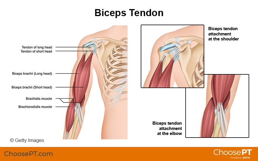
Physical Therapy Guide to Spondylolysis and Spondylolisthesis (Fracture of the Lumbar Spine and Slipped Vertebra)

Spondylolysis (spon-dee-low-lye-sis), or lumbar stress fracture, is a stress fracture of a section of the lumbar spine. The area of the fifth lumbar vertebra is most often affected. The injury can occur on the left or right of the vertebra. Lumbar stress fractures occur in up to 11.5% of the general population in the United States. It is a common cause of low back pain in older children and youth, most often young males. It is a common cause of low back pain in older children and youth. It most often occurs in young males, but also can occur in girls. Highly active teens who engage in activities like lifting heavy loads, repeated backward bending, or twisting of the trunk, are most at risk. Activities like football, hockey, gymnastics, or dance put athletes at higher risk. Only a small percentage of cases require surgery. The majority (85% to 90%) of young patients recover in three to six months with proper treatment. Recovery time can be longer and is different for each person.

Spondylolisthesis (spon-dee-low-lis-thee-sis), or slipped vertebra, is a condition that involves the forward slippage of one vertebra over the one under it. If a crack or stress fracture occurs on both sides of the vertebra it is called spondylolisthesis.

Lumbar stress fracture and slipped vertebra are often described together because they are similar in:

  • The mode of injury.

  • Age of the patient.

  • Symptoms.

  • Treatment.

What Are Stress Fracture and Slipped Vertebra of the Lumbar Spine?

Lumbar stress fracture is a fracture of the part of the bony ring that connects the front part of the spinal column to the back part. The fracture occurs between the part of the bone that sticks out of the back of the spine and the part that sticks out of the side of the spine. Doctors sometimes refer to this condition as a "pars defect." Strain on the lumbar spine due to repeated activities in a growing child can cause this type of injury. It results in low back pain.

Slipped vertebra is the forward slip of a defective, unstable vertebra. There are five grades of slips, with grade I being the smallest amount of slippage and grade V being a slippage of 100%. With milder slippage, and a defect on just one side of the vertebra, physical therapy treatment is effective. Young athletes whose teenage growth spurt has not yet occurred are at greater risk for continued slippage. These athletes and are monitored until they are fully grown.

Key points to understand about lumbar stress fracture and slipped vertebra:

  • Early detection and proper diagnosis of these conditions are important. With early diagnosis and treatment, people with these conditions can safely return to sport or an active lifestyle. If symptoms last a long time and you wait to get help, healing may take much longer.

  • The majority of symptoms can resolve with rest and the help of a physical therapist.

  • Surgery may be needed when treatment of more than six months fails, and symptoms persist.

  • Both of these conditions need to be ruled out in a young athlete who is has low back pain that lasts for more than a few weeks. Active young athletes in sports such as football, hockey, gymnastics, and dance are at the greatest risk. This is especially true while the athlete is still growing.

  • If an X-ray does not show a fracture, but a clinical exam suggests a high likelihood of lumbar stress fracture, your doctor may order an MRI to rule it out.

  • These conditions are not a major cause of low back pain in adults. It can, however, occur high-level adult athletes who take part in high-risk sports.


Signs and Symptoms

Lumbar stress fracture or slipped vertebra may be present if you experience:

  • Low back pain with or without buttock or leg pain. If leg pain is present, it is felt into the thigh, but generally not below the knee.

  • Muscle spasms in your low back, buttocks, and thighs.

  • Difficulty or pain with walking or standing for long periods.

  • Symptoms that are relieved by sitting, slouching, or bending forward.

  • Pain with sports or manual labor.

  • Pain with bending backward, twisting the spine, or with throwing.

  • Decreased flexibility of the leg muscles.


How Is It Diagnosed?

Your physical therapist will conduct a thorough evaluation that includes questions about your health history. Their goal is to assess the degree of your injury and to determine the cause and contributing factors. Your physical therapist also may gather information from forms you fill out before your first session. Their questions may include:

  • How did your injury occur? Was there a single episode or did your condition become worse over time?

  • How have you taken care of your condition? Have you seen other health care providers? Have you had imaging (e.g., X-ray, MRI) or other tests, and do you have the results of those tests?

  • How long have you had pain? Did it come on suddenly or gradually?

  • Does your pain occur with activity, at rest, or during the day or night?

  • What activities or positions make your pain better or worse?

  • Do you take part in activities like football, hockey, gymnastics, or competitive dance?

  • Can you point with one finger to the area on your back that is painful?

  • Do you have any other symptoms, such as fever, chills, or night sweats?

  • Do you have trouble with bowel and bladder control?


After your physical therapist learns the specifics of your condition they will conduct a physical exam.

The physical exam most often will begin with watching some of the movements that were discussed in the interview. It will include the area of your main symptoms as well as other areas that may be involved, such as your hip. Your physical therapist may:

  • Watch you walk.

  • Have you bend forward to try to touch your toes, and bend back as far as you can.

  • Ask you to stand on one leg and bend back.

  • Ask you to turn your trunk from side to side.


Your physical therapist uses these tests to assess your leg and spine flexibility as well as your core strength. They may ask you if the testing changes your symptoms. They may gently but skillfully press specific areas of your low back and pelvis to see if they are painful. This information helps your physical therapist determine the cause of your pain, exactly where your pain is, and the best treatment to resolve your symptoms.

After the interview and physical exam, your physical therapist will discuss the findings with you. If your physical therapist suspects a stress fracture, they may refer you to an orthopedic or sports medicine doctor familiar with back injuries. The doctor may order imaging tests (X-ray, MRI) to confirm a diagnosis and rule out other conditions.


How Can a Physical Therapist Help?

Spondylolisis and listhesisGettyImages-1139743540.jpg


Your physical therapist will design a targeted treatment program based on your condition and goals. It will be designed to safely return you to sport or daily activities. Your treatment plan may include:

Patient education. Your physical therapist will work with you to identify and change any external factors causing your pain. These factors can include the type and amount of exercise you do, your athletic activities, footwear, or the surfaces on which you practice and play. They may recommend changes in your daily activities.

Pain management. Your physical therapist will design a program to address your pain. This may include applying ice to the affected area. Applying heat also is helpful in some cases. Electrical stimulation gently targets nerve fibers that send pain signals to the brain. It also may be used together with ice to help relieve your pain. Your physical therapist also may recommend decreasing some activities that cause pain. Physical therapists are experts in prescribing pain-management techniques that reduce or avoid the need for medicines, including opioids.

Body mechanics. How you move and use your body for work and other activities can contribute to lumbar spine problems and pain. Your physical therapist will teach you how to improve your movements or body mechanics based on your daily activities. They also may make recommendations to improve the way you do certain activities, such as lifting and carrying objects.

Manual therapy. Often, manual therapy for lumbar stress fracture and slipped vertebra includes "soft tissue release" or massage for tight and sore muscle groups. These hands-on techniques may be used to correct tightness of muscles to promote normal movement.

Stretching exercises. Stretching exercises can help improve the flexibility of tight muscles. They also may help to improve movement in the spine and lower extremities and help decrease stress on the lumbar spine during daily activities.

Strengthening exercises. Strengthening helps to make the lumbar spine, pelvis, and hip joints more stable. This, in turn, helps to reduce strain on tissues, and pain. These movements are focused on weak muscles, including the lower abdominal, pelvic floor, and buttocks muscles.

Functional training. Once your pain, strength, and motion improve you will need to safely move back into more demanding activities. To lessen your risk of repeated injury, it is important to learn safe, controlled movements. Based on your unique movement assessment and goals, your physical therapist will create a series of activities to help you use and move your body more correctly and safely for years to come.


Can This Injury or Condition Be Prevented?

Lumbar stress fracture and some types of slipped vertebra may be preventable by educating individuals who are at higher risk of injury.

For the growing young athlete, it is necessary to manage how much, how intensely, and how often you exercise. Parents and coaches should:

  • Limit a child’s participation to one high-risk sport at a time during a season.

  • Limit participation to only one team at a time during a season.

  • Require and enforce one to two days of rest from training per week.

  • Gradually increase training volume, intensity, and frequency when a person is starting a new sport or activity.


What Kind of Physical Therapist Do I Need?

All physical therapists are prepared through education and experience to treat lumbar stress fracture and slipped vertebra. However, you may want to consider:

  • A physical therapist who is experienced in treating people with spine injuries and/or athletes. Some physical therapists have a practice with an orthopaedic or sports physical therapy focus.

  • A physical therapist who is a board-certified clinical specialist or who completed a residency or fellowship in orthopaedic and/or sports physical therapy. This physical therapist has advanced knowledge, experience, and skills that may apply to your condition.

General tips when you are looking for a physical therapist (or any other health care provider):

  • Get recommendations from family, friends, or from other health care providers.

  • When you contact a physical therapy clinic for an appointment, ask about the physical therapists' experience in helping people who are athletes or active individuals with lumbar stress fracture.

  • During your first visit with the physical therapist, be prepared to describe your symptoms in as much detail as possible, and say what makes your symptoms worse.


Further Reading

The American Physical Therapy Association believes that consumers should have access to information that could help them make health care decisions and also prepare them for a visit with their health care provider.

The following articles provide some of the best scientific evidence related to physical therapy treatment of lumbar stress fracture and slipped vertebra. The articles report recent research and give an overview of the standards of practice both in the United States and internationally. The article titles are linked either to a PubMed* abstract of the article or to free full text, so that you can read it or print out a copy to bring with you to your health care provider.

Iwaki K, Sakai T, Hatayama D, et al. Physical features of pediatric patients with lumbar spondylolysis and effectiveness of rehabilitation. J Med Invest. 2018;65(3.4):177–83. Article Summary in PubMed.

Lawrence KJ, Elsar T, Stromberg R. Lumbar spondylolysis in the adolescent athlete. Phys Ther Sport. 2016;20:56–60. Article Summary in PubMed.

Schroeder GD, LaBelle CR, Mendoza M. The role of intense athletic activity on structural lumbar abnormalities in adolescent patients with symptomatic low back pain. Eur Spine J. 2016;25:2842–2848. Article Summary in PubMed.

Kim HJ, Green DW. Spondylolysis in the adolescent athlete. Curr Opin Pediatr. 2011;23(1):68–72. Article Summary in PubMed.

Kalichman L, Kim DH, Li L, et al. Spondylolysis and Spondylolisthesis: prevalence and association with low back pain in the adult community-based population. Spine (Phila Pa 1976). 2009;34(2):199–205. Article Summary in PubMed.

*PubMed is a free online resource developed by the National Center for Biotechnology Information. PubMed contains millions of citations to biomedical literature, including citations in the National Library of Medicine’s MEDLINE database.


Revised in 2020 by Susan Reischl, PT, DPT, board-certified clinical specialist in orthopaedic physical therapy, and reviewed by Stephen Reischl, PT, DPT, board-certified clinical specialist in orthopaedic physical therapy, on behalf of the Academy of Orthopaedic Physical Therapy. Authored in 2014 by Donna Merkel, PT.


9 Things You Should Know About Pain

Here are nine things physical therapists want you to know about pain. 

WomanShoulderPain.jpg

1. Pain is output from the brain.

While we used to believe that pain originated within the tissues of our body, we now understand that pain does not exist until the brain determines it does. The brain uses a virtual "road map" to direct an output of pain to tissues that it suspects may be in danger. This process acts as a means of communication between the brain and the tissues of the body, to serve as a defense against possible injury or disease.

2. The degree of injury does not always equal the degree of pain.

Research has demonstrated that we all experience pain in individual ways. While some of us experience major injuries with little pain, others experience minor injuries with a lot of pain (think of a paper cut).

3. Despite what diagnostic imaging (MRIs, x-rays, CT scans) shows us, the finding(s) may not be the cause of your pain.

A study performed on individuals 60 years or older who had no symptoms of low back pain found that 36% had a herniated disc, 21% had spinal stenosis, and more than 90% had a degenerated or bulging disc, upon diagnostic imaging.

4. Psychological factors, such as depression and anxiety, can make your pain worse.

Pain can be influenced by many different factors, such as psychological conditions. A recent study in the Journal of Pain showed that psychological variables that existed before a total knee replacement were related to a patient's experience of long-term pain following the operation.

5. Your social environment may influence your perception of pain.

Many patients state their pain increases when they are at work or in a stressful situation. Pain messages can be generated when an individual is in an environment or situation that the brain interprets as unsafe. It is a fundamental form of self-protection.

6. Understanding pain through education may reduce your need for care.

A large study conducted with military personnel demonstrated that those who were given a 45-minute educational session about pain sought care for low back pain less than their counterparts.

7. Our brains can be tricked into developing pain in prosthetic limbs.

Studies have shown that our brains can be tricked into developing a "referred" sensation in a limb that has been amputated, causing a feeling of pain that seems to come from the prosthetic limb – or the "phantom" limb. The sensation is generated by the association of the brain's perception of what the body is from birth (whole and complete) and what it currently is (postamputation).

8. The ability to determine left from right may be altered when you experience pain.

Networks within the brain that assist you in determining left from right can be affected when you experience severe pain. If you have been experiencing pain, and have noticed your sense of direction is a bit off, it may be because a "roadmap" within the brain that details a path to each part of the body may be a bit "smudged." (This is a term we use to describe a part of the brain's virtual roadmap that isn’t clear. Imagine spilling ink onto part of a roadmap and then trying to use that map to get to your destination.)

9. There is no way of knowing whether you have a high tolerance for pain or not. Science has yet to determine whether we all experience pain in the same way.

While some people claim to have a "high tolerance" for pain, there is no accurate way to measure or compare pain tolerance among individuals. While some tools exist to measure how much force you can resist before experiencing pain, it can’t be determined what your pain "feels like."

If you have pain that limits your movement or keeps you from taking part in work, daily living, and other activities, a physical therapist can help. 

Author: Joseph Brence, PT, DPT

Bibliography

Allegri M, Montella S, Salici F, et al. Mechanisms of low back pain: a guide for diagnosis and therapy [revised]. F1000Res. 2016;5:F1000 Faculty Rev-1530. doi: 10.12688/f1000research.8105.2.

George SZ, Childs JD, Teyhen DS, et al. Brief psychosocial education, not core stabilization, reduced incidence of low back pain: results from the Prevention of Low Back Pain in the Military (POLM) cluster randomized trial. BMC Med. 2011;9:128.

Carroll I, Wang J, Wang M, et al. Psychological impairment influences pain duration following surgical injury. J Pain. 2008;9 (Suppl 2):21.

Physical Therapy Guide to Shoulder Dislocation: Overview

The shoulder is the most mobile joint in the body and is the most likely joint to dislocate. A dislocation is the separation of 2 bones where they meet at a joint. Shoulder dislocations most often occur during contact sports, but everyday accidents, such as falls, also can cause the joint to dislocate. Athletes, nonathletes, children, and adults can all dislocate their shoulders. A dislocated shoulder usually requires the assistance of a health care professional to guide the joint back into place. After the joint is realigned, a physical therapist directs the rehabilitation of the shoulder, and helps the affected individual prevent reinjury.

CAUTION: A shoulder dislocation requires immediate medical attention, especially if you experience:

  • Numbness in your arm or hand

  • Discoloration of your arm or hand

  • A feeling of coldness in your arm

Any of these conditions may indicate injury to a nerve or blood vessel. Seek medical help immediately.

What is a Shoulder Dislocation?

The shoulder includes the clavicle (collar bone), scapula (shoulder blade), and humerus (upper-arm bone). The rounded top of the humerus and the cup-like end of the scapula fit together like a ball and socket. A shoulder dislocation can occur with an injury, such as when you "fall the wrong way" on your shoulder or outstretched arm, forcing the shoulder beyond its normal range of movement and causing the humerus to come out of the socket. Dislocation can result in damage to many parts of the shoulder, including the bones, the ligaments, the labrum (the ring of cartilage that surrounds the socket), and the muscles and tendons around the shoulder joint.

ShoulderDislocation_SM.jpg


Joints may dislocate when a sudden impact causes the bones in the joint to shift out of place. Dislocations are among the most common traumatic injuries affecting the shoulder.


How Does It Feel?

With most shoulder dislocations, you will feel the humerus coming out of the socket, followed by:

  • Pain

  • Inability to move the arm

  • Awkward appearance of the shoulder

If you have any signs or symptoms of a nerve or blood vessel injury, as listed above, seek immediate medical attention.

The humerus usually remains out of the socket until a physician guides it back into place. X-rays are routinely taken after the dislocation is moved back into place to make sure that you don’t have a fracture.

Occasionally, the shoulder may go back into place on its own. You might not even realize that you have dislocated your shoulder; you may only feel that you have injured it. If you have injured your shoulder and have pain, seek medical attention.


How Can a Physical Therapist Help?

After the dislocated humerus has been moved back into position, your arm will be placed in a sling to protect you from reinjury and to make your shoulder more comfortable. Your physical therapist can review your health and injury history and conduct a physical examination to determine your rehabilitation needs. Based on the results of the examination and your goals, your physical therapist will guide you through a rehabilitation program to restore your mobility, strength, joint awareness, and sport-specific skills. Your therapist also will show you how to control your pain and relieve any inflammation.

Your treatment program may include:

Range-of-motion exercises. Swelling and pain can reduce your shoulder movement. Your physical therapist will teach you how to perform safe and effective exercises to restore full range of motion to your shoulder. Your physical therapist might apply manual (hands-on) therapy to help decrease pain in the shoulder.

Strengthening exercises. Poor muscle strength can cause the shoulder joint to remain unstable and possibly reinjure it. Based on how severe your injury is and where you are on the path to recovery, your physical therapist can determine which strengthening exercises are right for the rehabilitation of your shoulder.

Joint awareness and muscle retraining. Specialized exercises help your shoulder muscles relearn how to respond to sudden forces. Your physical therapist will design individualized exercises to help you return to your regular activities.

Activity- or sport-specific training. Depending on the requirements of your job or the type of sports you play, you might need additional rehabilitation tailored to the demands your activities place on your shoulder. Your physical therapist can develop a program that takes all of these demands (as well as your specific injury) into account. For example, if you are an overhead thrower, such as a baseball pitcher, your physical therapist will guide you through a throwing progression and pay specific attention to your throwing mechanics.


Can This Injury or Condition Be Prevented?

Shoulder dislocations are dependent on how loose the shoulder is, and are more likely to occur during sports or aggressive activities. Your physical therapist can advise you about the positions that frequently cause dislocations, and teach you ways to reduce your risk of dislocation. See your physical therapist if you:

  • Have pain in your shoulder, especially when performing forceful activities

  • Have symptoms that feel as though your shoulder is "slipping," “shifting,” or "moving"

  • Hear a popping sound in your shoulder accompanied by pain

If you already have a history of shoulder dislocation, you are at a greater risk for reinjury if your shoulder does not heal properly or if you do not regain your normal shoulder strength or joint awareness. Research shows that a high percentage of dislocated shoulders will dislocate again. Physical therapists play an important role in helping people prevent recurring shoulder problems.

If you return to sports or activities too soon following injury, you could cause a reinjury. Your physical therapist can determine when you are ready to return to your activities and sports by making sure that your shoulder is strong and ready for action. Your physical therapist may recommend a shoulder brace to allow you to gradually and safely return to your previous activities.


Real Life Experiences

Bob is a 25-year-old salesman and a competitive snowboarder. Recently, he spent the day snowboarding with friends. Toward the end of the day, the front edge of his snowboard caught in the snow, throwing him off-balance. As he fell, he reached out his right arm to break the fall. He felt a pop in his shoulder, and a sharp pain. He felt like his shoulder was out of place. His friend called the ski patrol, who guided him safely down the mountain and took him to the local emergency department. X-rays showed that Bob’s shoulder was dislocated. The emergency physician put Bob’s shoulder back into place and secured it in a sling. He recommended that Bob see a physical therapist for rehabilitation.

At his first visit, Bob’s physical therapist reviews the history of his injury and his general health and performs a thorough examination. Because Bob's goal is to return to an active lifestyle as soon as possible, she develops a rehabilitation plan of care to restore the mobility, strength, and function of Bob's shoulder.

Bob’s physical therapist applies a cold pack to relieve his pain, and performs gentle hands-on range-of-motion exercises to the shoulder area. She also teaches Bob a few gentle movement and strengthening exercises he can do himself.

Once Bob's pain has decreased, his rehabilitation focuses on restoring the dynamic stability of his shoulder through movement re-education and drills, and strengthening of the shoulder area. Bob's physical therapist chooses specific movements and exercises to gently restore his shoulder range of motion, while allowing the shoulder to heal. Particular emphasis is placed on educating Bob and avoiding stretches and activities that put too much stress on the injured parts of his shoulder.

Working with his physical therapist, Bob steadily increases his shoulder strength and range of motion.

At 12 weeks postinjury, Bob’s physical therapist informs him he is ready to return to training for competition. She advises him to be aware of his shoulder movements and to use the techniques he learned in physical therapy to avoid reinjury.

Bob begins to train for the next season’s snowboarding competitions and is able to practice at his previous level—with his shoulder returned to its full strength and mobility.

This story was based on a real-life case. Your case may be different. Your physical therapist will tailor a treatment program to your specific case.


What Kind of Physical Therapist Do I Need?

All physical therapists are prepared through education and experience to treat patients who have a dislocated shoulder. You may want to consider:

  • A physical therapist who is experienced in treating people with musculoskeletal problems. Some physical therapists have a practice with an orthopedic focus.

  • A physical therapist who is a board-certified clinical specialist or who completed a residency or fellowship in orthopedic or sports physical therapy. This physical therapist has advanced knowledge, experience, and skills that may apply to your condition.

You can find physical therapists who have these and other credentials by using Find a PT, the online tool built by the American Physical Therapy Association to help you search for physical therapists with specific clinical expertise in your geographic area.

General tips when you're looking for a physical therapist (or any other health care provider):

  • Get recommendations from family and friends or from other health care providers.

  • When you contact a physical therapy clinic for an appointment, ask about the physical therapists' experience in helping people with shoulder dislocation.

  • During your first visit with the physical therapist, be prepared to describe your symptoms in as much detail as possible, and say what makes your symptoms worse.


Further Reading

The American Physical Therapy Association (APTA) believes that consumers should have access to information that could help them make health care decisions and also prepare them for their visit with their health care provider.

APTA has determined that the following articles provide some of the best scientific evidence for how to treat shoulder dislocation. The articles report recent research and give an overview of the standards of practice for treatment both in the United States and internationally. The article titles are linked either to a PubMed* abstract of the article or to free access of the full article, so that you can read it or print out a copy to bring with you to your health care provider.

Khiami F, Gerometta A, Loriaut P. Management of recent first-time anterior shoulder dislocations. Orthop Traumatol Surg Res. 2015;101(1 Suppl):S551–S557. Article Summary on PubMed.

Robinson CM, Seah M, Akhtar MA. The epidemiology, risk of recurrence, and functional outcome after an acute traumatic posterior dislocation of the shoulder. J Bone Joint Surg Am.2011;93(17):1605–1613. Article Summary on PubMed.

Godin J, Sekiya JK. Systematic review of rehabilitation versus operative stabilization for the treatment of first-time anterior shoulder dislocations. Sports Health. 2010;2:156–165. Free Article.

Brumitt J, Sproul A, Lentz P, et al. In-season rehabilitation of a division III female wrestler after a glenohumeral dislocation. Phys Ther Sport. 2009;10:112–117. Article Summary on PubMed.

Hovelius L, Olofsson A, Sandström B, et al. Nonoperative treatment of primary anterior shoulder dislocation in patients forty years of age and younger: a prospective twenty-five year follow up. J Bone Joint Surg Am. 2008;90:945–952. Article Summary on PubMed.

Robinson CM, Howes J, Murdoch H, Will E, Graham C. Functional outcome and risk of recurrent instability after primary traumatic anterior shoulder dislocation in young patients. J Bone Joint Surg Am. 2006;88(11):2326–2336. Article Summary on PubMed.

Robinson CM, Howes J, Murdoch H, Will E, Graham C. Functional outcome and risk of recurrent instability after primary traumatic anterior shoulder dislocation in young patients. J Bone Joint Surg Am. 2006;88:2326–2336. Article Summary on PubMed.

Millar AL, Lasheway PA, Eaton W, Christensen F. A retrospective, descriptive study of shoulder outcomes in outpatient physical therapy. J Orthop Sports Phys Ther. 2006;36:403–414. Article Summary on PubMed.

Buss DD, Lynch GP, Meyer CP, Huber SM, Freehill MO. Nonoperative management for in-season athletes with anterior shoulder instability [erratum in: Am J Sports Med. 2004;32:1780]. Am J Sports Med. 2004;32:1430–1433. Article Summary on PubMed.

Gibson K, Growse A, Korda L, Wray E, MacDermid JC. The effectiveness of rehabilitation for nonoperative management of shoulder instability: a systematic review. J Hand Ther. 2004;17:229–242. Article Summary on PubMed.

*PubMed is a free online resource developed by the National Center for Biotechnology Information (NCBI). PubMed contains millions of citations to biomedical literature, including citations in the National Library of Medicine’s MEDLINE database.

Authored by Jason Lunden, PT, DPT, board-certified sports clinical specialist. Reviewed by the editorial board.

6 Tips to Prevent New Parenting Injuries

Spot_Parents_750x419.jpg

The months following the birth of a child are some of the most rewarding for new parents—and the most challenging to a new parent’s body. Lifting and carrying a child, picking up toys off of the floor, and pushing a stroller are normal daily tasks for moms and dads.  

Here are some tips on how using proper body mechanics to help ease the strains and stresses of parenting:

1. Lifting Your Child From the Floor 
When picking up your child from the floor, you should use a half-kneel lift:

First, stand close to your child on the floor. While keeping your back straight, place one foot slightly forward of the other foot, and bend your hips and knees to lower yourself onto one knee. Once down on the floor, grasp your child with both arms and hold him or her close to your body. Tighten your stomach muscles, push with your legs, and slowly return to the standing position.

To place your child onto the floor, the same half-kneel technique should be performed.

2. Carrying/Holding Your Child 
When holding or carrying your child, you should always hold him or her close to your body and balanced in the center of your body. Avoid holding your child in one arm and balanced on your hip. When using a child carrier, be sure to keep your back straight and your shoulders back to avoid straining your back and neck.

3. Picking up Toys From the Floor 
While straightening up and picking items off the floor, keep your head and back straight, and while bending at your waist, extend one leg off the floor straight behind you. You can also use the half-kneel technique discussed above, if several toys are within the same space.

4. Lifting Your Child Out of the Crib 
As you lift your child out of the crib, keep your feet shoulder-width apart and knees slightly bent. Arch your low back and, while keeping your head up, bend at your hips. With both arms, grasp your child and hold him or her close to your chest. Straighten your hips so you are in an upright position, and then extend your knees to return to a full stand. To return your child to the crib, use the same technique and always remember to keep your child close to your chest.

5. The Stroller 
When you are lifting your child from a stroller, stand directly in front of the child to avoid twisting your back. It is important to bend from your hips rather than from your lower back, much like rising from a squatting position.

When walking your child in a stroller, you will want to stay as close to the stroller as possible, allowing your back to remain straight and your shoulders back. The force to push the stroller should come from your entire body, not just your arms. Avoid pushing the stroller too far ahead of you because this will cause you to hunch your back and round your shoulders forward.

6. The Changing Table 
Before placing the baby on the changing table, it is essential to keep him or her at the center of your body. The table should be at the appropriate height for parental use. When changing your baby's diaper, the best table placement and height is directly in front of and slightly below the elbows. This helps avoid the type of bending and twisting that can cause injury.

Other tips:

  • Place all diaper-changing materials within arm’s reach—for instance, in wide-set drawers directly below the changing area.

  • You may wish to place one leg on a stool when you are using the changing table. This can help take strain off your back and neck.

Physical Therapist's Guide to Patellofemoral Pain

What Is Patellofemoral Pain?

PFP may occur after a sudden increase in activities like running or jumping. Research suggests that PFP results from activity levels that are increased faster than the knee can adapt. Other contributing factors to PFP may include:

  • Weakness of the thigh muscles.

  • Specializing in a single sport, which requires repeating the same movements again and again.

  • Certain hip and knee coordination patterns during running and jumping activities.

PFP does not go away on its own. If you have symptoms of PFP, it’s important to seek care from a physical therapist so you can return to the activities that you enjoy.

How Does it Feel?

People with PFP may experience pain:

  • When walking up or down stairs or hills.

  • When playing a sport.

  • With deep knee bending (squatting).

  • When walking on uneven surfaces.

  • With activity, but improving with rest.

  • After sitting for long periods of time with the knee bent.

How Is It Diagnosed?

APTA-Knee-Joint-Patella-Pain_750x419.jpg

Your physical therapist will review your health history and conduct a series of tests to evaluate you and your knee. PFP is diagnosed by analyzing any movement that causes pain, and ruling out other possible conditions.

Your physical therapist may analyze your walking and running patterns. They may test the strength of your hip and thigh muscles to find out if weakness is contributing to your pain. Medical imaging, such as an X-ray or MRI, is not helpful in diagnosing PFP. However, your physical therapist may consult with an orthopedic physician who may order imaging to rule out other conditions.

How Can a Physical Therapist Help?

If PFP is diagnosed, your physical therapist will develop an exercise and rehabilitation program just for you. Your program may include:

Strengthening exercises. Your physical therapist will teach you exercises to help strengthen the muscles around the hip and the knee itself. Research shows that this type of exercise therapy is the best approach to managing PFP.

Taping. Your physical therapist may teach you how to apply tape to your knee, which may improve your ability to perform exercises that would normally be painful. However, taping alone will not resolve PFP. It must only be used along with your exercise program.

Shoe inserts. Your physical therapist may recommend shoe inserts to help reduce your pain when exercising. But inserts alone, like taping, will not treat PFP. Your physical therapist will design an exercise program to fit your specific needs and goals.

Coordination training. Based on your activity level, your physical therapist may help retrain your hip and knee movement patterns to reduce your knee pain.

This type of training is effective for athletes, in particular, and may focus on movements like:

  • Stair climbing.

  • Squatting.

  • Running and jumping.

Cross-training guidance. Physical therapists help athletes and active people perform different movements (cross-training). This helps them stay active until they can return to a favorite activity.

Return to full activity. Your physical therapist will help guide a gradual return to your favorite activities, such as running and jumping, and will teach you good overall exercise habits to help maximize the health of your knee.

Treatments That Do Not Work for PFP

While these can be appropriate for the treatment of other injuries or conditions, the following do notwork for PFP:

Quick fixes. “Passive” treatments like dry needling, ultrasound, laser, or electrical stimulation are not helpful for people with PFP. The most effective treatment for PFP is an exercise program that targets the hip and knee muscles.

Rest. If you are experiencing PFP, it is important to understand that rest only helps temporarily. Your pain will likely return when you go back to your normal activity. Rest is not helpful in the long term. A movement program guided by your physical therapist is your best treatment option.

Can this Injury or Condition be Prevented?

Current research shows that a person’s age, height, body weight, or foot alignment may not contribute to the risk of developing PFP at all. A knock-kneed posture also does not increase the risk of developing PFP.

However, a few preventive measures can be effective. To help reduce your risk of developing PFP:

  • Keep your thigh muscles strong.

  • Maintain good exercise habits.

  • Avoid rapid spikes in activity levels.

  • Participate in a variety of sports, rather than just repeating the same movements again and again.

What Kind of Physical Therapist Do I Need?

All physical therapists are prepared through education and experience to treat a variety of conditions or injuries including PFP. However, you may want to consider:

  • A physical therapist who is experienced in treating people with orthopedic, or musculoskeletal, problems.

  • A physical therapist who is a board-certified clinical specialist or who has completed a residency or fellowship in orthopedic physical therapy. This physical therapist will have advanced knowledge, experience, and skills that may apply to your condition.

  • While it may be tempting to seek quick fixes for your knee pain, there is no evidence that passive treatments work for persons with PFP. If you have PFP, seek care from a physical therapist who uses progressive exercise therapy for the treatment of this condition.

You can find physical therapists who have these and other credentials by using Find a PT, an online tool provided by the American Physical Therapy Association. You can search for physical therapists with specific clinical expertise in your geographic area.

General tips when you're looking for a physical therapist:

  • Get recommendations from family and friends or from other health care providers.

  • When you contact a physical therapy clinic for an appointment, ask about the physical therapist's experience in helping people with patellofemoral pain (PFP).

  • During your first visit with a physical therapist, you will be asked to describe your symptoms in as much detail as possible, and say what makes your symptoms worse. Here are some tips to prepare for your visit.

Further Reading

The American Physical Therapy Association (APTA) believes that consumers should have access to information that could help them make health care decisions and prepare for a visit with their health care provider.

The following articles provide some of the best scientific evidence related to physical therapy treatment of patellofemoral pain syndrome. The articles report recent research and give an overview of the standards of practice both in the United States and internationally. The article titles are linked either to a PubMed* abstract of the article or to free access of the full article, so you can read it or print out a copy to bring with you to your health care provider.

Patellofemoral pain: treating painful kneecaps. J Orthop Sports Phys Ther. 2019;49(9):633. Free Article.

Willy RW, Hoglund LT, Barton CJ, et al. Patellofemoral Pain. J Orthop Sports Phys Ther.2019;49(9):CPG1–CPG95. Free Article.

Neal BS, Lack SD, Lankhorst NE, Raye A, Morrissey D, van Middelkoop M. Risk factors for patellofemoral pain: a systematic review and meta-analysis. Br J Sports Med. 2019;53(5):270–281. Free Article.

Lack S, Barton C, Sohan O, Crossley K, Morrissey D. Proximal muscle rehabilitation is effective for patellofemoral pain: a systematic review with meta-analysis. Br J Sports Med. 2019;49(21):1365-1376. Free Article.

* PubMed is a free online resource developed by the National Center for Biotechnology Information (NCBI). PubMed contains millions of citations to biomedical literature, including citations in the National Library of Medicine’s MEDLINE database.

Authored by Christopher Bise, PT, DPT, MS. Revised and reviewed by Richard Willy, PT, PhD.

Hip Labral Tears

What is a Hip Labral Tear?

A hip labral tear occurs when there is damage to the labrum (ring of cartilage) within the hip joint. The hip joint is where the thigh bone (femur) meets the pelvis (ilium). It is described as a ball-and-socket joint. This design allows the hip to move in several directions. The bony hip socket is surrounded by the labrum, which provides additional stability and shock absorption to the hip joint.

A labral tear results when a part of the labrum separates or is pulled away from the socket. Most commonly, a labral tear is the result of repetitive stress (loading) causing irritation to the hip, often due to long-distance running or performing repeated, sharp, sports movements, such as twisting and cutting.

Repetitive loading is more likely to result in injury to the labrum when there are bony abnormalities at the hip joint. For example, hip impingement is a condition resulting in hip pain due to abnormal bony contact between the ball and socket. As the hip is moved into specific positions, this bony contact can place greater stress on the labrum.

Hip labral tears may result from a combination of several different variables, including:

  • Bony abnormalities in the hip joint (hip impingement)

  • Hip muscle tightness

  • Hip muscle weakness

  • An unstable hip joint

  • Improper technique when performing repetitive activities

  • Participation in sports that require distance running, or repetitive twisting and cutting

  • Typical wear-and-tear over time

Once torn, the labral tissue in the hip does not have the ability to heal on its own. There are surgical procedures to remove or repair torn labral tissue; however, treatment for a labral tear often begins with a course of physical therapy.

Nonsurgical treatment efforts are focused on addressing symptoms by maximizing the strength and mobility of the hip to minimize the stress placed on the injured area. In some cases, patients are able to achieve a satisfactory level of activity without surgery.

Surgical interventions are available to clean out the hip joint, and repair or reconstruct the torn labral tissue. Following surgery, patients will complete several months of physical therapy to regain function of the hip.


HipLabralTear-SM.jpg


How Does it Feel?

Many people have labral tears in the hip and do not experience symptoms; however, some labral tears can result in significant pain or limitations. Pain in the front of the hip or in the groin resulting from a hip labral tear can cause an individual to have limited ability to stand, walk, climb stairs, squat, or participate in recreational activities.

With a labral tear, you may experience:

  • A deep ache in the front of your hip or groin, often described by the "C sign." (People make a "C" with the thumb and hand, and place it on the fold at the front and side of the hip to locate their pain.)

  • Painful clicking or "catching" with hip movements; the feeling of something painful stuck in the hip or blocking hip motion.

  • Pain that increases with prolonged sitting or walking.

  • A sharp pain in the hip or groin when squatting.

  • Pain that comes on gradually rather than with one specific episode.

  • Weakness in the muscles surrounding the hip, or a feeling of the hip “giving way.”

  • Stiffness in the hip.


How Is It Diagnosed?

Your physical therapist will begin your evaluation by gathering information about your condition and medical history. Although a hip labral tear may be the result of a single injury, it most likely is a condition that develops as a consequence of repetitive irritation in the hip. Your physical therapist may ask you to describe:

  • Your current symptoms and how they affect your activities in a typical day

  • Any pain you are experiencing, its intensity and location, and how it may vary during the day

  • What activities you may be unable to do or have difficulty completing

  • What activities aggravate your symptoms, and how you reduce the level of your discomfort

  • Prior injury occurrences before your symptoms began

  • Other health care professional visits and any tests received


Your physical examination will focus on the region where your symptoms are occurring, but also include other areas that may have been affected as your body adjusted to pain. Your physical therapist may watch you walk, step onto a stair, squat, or balance on one leg.

Your physical therapist will gently but skillfully palpate (touch) the front, side, and back of your hip to determine exactly where it is most painful. The therapist will assess the mobility and strength of your hip and other regions of the body to determine the areas that require treatment.

Following the interview and physical examination, your physical therapist will discuss the findings with you and, through mutual collaboration, develop an individualized treatment program to begin your recovery.

Your physical therapist also may refer you to an orthopedic physician who specializes in hip injuries for diagnostic imaging (ie, X-ray, MRI). An X-ray helps to identify any bony abnormalities, such as those that occur with hip impingement, which may be contributing to your pain. An MRI helps to identify a labral tear.


How Can a Physical Therapist Help?

When you have been diagnosed with a hip labral tear, your physical therapist will work with you to develop a plan to help achieve your specific goals. To do so, your therapist will select treatment strategies in any or all of the following areas:

Education. Your physical therapist will work with you to identify and change any external factors causing your pain, such as exercise selection, footwear, or the amount of exercises you perform.

Pain management. Many pain-relief strategies may be implemented; the most beneficial strategy to alleviate hip pain is to apply ice to the area and to decrease or eliminate specific activities causing your symptoms. Your physical therapist will identify specific movements that aggravate the inside of your hip joint, and design an individualized treatment plan for you, beginning with a period of rest, and gradually adding a return to certain activities as appropriate. Physical therapists are experts in prescribing pain-management techniques that reduce or eliminate the need for medication, including opioids.

Manual therapy. Your physical therapist may apply hands-on treatments to gently move your muscles and joints to decrease your pain and improve motion and strength. These techniques often address areas that are difficult to treat on your own.

Movement reeducation. Your back and hip may be moving improperly, causing increased tension at the hip joint. Your physical therapist may teach you self-stretching techniques for the lower body to decrease tension and help restore normal motion in the back, hip, and leg. There are, however, certain hip motions to avoid following an injury to the hip labrum. Your physical therapist will carefully prescribe exercises that improve your range of motion while protecting the area that has the labral tear.

Muscle strengthening. Muscle weaknesses or imbalances can be the cause or the result of hip pain. Based on your specific condition, your physical therapist will design a safe, individualized, progressive resistance program for you, likely including your core (midsection) and lower extremity. You may begin by performing strengthening exercises while lying down, and advance to performing exercises in a standing position. Your physical therapist will choose what exercises are right for you.

Functional training. Once your pain, strength, and motion improve you will be able to safely transition back into more demanding activities. To minimize tension on the hip, it is important to teach your body safe, controlled movements. Based on your own unique movement assessment and goals, your physical therapist will create a series of activities to help you learn how to use and move your body correctly and safely. Your therapist also will discuss specific positions and activities that should be avoided or modified to protect your hip.


Can this Injury or Condition be Prevented?

Repetitive motion, such as sports or long-distance running, can create the risk of sustaining a labral injury. It is imperative to be aware of any hip pain that you experience, particularly with sitting and squatting, as these are signs of a potential hip injury. Identifying and addressing these injuries early is helpful in their treatment. A physical therapist can help an active individual learn proper body movements to lessen the possibility of injury.

After recovering from a hip labral tear, it is important to continue the lower-extremity mobility and muscle strengthening practices taught to you by your physical therapist, to help reduce the risk of further irritation or injury. In some cases, complete avoidance of the activity that contributed to the symptoms may be recommended.


Real Life Experiences

Erin is a 27-year-old accountant who is training for an upcoming half-marathon. She runs 5 days a week and also enjoys performing weight training and strengthening exercises 2 to 3 days a week. Over the past 2 weeks, Erin has begun to experience an achy pain in the front of her right hip. Her pain is worse after running, and while sitting in her car and at her desk. She also experiences occasional "catching" in her hip when reaching forward to pick up her 1-year-old daughter.

Erin is concerned about the pain she feels between runs and her inability to sit without discomfort. She is worried about her ability to perform daily activities, care for her daughter, and train for her upcoming race. She consults her physical therapist.

Erin’s physical therapist conducts a comprehensive assessment of her current symptoms and her health history. She assesses Erin’s motion, strength, balance, movement, and running mechanics. She skillfully palpates (touches) the front, side, and back of Erin’s hip to determine the precise location of her pain. Erin describes her typical daily running routine, her stretching routine, and her footwear. Based on these findings, her physical therapist suspects an injury to her labrum within her hip joint.

Because Erin’s hip is so tender, her physical therapist refers her to an orthopedic surgeon. The surgeon confirms the diagnosis of a hip labral tear. Erin and her surgeon discuss treatment options; the decision is made for nonoperative management of the condition, with a 2-month period of physical therapy.

Erin and her physical therapist work together to establish short- and long-term goals and identify immediate treatment priorities, including icing and activity modification to decrease her pain as well as gentle hip-strengthening exercises. Her physical therapist also teaches her a home-exercise program to perform daily to help speed her recovery.

Together, they outline a 4-week rehabilitation program. Erin sees her physical therapist 1 to 2 times each week; she assesses Erin’s progress, performs manual therapy techniques, and advances her exercise program as appropriate. She advises Erin on exercise and activity modifications that will enhance her recovery. Erin maintains her daily exercise routine at home.

After 6 weeks, Erin's hip no longer "catches" when she bends forward, and she only experiences periodic mild discomfort when sitting or running.

On the day of the half-marathon, Erin runs pain free—and is proud to high five her husband and her little daughter at the finish line!


What Kind of Physical Therapist Do I Need?

All physical therapists are prepared through education and experience to treat a labral injury in the hip. However, you may want to consider:

  • A physical therapist who is experienced in treating people with hip labral injuries or tears, and hip impingement. Some physical therapists have a practice with an orthopedic or musculoskeletal focus.

  • A physical therapist who is a board-certified clinical specialist, or who completed a residency or fellowship in orthopedic or sports physical therapy. This physical therapist has advanced knowledge, experience, and skills that may apply to your condition.

You can find physical therapists who have these and other credentials by using Find a PT, the online tool built by the American Physical Therapy Association to help you search for physical therapists with specific clinical expertise in your geographic area.

General tips when you're looking for a physical therapist (or any other health care provider):

  • Get recommendations from family and friends or from other health care providers.

  • When you contact a physical therapy clinic for an appointment, ask about the physical therapists' experience in helping people who have hip labral injury or hip impingement.

  • During your first visit with the physical therapist, be prepared to describe your symptoms in as much detail as possible, and describe what makes your symptoms worse.


Further Reading

The American Physical Therapy Association (APTA) believes that consumers should have access to information that could help them make health care decisions and also prepare them for a visit with their health care provider.

The following articles provide some of the best scientific evidence related to physical therapy treatment of labral tears in the hip. The articles report recent research and give an overview of the standards of practice both in the United States and internationally. The article titles are linked either to a PubMed* abstract of the article or to free full text, so that you can read it or print out a copy to bring with you to your health care provider.

McGovern RP, Martin RR, Kivlan BR, Christoforetti JJ. Non-operative management of individuals with non-arthritic hip pain: a literature review. Int J Sport Phys Ther. 2019;14(1): 135–147. Free Article.

Pennock AT, Bomar AD, Johnson KP, Randich K, Upasani W. Nonoperative management of femoroacetabular impingement: a prospective study. Am J Sports Med. 2018;46(14):3415–3422. Article Summary in PubMed.

Griffin DR, Dickenson EJ, O’Donnell J, et al. The Warwick Agreement on femoroacetabular impingement syndrome (FAI syndrome): an international consensus statement. Br J Sports Med. 2016;50:1169–1176. Free Article.

*PubMed is a free online resource developed by the National Center for Biotechnology Information (NCBI). PubMed contains millions of citations to biomedical literature, including citations in the National Library of Medicine’s MEDLINE database

Authored by Allison Mumbleau, PT, DPT, SCS. Revised by Jennifer Bagwell, PT, PhD, DPT, member of APTA's Academy of Orthopaedic Physical Therapy. Reviewed by an APTA section liaison. 


Guide to Anterior Cruciate Ligament (ACL) Tear

An anterior cruciate ligament (ACL) tear is an injury to the knee commonly affecting athletes, such as soccer players, basketball players, skiers, and gymnasts. Nonathletes can also experience an ACL tear due to injury or accident. Approximately 200,000 ACL injuries are diagnosed in the United States each year. It is estimated that there are 95,000 ruptures of the ACL and 100,000 ACL reconstructions performed per year in the United States. Approximately 70% of ACL tears in sports are the result of noncontact injuries, and 30% are the result of direct contact (player-to-player, player-to-object). Women are more likely than men to experience an ACL tear. Physical therapists are trained to help individuals with ACL tears reduce pain and swelling, regain strength and movement, and return to desired activities.

What is an ACL Tear?

The ACL is one of the major bands of tissue (ligaments) connecting the thigh bone (femur) to the shin bone (tibia) at the knee joint. It can tear if you:

  • Twist your knee while keeping your foot planted on the ground.

  • Stop suddenly while running.

  • Suddenly shift your weight from one leg to the other.

  • Jump and land on an extended (straightened) knee.

  • Stretch the knee farther than its usual range of movement.

  • Experience a direct hit to the knee.

ACLAttachment_Small.jpg

ACL Attachment: See More Detail

Back to Top

How Does it Feel?

When you tear the ACL, you may feel a sharp, intense pain or hear a loud "pop" or snap. You might not be able to walk on the injured leg because you can’t support your weight through your knee joint. Usually, the knee will swell immediately (within minutes to a few hours), and you might feel that your knee "gives way" when you walk or put weight on it.

Back to Top

How Is It Diagnosed?

Immediately following an injury, you may be examined by a physical therapist, athletic trainer, or orthopedic surgeon. If you see your physical therapist first, your therapist will conduct a thorough evaluation that includes reviewing your health history. Your physical therapist will ask:

  • What you were doing when the injury occurred.

  • If you felt pain or heard a "pop" when the injury occurred.

  • If you experienced swelling around the knee in the first 2 to 3 hours following the injury.

  • If you felt your knee buckle or give out when you tried to get up from a chair, walk up or down stairs, or change direction while walking.

Your physical therapist may perform gentle "hands-on" tests to determine the likelihood that you have an ACL tear, and may use additional tests to assess possible damage to other parts of your knee.

An orthopedic surgeon may order further tests, including magnetic resonance imaging (MRI), to confirm the diagnosis and rule out other possible damage to the knee.

Surgery

Most people who sustain an ACL tear will undergo surgery to repair the tear; however, some people may avoid surgery by modifying their physical activity to relieve stress on the knee. A select group can actually return to vigorous physical activity following rehabilitation without having surgery.

Your physical therapist, together with your surgeon, can help you determine if nonoperative treatment (rehabilitation without surgery) is a reasonable option for you. If you elect to have surgery, your physical therapist will help you prepare both for surgery and to recover your strength and movement following surgery.


Back to Top

How Can a Physical Therapist Help?

Once an ACL tear has been diagnosed, you will work with your surgeon and physical therapist to decide if you should have surgery, or if you can recover without surgery. If you don’t have surgery, your physical therapist will work with you to restore your muscle strength, agility, and balance, so you can return to your regular activities. Your physical therapist may teach you ways to modify your physical activity in order to put less stress on your knee. If you decide to have surgery your physical therapist can help you before and after the procedure.

Treatment Without Surgery

Current research has identified a specific group of patients (called "copers") who have the potential for healing without surgery following an ACL tear. These patients have injured only the ACL, and have experienced no episodes of the knee "giving out" following the initial injury. If you fall into this category, based on the specific tests your physical therapist will conduct, your therapist will design an individualized physical therapy treatment program for you. It may include treatments such as gentle electrical stimulation applied to the quadriceps muscle, muscle strengthening, and balance training.

Treatment Before Surgery

If your orthopedic surgeon determines that surgery is necessary, your physical therapist can work with you before and after your surgery. Some surgeons refer their patients to a physical therapist for a short course of rehabilitation before surgery. Your physical therapist will help you decrease your swelling, increase the range of movement of your knee, and strengthen your thigh muscles (quadriceps).

Treatment After Surgery

Your orthopedic surgeon will provide postsurgery instructions to your physical therapist, who will design an individualized treatment program based on your specific needs and goals. Your treatment program may include:

Bearing weight. Following surgery, you will use crutches to walk. The amount of weight you are allowed to put on your leg and how long you use the crutches will depend on the type of reconstructive surgery you have received. Your physical therapist will design a treatment program to meet your needs and gently guide you toward full weight bearing.

Icing and compression. Immediately following surgery, your physical therapist will control your swelling with a cold application, such as an ice sleeve, that fits around your knee and compresses it.

Bracing. Some surgeons will give you a brace to limit your knee movement (range of motion) following surgery. Your physical therapist will fit you with the brace and teach you how to use it safely. Some athletes will be fitted for braces as they recover and begin to return to their sports activities.

Movement exercises. During your first week following surgery, your physical therapist will help you begin to regain motion in the knee area, and teach you gentle exercises you can do at home. The focus will be on regaining full movement of your knee. The early exercises help with increasing blood flow, which also helps reduce swelling.

Electrical stimulation. Your physical therapist may use electrical stimulation to help restore your thigh muscle strength, and help you achieve those last few degrees of knee motion.

Strengthening exercises. In the first 4 weeks after surgery, your physical therapist will help you increase your ability to put weight on your knee, using a combination of weight-bearing and non-weight-bearing exercises. The exercises will focus on your thigh muscles (quadriceps and hamstrings) and might be limited to a specific range of motion to protect the new ACL. During subsequent weeks, your physical therapist may increase the intensity of your exercises and add balance exercises to your program.

Balance exercises. Your physical therapist will guide you through exercises on varied surfaces to help restore your balance. Initially, the exercises will help you gently shift your weight on to the surgery leg. These activities will progress to standing on the surgery leg, while on firm and unsteady surfaces to challenge your balance.

Return to sport or activities. As athletes regain strength and balance, they may begin running, jumping, hopping, and other exercises specific to their individual sport. This phase varies greatly from person-to-person. Physical therapists design return-to-sport treatment programs to fit individual needs and goals.

Back to Top

Can this Injury or Condition be Prevented?

Much of the research on ACL tears has been conducted with female collegiate athletes, because women are 4 to 6 times more likely to experience the injury. Preventive physical therapy programs have proven to lower ACL injury rates by 41% for female soccer players. Researchers have made the following recommendations for a preventive exercise program:

  • The program should be designed to improve balance, strength, and sports performance. Strengthening your core (abdominal) muscles is key to preventing injury, in addition to strengthening your thigh and leg muscles.

  • Exercises should be performed 2 or 3 times per week and should include sport-specific exercises.

  • The program should last no fewer than 6 weeks.

Although most exercise studies have been conducted with female athletes, the findings may benefit male athletes as well.

Back to Top

Real Life Experiences

Anita is a 20-year-old student at a local university, and a star basketball player. Her team is off to a great start this year; the buzz around campus is that this could be a dream team!

But tonight, when Anita goes up for a rebound and lands off-balance, she hears a "pop" in her left knee and feels a sharp pain. When she tries to walk, she realizes that she can't put weight on her left leg. She's led back to the training room, where the school physical therapist conducts an evaluation. The test results indicate injury, and the physical therapist notices an increase in swelling around the knee just 30 minutes after the incident. She suspects an ACL tear, and refers Anita to an orthopedic surgeon. The next day, the surgeon confirms the diagnosis of an ACL tear, and tells Anita that her injury requires surgery.

After a short course of treatment by her new local physical therapist, including pain and swelling management, manual (hands-on) therapy, and knee range-of-motion and strengthening exercises, Anita has surgery the following month. Her surgeon schedules her to receive physical therapy 3 days after her surgery. She is advised to ice and elevate the knee several times per day.

Three days after surgery, Anita returns to her local physical therapist to begin her rehabilitation. He shows her how to use her crutches properly to gently begin to put weight on the operative knee. He guides her to contract/tighten the quadriceps muscle, and gently performs manual (hands-on) stretches for her to straighten the knee.

Over the next few weeks, Anita is able to gradually stop using her crutches, and begins to put her full weight on her left leg. She can also fully straighten her knee and tighten her quadriceps muscle without help from her physical therapist. She learns exercises she can safely perform at home.

After 5 weeks, Anita is able to walk normally, fully extending her knee with no pain or feelings of instability. During the next 2 months, she and her physical therapist work on her strength and balance. She finds the hardest exercises are the balance exercises, which require her to balance on a piece of foam or a rocker board while throwing a ball.

About 4 months after surgery, Anita's physical therapist designs a gentle jogging program for her. At 5 months, he allows her to begin a running program. He also adds exercises during Anita's physical therapy sessions that mimic basketball activities such as rebounding or taking a jump shot. During these activities, Anita’s physical therapist teaches her proper landing techniques to lessen the chance of reinjuring her knee when she returns to play.

After 8 months, Anita is allowed to practice with her team. They are thrilled and excited to see their star player is back. Last year was a good year for the team, but it ended in the first round of the playoffs.

Anita and her team begin a new year of full competition 11 months after her surgery. With Anita back in top form, they make the playoffs, blast through to the finals – and bring home the trophy!

This story was based on a real-life case. Your case may be different. Your physical therapist will tailor a treatment program to your specific case.

Back to Top

What Kind of Physical Therapist Do I Need?

Although all physical therapists are prepared through education and experience to treat a variety of conditions or injuries, you may want to consider:

  • A physical therapist who is experienced in treating people with orthopedic (musculoskeletal) problems.

  • A physical therapist who is a board-certified clinical specialist or who has completed a residency or fellowship in orthopedic physical therapy and has advanced knowledge, experience, and skills that may apply to your condition.

You can find physical therapists with these and other credentials by using Find a PT, the online tool built by the American Physical Therapy Association to help you search for physical therapists with specific clinical expertise in your geographic area.

General tips when you're looking for a physical therapist:

  • Get recommendations from family and friends or from other health care providers.

  • When you contact a physical therapy clinic for an appointment, ask about the physical therapist's experience in helping people with ACL tears.

During your first visit with the physical therapist, be prepared to describe your symptoms in as much detail as possible, and say what makes your symptoms worse.

Back to Top

Further Reading

The American Physical Therapy Association (APTA) believes that consumers should have access to information that could help them make health care decisions and also prepare them for their visit with their health care provider.

The following articles provide some of the best scientific evidence related to physical therapy treatment of ACL tears. The articles report recent research and give an overview of the standards of practice for treatment both in the United States and internationally. The article titles are listed by year and are linked either to a PubMed* abstract of the article or to free access of the full article, so that you can read it or print out a copy to bring with you to your health care provider.

Nyland J, Mattocks A, Kibbe S, Kalloub A, Greene JW, Caborn DN. Anterior cruciate ligament reconstruction, rehabilitation, and return to play: 2015 update. Open Access J Sports Med. 2016;7:21–32. Free Article.

Anderson MJ, Browning WM III, Urband CE, Kluczynski MA, Bisson LJ. A systematic summary of the systematic reviews on the topic of the anterior cruciate ligament. Orthop J Sports Med. 2016;4:2325967116634074. Free Article.

Anterior cruciate ligament injury. Medscape website. Accessed June 16, 2016.

Logerstedt DS, Snyder-Mackler L, Ritter RC, Axe MJ, Godges JJ; Orthopaedic Section of the American Physical Therapy Association. Knee stability and movement coordination impairments: knee ligament sprain. J Orthop Sports Phys Ther. 2010;40:A1–A37. Free Article.

Eitzen I, Moksnes H, Snyder-Mackler L, Risberg MA. A progressive 5-week exercise therapy program leads to significant improvement in knee function early after anterior cruciate ligament injury. J Orthop Sports Phys Ther. 2010;40:705-721. Free Article.

Nyland J, Brand E, Fisher B. Update on rehabilitation following ACL reconstruction. Open Access J Sports Med. 2010;1:151–166. Free Article.

Risberg MA, Holm I. The long-term effect of 2 postoperative rehabilitation programs after anterior cruciate ligament reconstruction: a randomized controlled clinical trial with 2 years of follow-up. Am J Sports Med. 2009;37:1958–1966. Free Article.

Gilchrist J, Mandelbaum BR, Melancon H, et al. A randomized controlled trial to prevent noncontact anterior cruciate ligament injury in female collegiate soccer players. Am J Sports Med. 2008;36:1476–1483. Article Summary on PubMed.

Hurd WJ, Axe MJ, Snyder-Mackler L. A 10-year prospective trial of a patient management algorithm and screening examination for highly active individuals with anterior cruciate ligament injury: Part 1, outcomes. Am J Sports Med. 2008;36:40-47. Free Article.

Benjaminse A, Gokeler A, van der Schans CP. Clinical diagnosis of an anterior cruciate ligament rupture: a meta-analysis. J Orthop Sports Phys Ther. 2006;36:267–288. Article Summary on PubMed.

Hewett TE, Ford KR, Myer GD. Anterior cruciate ligament injuries in female athletes: part 2, a meta-analysis of neuromuscular interventions aimed at injury prevention. Am J Sports Med. 2006;34:490–498. Article Summary on PubMed.

Beynnon BD, Johnson RJ, Abate JA, Fleming BC, Nichol CE. Treatment of anterior cruciate ligament injuries, part 2. Am J Sports Med. 2005;33:1751–1767. Article Summary on PubMed.

Fitzgerald GK, Piva SR, Irrgang JJ. A modified neuromuscular electrical stimulation protocol for quadriceps strength training following anterior cruciate ligament reconstruction. J Orthop Sports Phys Ther. 2003;33:492–501. Article Summary on PubMed.

*PubMed is a free online resource developed by the National Center for Biotechnology Information (NCBI). PubMed contains millions of citations to biomedical literature, including citations in the National Library of Medicine’s MEDLINE database.

Authored by Christopher Bise, PT, DPT, MS. Revised by Julie Mulcahy, PT. Reviewed by the editorial board.

Posterior Cruciate Ligament (PCL) Injury

The posterior cruciate ligament (PCL) is a thick band of tissue deep inside the knee that connects the thighbone to the shinbone. The PCL prevents the shinbone from sliding too far backward under the thighbone. Any force that pushes the shinbone backward under the thighbone can cause a PCL injury. The PCL may be stretched, partially torn, or completely torn.

Knee injuries that tear the PCL often damage other ligaments or cartilage in the knee. A PCL injury can also break a piece of bone loose within the knee. PCL injuries can occur quickly—with a blunt force injury to the knee—or slowly, as the ligament is stressed over time.

download.jpg


How Does it Feel?

With an injured PCL, you may experience:

  • Sharp or dull pain deep inside the knee joint or the back of the knee

  • Pain in the knee when lifting a heavy load

  • Pain when walking longer distances

  • Swelling throughout the knee

  • Stiffness in the knee

  • A wobbly feeling in the knee

  • Difficulty walking on the injured leg

  • Difficulty going up or down stairs

  • Difficulty when starting to run

PCL injuries sometimes do not cause a "popping" sound or sensation when they first occur, and for that reason, people may not be sure of the exact time of their injury.

How Is It Diagnosed?

If you see your physical therapist first, the therapist will conduct a thorough evaluation that includes taking your health history. Your physical therapist will also ask you detailed questions about your injury, such as:

  • How and when did you notice the pain?

  • Did you feel pain or hear a "pop" when you injured your knee?

  • Did your knee straighten out very quickly, past its normal position?

  • Did your knee get forcefully bent, past its normal position?

  • Did you receive a direct hit to the leg while the knee was bent?

  • Did you fall on a bent knee with the foot pointed downward?

  • Did you see swelling around the knee in the first 2 to 3 hours following the injury?

  • Does your knee feel like it is wobbling, locking, catching, buckling, or giving away when you try to use it?

Your physical therapist also will perform special tests to help determine the likelihood that you have a PCL injury. Your physical therapist will gently press on the front of your knee to determine if the ligament feels loose, and may perform additional tests to determine if other parts of your knee are injured. Your physical therapist will also observe how you are walking.

To provide a definitive diagnosis, your physical therapist may collaborate with an orthopedic physician or other health care provider, who may order further tests, such as an x-ray, to confirm the diagnosis and to rule out other damage to the knee, including fracture.

How Can a Physical Therapist Help?

Your physical therapist will work with you to design a specific treatment program that will speed your recovery, including exercises you can do at home. Physical therapy will help you return to your normal lifestyle and activities. The time it takes to heal the condition varies, but improvement is generally noted in 2 to 12 weeks.

During the first 24 to 48 hours following your injury, your physical therapist may advise you to:

  • Rest your knee by using crutches or a brace, reducing the amount of weight you put on your injured leg, and avoiding any activity that causes pain.

  • Apply ice packs to the area for 15– to 20 minutes every 2 hours.

  • Consult with a physician for further services such as medication or diagnostic tests.

Your physical therapist will work with you over time to:

Reduce Pain and Swelling

Your physical therapist may use a variety of treatments and technologies to control and reduce your pain and swelling, which may include ice, heat, ultrasound, electrical stimulation, taping, exercises, and hands-on therapy, such as massage.

Improve Motion

Your physical therapist will choose specific activities and treatments to help restore normal movement in the knee and leg. These might begin with passive motions that your physical therapist performs for you to gently move your leg and knee joint, and progress to active exercises and stretches that you do yourself.

Improve Flexibility

Your physical therapist will determine if any of your leg muscles are tight, and teach you how to stretch them with gentle exercise.

Improve Strength

Certain exercises will aid healing at each stage of recovery; your physical therapist will choose and teach you the correct exercises and equipment to steadily restore your strength and agility. These may include the use of cuff weights, stretch bands, weight-lifting equipment, and cardio-exercise equipment, such as treadmills or stationary bicycles.

Improve Balance

Regaining your sense of balance is important after an injury. Your physical therapist will teach you exercises to improve your balance skills.

Restore Agility

Speed and accuracy of leg movement is important in athletic activities. Your physical therapist will help you regain these skills in preparation for a return to sports activities.

Speed Recovery Time

Your physical therapist is trained and experienced in choosing the best treatments and exercises to help you heal, return to your normal lifestyle, and reach your goals faster than you are likely to do on your own.

Your physical therapist will work with you to set your work, sport, and home-life recovery goals. Your treatment program will help you reach those goals in the safest, fastest, and most effective way possible. Your physical therapist will teach you exercises, work retraining activities, and sport-specific techniques and drills to help you return to your regular activities.

If Surgery Is Necessary

Surgery is not usually required to treat a mild PCL injury. However, it may be needed if:

  • The PCL is completely torn

  • A piece of bone has broken loose

  • There are other ligament injuries

  • You constantly feel like your knee is going to buckle beneath you

Athletes may elect to undergo surgical replacement of the PCL to improve the stability of the knee during sports activities. If other parts of the knee are injured at the same time as the PCL, you may need different treatment for those injuries, including surgery. If surgery is needed, you will follow a recovery program over several weeks guided by your physical therapist. Your physical therapist will help you minimize pain, regain motion and strength, and return to normal activities in the speediest manner possible.

Can this Injury or Condition be Prevented?

Your physical therapist can recommend a home-exercise program to strengthen and stretch the muscles around your knee, upper leg, and abdomen to help prevent future injury. These may include strength and flexibility exercises for the leg, knee, and core muscles.

To help prevent a recurrence of the injury, your physical therapist may advise you to:

  • Always use a seat belt to help prevent injury during a car accident.

  • Position your car seat so it is not too close to the dashboard.

  • Avoid intentionally landing on the front of your shinbone or on your knees.

  • Always warm up before starting a sport or heavy physical activity.

  • Maintain or improve sport-specific conditioning and techniques that are right for your level of sport activity.

  • Wear shoes that are in good condition and fit well.

  • Maintain a healthy weight.

Real Life Experiences

Steven is a 35-year-old nurse. While driving home one day, he is involved in an accident. His knees hit his car's dashboard very hard. His left knee is bruised but otherwise fine;  the right knee feels painful and swells overnight. After a few days, the right knee does not feel better and starts to feel wobbly. He is not able work. He calls his physical therapist. 

Steven's physical therapist is able to see him immediately, and thoroughly examines the knee. She can see there is swelling throughout the knee-joint area. The bruised areas are painful when she gently touches them. She tests the ligaments of the knee and finds that the PCL is noticeably looser than the PCL on the other leg. She determines that the PCL is moderately sprained—overstretched, but not torn. She applies an ice pack and electrical stimulation to help reduce the pain and swelling, and a brace to support the knee and limit bending. She teaches Steven how to use crutches, and advises him to consult with his personal physician to rule out a bone fracture. His physician confirms the diagnosis of PCL sprain.

When Steven returns for his next session, his physical therapist teaches him gentle motion and strengthening exercises. Over the next few weeks, she helps him progress through a rehabilitation program that includes strengthening, stretching, and balance exercises. The physical therapy program reduces the swelling and restores the knee's motion and strength, so that Steven no longer needs crutches after 2 weeks, and he is able to stop using the brace after 3 weeks. After about 6 weeks, Steven is able to resume all of his normal daily activities, including his full-time duties as a nurse.

What Kind of Physical Therapist Do I Need?

All physical therapists are prepared through education and experience to treat PCL sprains. However, you may want to consider:

  • A physical therapist who is experienced in treating people with orthopedic injuries. Some physical therapists have a practice with an orthopedic focus.

  • A physical therapist who is a board-certified clinical specialist or who completed a residency or fellowship in orthopedic or sports physical therapy. This therapist has advanced knowledge, experience, and skills that may apply to your condition.

You can find physical therapists that have these and other credentials by using Find a PT, the online tool built by the American Physical Therapy Association to help you search for physical therapists with specific clinical expertise in your geographic area.

General tips when you're looking for a physical therapist (or any other health care provider):

  • Get recommendations from family and friends or from other health care providers.

  • When you contact a physical therapy clinic for an appointment, ask about the physical therapists' experience in helping people who have your type of injury.

  • During your first visit with the physical therapist, be prepared to describe your symptoms in as much detail as possible, and say what makes your symptoms worse.

Further Reading

The American Physical Therapy Association (APTA) believes that consumers should have access to information that could help them make health care decisions and also prepare them for their visit with their health care provider.

The following articles provide some of the best scientific evidence related to physical therapy treatment of your injury. The articles report recent research and give an overview of the standards of practice both in the United States and internationally. The article titles are linked either to a PubMed* abstract of the article or to free full text, so that you can read it or print out a copy to bring with you to your health care provider.

Kopkow C, Freiberg A, Kirschner S, et al. Physical examination tests for the diagnosis of posterior cruciate ligament rupture: a systematic review. J Orthop Sports PhysTher. 2013;43:804-813. Article Summary on PubMed.

Kim JG, Lee YS, Yang BS, et al. Rehabilitation after posterior cruciate ligament reconstruction: a review of the literature and theoretical support. Arch Orthop Trauma Surg. 2013;133:1687-1695. Article Summary on PubMed.

Jansson KS, Costello KE, O'Brien L, et al. A historical perspective of PCL bracing. Knee Surg Sports Traumatol Arthrosc. 2013;21:1064-1070. Article Summary on PubMed.

Pierce CM, O'Brien L, Griffin LW, Laprade RF. Posterior cruciate ligament tears: functional and postoperative rehabilitation. Knee Surg Sports Traumatol Arthrosc. 2013;21:1071-1084. Article Summary on PubMed.

Rosenthal MD, Rainey CE, Tognoni A, Worms R. Evaluation and management of posterior cruciate ligament injuries. Phys Ther Sport. 2012;13:196-208. Article Summary on PubMed.

Chandrasekaran S, Ma D, Scarvell JM, et al. A review of the anatomical, biomechanical and kinematic findings of posterior cruciate ligament injury with respect to non-operative management. Knee. 2012;19:738-745. Article Summary on PubMed.

*PubMed is a free online resource developed by the National Center for Biotechnology Information (NCBI). PubMed contains millions of citations to biomedical literature, including citations in the National Library of Medicine’s MEDLINE database.

Authored by Andrea Avruskin, PT, DPT. Reviewed by the editorial board.

Pes Anserine Bursitis

Pes anserine bursitis is a condition that produces pain on the inside of the knee and lower leg. It occurs most commonly in young people involved in sports (eg, running or swimming the breaststroke), middle-aged women who are overweight, and people aged 50 to 80 years who have osteoarthritis of the knee. Up to 75% of people who have osteoarthritis of the knee have symptoms of pes anserine bursitis. The condition is also commonly associated with type 2 diabetes; 24% to 34% of patients with type 2 diabetes who report knee pain are found to have pes anserine bursitis. However, in some cases no direct cause can be identified. Physical therapists treat people with pes anserine bursitis to reduce pain, swelling, stiffness, and weakness, as well as identify and treat the underlying cause of the condition.

download.jpg

What is Pes Anserine Bursitis?

The pes anserine bursa is a small, fluid-filled sac located 2 to 3 inches below the knee joint on the inside of the lower leg. It lies beneath 3 tendons that attach to muscles of the thigh, and prevents the tendons from rubbing on the shinbone (tibia). The term “bursitis” describes a condition where the bursa has become irritated and inflamed. This condition is usually painful and associated with some swelling in the affected area. Certain positions, motions, or disease processes can cause increased friction or stress on the bursa, leading to the development of bursitis.

Pes anserine bursitis can be caused by:

  • Repetitive activities, like squatting, stair climbing, and other work or household activities that are often repeated

  • Incorrect sports training techniques, such as a lack of stretching, sudden increases in run distances, or too much uphill running

  • Obesity

  • Osteoarthritis of the knee

  • Valgus positioning of the knee (ie, a “knock-knee” position where the knees angle inward)

  • Turning the leg sharply with the foot planted on the ground

  • Injury, such as a direct hit to the leg

  • Tight hamstring (back of the thigh) muscles

  • A tear in the cartilage of the knee

  • Flat feet

How Does it Feel?

With pes anserine bursitis, you may experience:

  • Pain and swelling on the inside of the lower leg, 2 to 3 inches below the knee joint; this pain may also extend to the front of the knee and down the lower leg

  • Pain when touching the inside of the lower leg, 2 to 3 inches below the knee joint

  • Pain when bending or straightening the knee

  • Pain or difficulty walking, sitting down, rising from a chair, or climbing stairs

How Is It Diagnosed?

Your physical therapist will conduct a thorough examination that includes taking your health history as well as asking you detailed questions about your injury, such as:

  • How and when did you notice the pain?

  • Did you feel pain or hear a "pop" when you injured your leg?

  • Did you turn your leg with your foot planted on the ground?

  • Did you change direction quickly while running?

  • Did you receive a direct hit to the leg while your foot was planted on the ground?

  • Did you see swelling around the knee in the first 2 to 3 hours following the injury?

  • Does your knee feel like it is buckling or “giving way” when you try to use it?

Your physical therapist also will perform special tests to help determine the likelihood that you have pes anserine bursitis. Your therapist may:

  • Gently press on the inner side of your knee to see if it is painful to the touch

  • Assess the range of motion you have at the knee and hip, as well as the strength of some of the muscles at these joints

  • Observe how you are walking, squatting, and performing other functional and sports-specific tasks as appropriate

To provide a definitive diagnosis, your physical therapist may collaborate with an orthopedic physician or other health care provider, who may order further tests, such as an x-ray, to confirm the diagnosis and to rule out other damage to the knee.

How Can a Physical Therapist Help?

Your physical therapist will work with you to design a specific treatment program that will speed your recovery, including exercises and treatments that you can do at home. Physical therapy will help you return to your normal lifestyle and activities.

The First 24 to 48 Hours

If you see a physical therapist within 24 to 48 hours of your injury, your therapist may advise you to:

  • Rest the area by avoiding walking or any activity that causes pain.

  • Apply ice packs to the area for 15 to 20 minutes every 2 hours.

  • Consult with a physician for further services, such as medication or diagnostic tests.

Individualized Treatment

Depending on your condition and goals, your individualized rehabilitation plan may include treatments to:

Reduce pain and swelling. Your physical therapist may use different types of treatments to control and reduce your pain and swelling, including ice, heat, ultrasound, electrical stimulation, taping, exercises, and hands-on therapy, such as massage.

Improve motion. Your physical therapist will choose specific activities and treatments to help restore normal movement in the knee and leg. These might begin with "passive" motions that the physical therapist performs for you to gently move your leg and knee joint, and then progress to active exercises that you do yourself.

Improve flexibility. Pes anserine bursitis is often related to tight hamstring (back of the thigh) muscles. Your physical therapist will determine if your hamstring muscles or any other leg muscles are tight, and teach you how to stretch them.

Improve strength. Certain exercises will aid healing at each stage of recovery. Your physical therapist will choose and teach you the correct exercises and equipment to steadily restore your muscle strength and power. These may include using cuff weights, stretch bands, weight-lifting equipment, and cardio-exercise equipment, such as treadmills or stationary bicycles.

Improve balance. Regaining your sense of balance is important after an injury. Your physical therapist may teach you exercises to improve your balance skills.

Speed recovery time. Your physical therapist is trained and experienced in choosing the best treatments and exercises to help you heal, return to a normal lifestyle, and reach your goals faster than you are likely to do on your own.

Return to activities. Initially, your physical therapist may recommend that you reduce or eliminate activities that aggravate your condition for a period of time. Your physical therapist will discuss your goals with you and set up a treatment program to help you meet them in the safest, fastest, and most effective way possible. You may learn specific exercises, work retraining activities, and sport-specific techniques and drills to help you achieve your own unique goals.

Other Treatment Options

Studies have shown that some patients who do not respond to conservative treatment, such as physical therapy, may benefit from medical therapy. Your physical therapist may recommend that you discuss other treatment options with your physician, including surgery. Although surgery is rarely prescribed for pes anserine bursitis, it sometimes is needed. If surgery is required for your condition, you will follow a recovery program over several weeks guided by your physical therapist. Your physical therapist will help you minimize pain, regain motion and strength, and return to normal activities in the safest and speediest manner possible.

Can this Injury or Condition be Prevented?

Your physical therapist can recommend a home-exercise program to strengthen and stretch the muscles around your knees, upper legs, and abdomen to help prevent the onset or recurrence of pes anserine bursitis. These may include strength and flexibility exercises for the legs, knees, and core muscles.

To help prevent a recurrence of the injury, or prevent its onset if you seek guidance before injury, your physical therapist may advise you to:

  • Learn correct knee positioning when participating in athletic activities.

  • Follow a consistent flexibility and strength exercise program, especially for the leg and hip muscles, to maintain good physical conditioning.

  • Practice balance and agility exercises and drills.

  • Always warm up before starting a sport or heavy physical activity.

  • Avoid sudden increases in running mileage or uphill running.

  • Wear shoes that are in good condition and fit well.

  • Maintain a healthy weight.

  • Treat and manage diabetes very closely.

  • Wear orthotics to reduce flat feet and valgus (knock-knee) positioning of the lower extremities.

  • Wear a knee brace to support the knee and reduce strain on the inside of the joint.

Real Life Experiences

Martha is a 40-year-old secretary who has become obese. Her goal is to lose 100 pounds with diet and exercise. To reach that goal, Martha recently joined a gym, and decided to try the latest craze—a Zumba class. Martha enjoyed the first week of classes, but when leaving the gym after the fourth class, she felt a sharp pain in the inner, lower side of her right knee. It got worse when she bent and straightened her knee and when she walked upstairs to go to bed that night. The next day, the pain was still there, making it hard for her to get to work. She contacted her physical therapist.

Martha’s physical therapist performed special tests on the tendons and muscles around the knee, and found that her hamstring (back of the thigh) muscles were extremely tight and her quadriceps (front of the thigh) muscles were weak. Martha’s knee was tender to the touch, and mildly swollen 2 to 3 inches below the knee joint on the inner side of the leg, where the pes anserine bursa is located.

Martha's physical therapist explained that her pes anserine bursa was irritated and swollen. He applied ice and electrical stimulation to the area for 20 minutes. He also applied some tape to gently support Martha's hamstring muscles and alleviate the swelling and pain. He showed her how to stretch her hamstring muscles at home, and how to apply ice every few hours. He recommended that she not attend her Zumba class until her symptoms cleared up.

When Martha returned for her next visit, her physical therapist taught her some exercises to improve the strength of the muscles of her legs and “core,” and to improve her balance. Martha and her physical therapist worked together consistently over the next few weeks. Her treatment program, both in the clinic and at home, as well as her return to activity, were carefully adjusted to help ensure her safe and effective recovery.

Martha received physical therapy treatments for 6 weeks, at which time she felt almost 100% pain free—and much stronger. Martha returned to the gym to perform the exercises and stretches she learned in physical therapy as well as a modified fitness program. By the fourth week, she was able to participate in half of the Zumba class and by the fifth week, to finish the full class.

Martha has continued to do the stretches and exercises she learned from her physical therapist, and is proud to report to her friends and family that she is now pain free—and losing weight!

What Kind of Physical Therapist Do I Need?

All physical therapists are prepared through education and experience to treat pes anserine bursitis. However, you may want to consider:

  • A physical therapist who is experienced in treating people with orthopedic injuries. Some physical therapists have a practice with an orthopedic focus.

  • A physical therapist who is a board-certified clinical specialist or who has completed a residency or fellowship in orthopedic or sports physical therapy. This physical therapist has advanced knowledge, experience, and skills that may apply to your condition.

You can find physical therapists who have these and other credentials by using Find a PT, the online tool built by the American Physical Therapy Association to help you search for physical therapists with specific clinical expertise in your geographic area.

General tips when you're looking for a physical therapist (or any other health care provider):

  • Get recommendations from family and friends, or from other health care providers.

  • When you contact a physical therapy clinic for an appointment, ask about the physical therapists' experience in helping people who have your type of injury.

  • During your first visit with the physical therapist, be prepared to describe your symptoms in as much detail as possible, and say what makes your symptoms worse.

Further Reading

The American Physical Therapy Association (APTA) believes that consumers should have access to information that could help them make health care decisions and also prepare them for a visit with their health care provider.

The following articles provide some of the best scientific evidence related to physical therapy treatment of pes anserine bursitis. The articles report recent research and give an overview of the standards of practice both in the United States and internationally. The article titles are linked either to a PubMed* abstract of the article or to free full text, so that you can read it or print out a copy to bring with you to your health care provider.

Uysal F, Akbal A, Gökmen F, Adam G, Reşorlu M. Prevalence of pes anserine bursitis in symptomatic osteoarthritis patients: an ultrasonographic prospective study. Clin Rheumatol. 2015;34(3):529–533. Article Summary in PubMed.

Klontzas ME, Akoumianakis ID, Vagios I, Karantanas AH. MR imaging findings of medial tibial crest friction. Eur J Radiol. 2013;82(11):e703–e706. Article Summary in PubMed.

Chatra PS. Bursae around the knee joints. Indian J Radiol Imaging. 2012;22(1):27-30. Free Article.

Helfenstein M Jr, Kuromoto J. Anserine syndrome [article in English and Portuguese]. Rev Bras Reumatol. 2010;50(3):313–327. Free Article.

Alvarez-Nemegyei J. Risk factors for pes anserinus tendinitis/bursitis syndrome: a case control study. J Clin Rheumatol. 2007;13(2):63–65. Article Summary in PubMed

Rennie WJ, Saifuddin A. Pes anserine bursitis: incidence in symptomatic knees and clinical presentation. Skeletal Radiol. 2005;34:395–398. Article Summary in PubMed

Handy JR. Anserine bursitis: a brief review. South Med J. 1997;90(4):376–377. Article Summary on PubMed.

Butcher JD, Salzman KL, Lillegard WA. Lower extremity bursitis [published correction in: Am Fam Physician. 1996;54(2):468]. Am Fam Physician. 1996;53(7):2317-2324. Article Summary on PubMed.

Hemler DE, Ward WK, Karstetter KW, Bryant PM. Saphenous nerve entrapment caused by pes anserine bursitis mimicking stress fracture of the tibia. Arch Phys Med Rehabil. 1991;72(5):336–337. Article Summary on PubMed.

*PubMed is a free online resource developed by the National Center for Biotechnology Information (NCBI).  PubMed contains millions of citations to biomedical literature, including citations in the National Library of Medicine’s MEDLINE database.

Revised by Daniel Farwell, PT, DPT, a board-certified sports clinical specialist. Authored by Andrea Avruskin, PT. Reviewed by the editorial board.

Osteoporosis

What is Osteoporosis?

Osteoporosis is a bone disease characterized by low bone density (thickness of the bone), decreased bone strength, and a change in the bone structure, which can lead to an increased risk of fracture. The normal bone structure becomes thinned out and porous with poor nutrition, aging, or when osteoporosis develops, lessening the ability of the bone to withstand the typical forces that are applied in everyday living. Fractures from low bone density and osteoporosis can be serious, causing pain and affecting quality of life.

Bone is living tissue. Normally, one type of cell removes bone and another type of cell adds bone in a balanced, ongoing process. In osteoporosis, bones weaken when not enough new bone is formed and/or too much bone is lost. This imbalance commonly begins in women during the first 5 years of menopause. However, it can also occur in men and in children, often due to diseases that affect bone development, such as celiac disease, inflammatory bowel disease, rheumatoid arthritis, spina bifida, cystic fibrosis, or kidney disease. Some medicines, such as steroids, may increase the risk of developing osteoporosis. Athletes who are underweight during the time of peak bone development are also susceptible.

There are many factors that can cause a person to be at risk for developing osteoporosis. It is important to know your risks so that you can be diagnosed and proactive in your treatment.


Unknown.jpeg

Risk Factors for Osteoporosis

Noncontrollable risks

  • Female gender

  • Small frame

  • Advanced age

  • Hormone levels

  • Genetics

  • Predisposing medical conditions

Controllable risks

  • Cigarette smoking

  • Excessive alcohol intake

  • Inactive lifestyle

  • Excessive caffeine intake

  • Lack of weight-bearing exercise

  • Drugs (eg, steroids, heparin)

  • Poor health

  • Low weight

  • Calcium-poor diet

  • Low vitamin D levels


How Does it Feel?

Osteoporosis is a disease that can be "silent." There may be no outward symptoms until a fracture occurs. If you are middle-aged or older, you may notice a loss of height or the appearance of a humpback. You may also begin to experience pain between your shoulder blades or above the crest of the pelvis.

People with low bone density may experience fractures in everyday situations that would not occur in persons with healthy bones, such as breaking a hip or a wrist with a fall from a standing height, breaking a rib when opening a window or when receiving a hug, or breaking an ankle after stepping off a curb. These are called fragility fractures and are a red flag for bone disease. Spinal compression fractures, particularly those in the upper back or thoracic spine (area between the neck and the lower back), are the most common fractures, followed by hip and wrist fractures.


How Is It Diagnosed?

If you are seeing a physical therapist for back pain or other rehabilitation issues, the therapist will review your medical, family, medication, exercise, dietary, and hormonal history, conduct a complete physical examination, and determine your risk factors for osteoporosis. The assessment may lead the physical therapist to recommend further testing.

Osteoporosis is best diagnosed through a quick and painless specialized X-ray called the DXA, which measures bone density. The results are reported using T-scores and Z-scores.

  • The T-score compares your score to that of healthy 30-year-old adults. If you have a T-score of -1 or less, you have a greater risk of having a fracture.

  • If the T-score is -2.5 or less you will receive the diagnosis of osteoporosis.

  • The Z-score compares your bone mineral density to those of the same sex, weight, and age. It is used for those whose bone mass has not yet peaked, premenopausal women, and men older than 50.

Other methods of measuring bone density include X-ray, ultrasound, and CT scan. 


How Can a Physical Therapist Help?

Your physical therapist can develop a specific program based on your individual needs to help improve your overall bone health, keep your bones healthy, and help you avoid fracture. Your physical therapist may teach you:

  • Specific exercises to build bone or decrease the amount of bone loss

  • Proper posture to protect your spine from fracture

  • Proper alignment during activities of daily living

  • How to improve your balance so as to reduce your risk of falling

  • How to adjust your environment to protect your bone health

Healthy bone is built and maintained through a healthy lifestyle. Your physical therapist will teach you specific exercises to meet your particular needs.

The exercise component for bone building or slowing bone loss is very specific and similar for all ages. Bone grows when it is sufficiently and properly stressed, just as muscle grows when challenged by more than usual weight. Two types of exercise are optimal for bone health: weight-bearing and resistance.

It is best for a physical therapist to provide your individual bone-building prescription to ensure that you are neither overexercising nor underexercising. Typically, exercises are performed 2 to 3 times a week as part of an overall fitness program.

Weight-bearing exercises

  • Dancing

  • Jogging (if your bone density is higher than -3.0)

  • Racquet sports

  • Heel drops

  • Stomping

Resistance exercises

  • Weight lifting in proper spine and lower-extremity alignment

  • Use of exercise bands

  • Gravity resistance (eg, push-ups, prone trunk extension with cushion to protect lowest ribs, single-leg heel raises, squats, lunges, sustained standing yoga poses in neutral spine position)

  • Exercises that reduce or stabilize kyphosis (hunchback)

  • Balance exercises

If you are diagnosed with osteoporosis or low bone density, your physical therapist will work with you to:

  • Build bone or lessen the amount of bone loss at areas most vulnerable to fracture through exercise—hip, spine, shoulder, arms.

  • Improve your dynamic balance to avoid falls.

  • Improve your posture.

  • Adjust your work and living environments to limit risk.

  • Help you avoid exercises and movements that may contribute to spinal fracture, including any type of sit-up or crunch, and excessive spinal or hip twisting.

Conservative treatment of a fracture includes bed rest and appropriate pain treatment. Your physical therapist will work with you to:

  • Decrease your pain through positioning and other pain-relieving modalities. Individualized physical therapist regimens can help reduce pain without the need for medications, such as opioids.

  • Provide appropriate external devices, such as bracing, to promote healing and improve posture.

  • Decrease your risk of a fall, strengthen your muscles, and improve your postural alignment.

  • Avoid exercises that involve too much forward or side bending or twisting.

  • Avoid water or endurance exercises, as they have been shown to negatively affect bone density.

If your pain lasts longer than 6 weeks following a spinal fracture, you can discuss surgical options, such as vertebroplasty or kyphoplasty, with your physical therapist, primary care physician, and surgeon.

Children and adolescents. Physical therapists can educate families and youth groups on proper exercise and posture, and about the need to move daily to build bone strength and prevent bone loss. Children with health issues such as spina bifida, diabetes, Crohn's disease, and cerebral palsy are at a greater risk for bone disease and can particularly benefit from the guidance of a physical therapist. Proper physical conditioning is crucial for children and adolescents: the majority of bone is built during adolescence and peaks by the third decade of life.

Middle-aged and older adults. As people age, they may begin to notice postural, balance, and strength changes. Physical therapists work with middle-aged and older adults to:

  • Develop individualized exercise programs to promote bone growth or lessen bone loss

  • Improve dynamic balance to avoid falls

  • Improve posture

  • Improve the strength of back muscles

  • Improve hip strength and mobility


Can this Injury or Condition be Prevented?

Osteoporosis can be prevented by building adequate bone density through childhood, adolescence, and early adulthood. Building strong bones requires an adequate intake of calcium and vitamin D, and regular exercise.

There are steps to take to improve bone health at any age. An active lifestyle that includes resistance and weight-bearing exercise is important to maintain healthy bone. It is also important to avoid habits that promote bone loss, such as smoking, excessive alcohol consumption, and an inadequate intake of calcium in your diet. Maintaining good body mechanics and posture also contribute to good bone health. We have no control over the genetic tendencies we have inherited, but we can choose to manage osteoporosis through proper medication, diet, and appropriate exercise.

As with any health issue, an overall healthy lifestyle is important for staying well.


Real Life Experiences

Anna is a 69-year-old retired legal secretary. She has enjoyed her early years of retirement, taking long walks in beautiful settings across the United States. Two years into her retirement, however, she began having knee pain during some of her walks, which gradually grew worse. Last year, she had a total knee replacement due to arthritis. She now walks with a cane because of chronic knee and ankle pain, and has experienced a loss of balance. She also has developed a rounded upper back, and low back pain. She seeks the help of a physical therapist.

Anna's physical therapist performs an assessment that includes a medical review for osteoporosis risk factors and for other health issues. He evaluates her range of motion and strength, testing her arms, legs, and trunk—especially her upper back. He tests the flexibility of her spine and her balance, her walking ability, and her risk of falling. Anna's walking style is uneven and she leans heavily on her cane. A DXA scan reveals that Anna has lost bone density in her spine and both hips. A vertebral fracture assessment X-ray shows that she has painless compression fractures of her spine. Her physical therapist diagnoses osteoporosis of the spine.

Anna first works with her physical therapist to improve her posture and knee function through flexibility and strengthening exercises, so she can walk more normally while working on her balance to lower her fall risk. She tells him her main goal is to be able to take walks in the park again.

Anna’s physical therapist teaches her safe trunk movement to avoid spinal fracture. Anna agrees to wear a dynamic trunk brace 2 hours a day to help make her posture more upright. She practices weight-bearing exercises with considerations for her arthritis, and learns resistive strengthening exercises for her spine and hip. Anna's physical therapist designs a gentle home-exercise program for her as well.

By her last visit, the flexibility and strength of Anna’s trunk and legs and her tolerance of physical activity have improved. The quality of her walking and dynamic balance are measurably improved, and her risk of falling has decreased. Anna feels much more confident about managing her condition.

Just this past week, Anna joined a therapeutic senior walking group that meets at the local botanic garden twice a week. She is thrilled to be enjoying gentle walks in nature again, and looks forward to coordinating other activities with her new group of friends!

This story was based on a real-life case. Your case may be different. Your physical therapist will tailor a treatment program to your specific case.


What Kind of Physical Therapist Do I Need?

All physical therapists are prepared through education and experience to treat those with osteoporosis. However, if you have a diagnosis of osteoporosis or low bone density, you may want to consider:

  • A physical therapist who is a board-certified clinical specialist or who completed a residency or fellowship in orthopedic physical therapy or geriatric physical therapy. This physical therapist has advanced knowledge, experience, and skills that may apply to your condition.

  • A physical therapist who specializes in the treatment of osteoporosis.

You can find physical therapists with these and other credentials by using Find a PT, the online tool by the American Physical Therapy Association that can help you search for physical therapists with specific clinical expertise in your geographic area.

General tips when you're looking for a physical therapist (or any other health care provider):

  • Get recommendations from family and friends or from other health care providers.

  • When you contact a physical therapy clinic for an appointment, ask about the physical therapists' experience in helping people who have osteoporosis.


Further Reading

The American Physical Therapy Association (APTA) believes that consumers should have access to information that can help them make health care decisions and also prepare them for a visit with their health care provider.

The following websites are important and reputable resources to obtain more information about improving your bone health:

National Osteoporosis Foundation. Accessed March 28, 2018.   

American Bone Health. Accessed March 28, 2018.

American Bone Health. FORE fracture risk calculator. Accessed March 28, 2018.

Osteoporosis Canada. Accessed March 28, 2018.

Osteoporosis Canada. Too fit to fracture series. Accessed March 28, 2018.

National Bone Health Alliance. Accessed March 28, 2018.

Own the Bone. Accessed March 28, 2018.

National Osteoporosis Foundation and Pilates Anytime. Safe movement video series. Accessed March 28, 2018.

MedBridge. Osteoporosis education courses for physical therapists. Accessed March 28, 2018.

Office of the US Surgeon General. The 2004 Surgeon General’s report on bone health and osteoporosis. Accessed March 28, 2018. 

Physical Activity Guidelines Advisory Committee, US Dept of Health and Human Services. Physical Activity Guidelines Advisory Committee Report, 2008Washington, DC: US Department of Health and Human Services. Published June 2008. Accessed March 28, 2018. 

The following articles provide some of the best scientific evidence related to physical therapy treatment of osteoporosis and fracture prevention. The articles report recent research and give an overview of the standards of practice both in the United States and internationally. The article titles are linked either to a Pub Med* abstract of the article or to free full text, so that you can read it or print out a copy to bring with you to your health care provider.

Watson SL, Weks BK, Weis LJ, Harding AT, Horan SA, Beck BR. High-intensity resistance and impact training improves bone mineral density and physical function in postmenopausal women with osteopenia and osteoporosis: the LIFTMOR randomized controlled trial. J Bone Miner Res. 2018;33(2):211–220. Article Summary in PubMed.

Beck BR, Daly RM, Singh MA, Taaffe DR. Exercise and Sports Science Australia (ESSA) position statement on exercise prescription for the prevention and management of osteoporosis. J Sci Med Sport. 2017;20(5):438–445. Article Summary in PubMed.

Sözen T, Özışık L, Başaran NÇ. An overview and management of osteoporosis. Eur J Rheumatol. 2017;4(1):46–56. Free Article.

Giangregorio LM, McGill S, Wark JD, et al. Too fit to fracture: outcomes of a Delphi consensus process on physical activity and exercise recommendations for adults with osteoporosis with or without vertebral fractures. Osteoporos Int. 2015;26(3):891–910. Free Article.

Bansal S, Katzman WB, Giangregorio LM. Exercise for improving age-related hyperkyphotic posture: a systematic review. Arch Phys Med Rehabil. 2014;95(1):129–140. Free Article.

Clark EM, Carter L, Gould VC, Morrison L, Tobias JH. Vertebral fracture assessment (VFA) by lateral DXA scanning may be cost-effective when used as part of fracture liaison services or primary care screening. Osteoporos Int. 2014;25(3):953–964. Article Summary in PubMed.

Siris ES, Adler R, Bilezikian J, et al. The clinical diagnosis of osteoporosis: a position statement from the National Bone Health Alliance Working Group. Osteoporos Int. 2014;25(5):1439–1443. Free Article.

Silva BC, Boutroy S, Zhang C, et al. Trabecular bone score (TBS): a novel method to evaluate bone microarchitectural texture in patients with primary hyperparathyroidism. J Clin Endocrinol Metab. 2013;98(5):1963–1970. Free Article.

Cheung AM, Giangregorio L. Mechanical stimuli and bone health: what is the evidence? Curr Opin Rheumatol. 2012;24:561–566. Article Summary in PubMed.

Pfeifer M, Kohlwey L, Begerow B, Minne HW. Effects of two newly developed spinal orthoses on trunk muscle strength, posture, and quality-of-life in women with postmenopausal osteoporosis: a randomized trial. Am J Phys Med Rehabil. 2011;90:805–815. Article Summary on PubMed.

Kasukawa Y, Miyakoshi N, Hongo M, et al. Relationships between falls, spinal curvature, spinal mobility and back extensor strength in elderly people. J Bone Miner Metab. 2010;28:82–87. Article Summary in PubMed.

Nikander R, Kannus P, Dastidar M, et al. Targeted exercises against hip fragility. Osteoporos Int. 2009;20:1321–1328. Article Summary in PubMed.

Hongo M, Itoi E, Sinaki M, et al. Effect of low-intensity back exercise on quality of life and back extensor strength in patients with osteoporosis: a randomized controlled trial. Osteoporos Int. 2007;18:1389–1395. Article Summary in PubMed.

Vainionpaa A, Korpelainen R, Leppaluoto J, Jamsa T. Effects of high-impact exercise on bone mineral density: a randomized controlled trial in premenopausal women. Osteoporos Int. 2005;16:191–197. Article Summary in PubMed.

*PubMed is a free online resource developed by the National Center for Biotechnology Information (NCBI). PubMed contains millions of citations to biomedical literature, including citations in the National Library of Medicine's MEDLINE database.

Authored by Mary Saloka Morrison, PT, DScPT, MHS. Reviewed by the editorial board.