Physical Therapy Guide to Acromioclavicular Joint Injuries

The acromioclavicular, or AC, joint is part of the shoulder girdle (the collar bone and shoulder blade that support the shoulder joint). An AC joint injury describes an injury to the top of the shoulder. It occurs where the front of the shoulder blade (acromion) attaches to the collarbone (clavicle). Most often, trauma, such as a fall directly on the outside of the shoulder, causes an AC joint injury. Overuse (repeated lifting of heavy weights or objects overhead with poor mechanics) also can result in an AC joint injury.

AC joint injuries are most common in people younger than age 35. Males sustain five times more traumatic AC joint injuries than females. Younger athletes who take part in activities like football, biking, skiing, and hockey have the highest risk for this injury.

Physical therapists can identify and effectively treat AC joint injuries, often avoiding the need for surgery.


What Are Acromicioclavicular (AC) Joint Injuries?

Acromioclavicular Joint Injury_ChoosePT_frame.png

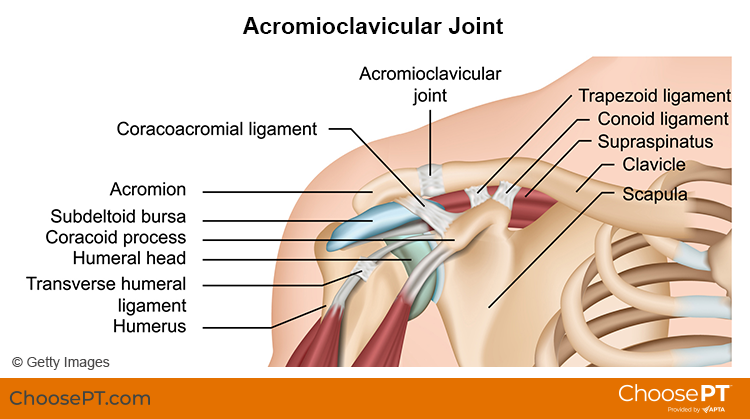
There are four ligaments that hold the two bones of the AC joint together. When an AC joint injury occurs, these ligaments are stressed. This stress results in some degree of joint separation. There are two types of injuries of the AC joint: traumatic and overuse injuries.

Traumatic AC joint injury. This type of injury occurs when the joint is disrupted. The ligaments that hold the two bones of the joint together get stretched too far. This is called a shoulder separation. It is different from a shoulder dislocation, which involves the ball-and-socket joint.

Traumatic AC joint injuries are most common in people who fall and land on the outside of the shoulder or hand. Examples include a:

  • Football player who is tackled.

  • Cyclist who crashes.

  • Worker who falls off a ladder.

Traumatic AC joint injuries can range from a mild to severe grade. Grading is based on the amount of joint separation involved. Mild cases can be treated by a physical therapist. More severe cases may require surgery followed by physical therapy.

Overuse AC joint injury. This type of injury occurs over time due to repeated and too much stress on the joint. Cartilage at the end of the acromion and clavicle bones protects the joint from daily wear and tear. Over time, the demand placed on this cartilage may be more than it can endure. The result is an overuse injury that can lead to major wearing of the cartilage and arthritis. Overuse AC joint injury is most common in people who do repeated tasks. Examples include:

  • Heavy weightlifting (bench and military presses).

  • Jobs that require physical work with the arms stretched over the head.

How Does It Feel?

With an AC joint injury, you may experience:

  • General shoulder pain and swelling.

  • Swelling and tenderness over the AC joint.

  • Loss of shoulder strength.

  • A visible bump above the shoulder.

  • Pain when lying on the involved side.

  • Loss of shoulder motion.

  • A “popping” sound or feeling that your shoulder “catches” with movement.

  • Discomfort with daily activities that stress the AC joint. Examples include lifting objects overhead, reaching across your body, or carrying heavy objects at your side.

How Is It Diagnosed?

Physical therapists can diagnose an AC joint injury through a shoulder exam. Your physical therapist will conduct a full evaluation to find out the degree of your injury and identify all the factors that may contribute to it.

They will begin by interviewing you to learn about your health history. They may be helped by forms you fill out before your first session. The interview will become more specific to the condition of your shoulder. Your physical therapist may ask you questions such as:

  • How did your injury occur?

  • How have you taken care of the condition, such as seeing other health care providers? Have you had imaging (X-ray, MRI) or other tests and received their results?

  • What are your current symptoms? How have they changed your typical day and activities?

  • Do you have pain, and if so, what is the location and intensity of your pain? Does the pain vary during the day?

  • Do you have trouble doing any activities? What activities are you unable to do?

This information allows the physical therapist to better understand what you are going through. It also helps determine the course of your physical exam.

The physical exam will vary depending on your interview. Most often it will begin with observing the region of your symptoms and any movements or positions that cause pain. Your physical therapist also may examine other areas of your body that may have changed due to problems with your shoulder function. They may:

  • Watch you move your arm and shoulder overhead, and while doing other reaching tasks.

  • Assess the mobility and strength of your shoulder.

  • Check other regions of the body as needed. This will help to determine if other areas also require treatment to improve your condition.

  • Gently, but skillfully, feel around your shoulder and the AC joint to find exactly where it is most painful.

Your physical therapist will discuss their findings with you. They will work with you to develop a program for your specific needs and goals and to help you heal.

In some cases, your physical therapist may refer you for diagnostic imaging. Ultrasound, X-ray, or MRI can help to confirm the diagnosis and find out how severe the injury is.

How Can a Physical Therapist Help?

Your physical therapist will design a treatment plan to help you safely return to your desired activities. Your treatment plan may include:

  • Patient education. Your physical therapist will educate you about your AC joint and shoulder injury. They will work with you to identify any external factors causing your pain, including the amount and type of exercises and activities you do. Your physical therapist will recommend improvements to your activities.

  • Pain management. Your physical therapist will address your pain. This may include applying ice to the affected area and other methods. They also may recommend changing some activities that cause pain. Physical therapists are experts in prescribing pain-management methods. These can reduce or eliminate the need for medicines, including opioids.

  • Range-of-motion exercise. The mobility of the AC joint and shoulder may be limited, causing increased stress on the shoulder. Your physical therapist may teach you self-stretching methods. These can decrease tension and help restore normal motion of your injured joints.

  • Manual therapy. Your physical therapist may apply hands-on treatments to gently move your muscles and joints. These techniques help to improve movement. Your physical therapist also may use manual therapy to guide your shoulder area into a less stressful movement pattern.

  • Muscle strength. Muscle weaknesses or imbalances can contribute to problems of the AC joint and shoulder. They also can cause continued symptoms. Based on the how serious your injury is, your physical therapist will design a safe resistance program to aid your recovery. Exercises may include using resistance machines in the clinic and doing exercises to strengthen your core (midsection). You may begin doing exercises while lying on a table or at home on the bed or floor. You then may advance to exercises done in a standing position. Your physical therapist will choose what exercises are right for you based on your diagnosis, age, and condition. They will determine when it is safe for you to exercise on your own at home or in a gym.

  • Functional training. Once your pain, strength, and motion improve, functional training can help you safely resume more demanding activities. To minimize the stress to the AC joint and shoulder it is important to teach your body safe, controlled movements. Your physical therapist will create a series of activities to help you learn how to use and move your body correctly and safely. These may include retraining your movements and positioning when throwing, swinging a racket, lifting objects overhead, or doing other daily activities.

Can This Injury or Condition Be Prevented?

Accidents happen and it can be difficult to prevent many traumatic AC joint injuries. However, much can be done to prevent the string of events that lead to overuse injuries. Physical therapists can help reduce overuse injuries by:

  • Teaching you how to properly lift objects overhead at work.

  • Demonstrating good form for overhead resistance training or sports activities.

  • Helping you maintain general shoulder strength and motion to safely perform tasks.

Consult a physical therapist as soon as possible if you have persistent constant or worsening symptoms.

What Kind of Physical Therapist Do I Need?

All physical therapists are prepared through education and clinical experience to treat a variety of conditions or injuries. You may want to consider:

  • A physical therapist who is experienced in treating people with orthopedic or musculoskeletal (muscle, bone, and joint injuries).

  • A physical therapist who is a board-certified specialist or who has completed a residency in orthopedic or sports physical therapy. This physical therapist will have advanced knowledge, experience, and skills that apply to those who are physically active.

You can find physical therapists who have these and other credentials by using Find a PT, the online tool built by the American Physical Therapy Association. This tool can help you search for physical therapists with specific clinical expertise in your area.

General tips when you're looking for a physical therapist (or any other health care provider):

  • Get recommendations from family, friends, or other health care providers.

  • When you contact a physical therapy clinic for an appointment, ask about the physical therapists' experience in helping patients with shoulder pain.

  • During your first visit with the physical therapist, be prepared to describe your symptoms in as much detail as possible, and report activities and movements that make your symptoms worse.

Further Reading

The American Physical Therapy Association believes that consumers should have access to information to help them make informed health care decisions and prepare them for their visit with a health care provider.

The following resources offer some of the best scientific evidence related to physical therapy treatment for AC joint injuries. They report recent research and give an overview of the standards of practice both in the United States and internationally. They link to a PubMed* abstract that also may offer free access to the full text, or to other resources. You can read them or print out a copy to bring with you to your health care provider.

Frank RM, Cotter EJ, Leroux TS, Romeo AA. Acromioclavicular joint injuries: evidence-based treatment. J Am Acad Orthop Surg. 2019;27(17):e775–e788. Article Summary in PubMed.

Li X, Ma R, Bedi A, Dines DM, Altchek DW, Dines JS. Management of acromioclavicular joint injuries. J Bone Joint Surg Am. 2014;96(1):73–84. Article Summary in PubMed .

Harris KD, Deyle GD, Gill NW, Howes RR. Manual physical therapy for injection-confirmed nonacute acromioclavicular joint pain. J Orthop Sports Phys Ther. 2012;42(2):66–80. Article Summary in PubMed.

Pallis M, Cameron KL, Svoboda SJ, Owens BD. Epidemiology of acromioclavicular joint injury in young athletes. Am J Sports Med. 2012;40(9):2072–2077. Article Summary in PubMed .

*PubMed is a free online resource developed by the National Center for Biotechnology Information. PubMed contains millions of citations to biomedical literature, including citations in the National Library of Medicine's MEDLINE database.


Reviewed and revised in 2021 by Erin Hayden, PT, DPT, board-certified clinical specialist in orthopedic physical therapy, and Stephen Reischl, PT, DPT, board-certified clinical specialist in orthopedic physical therapy, on behalf of the Academy of Orthopaedic Physical Therapy. Authored in 2014 by Allison Mumbleau, PT, DPT, board-certified clinical specialist in sports physical therapy.


Physical Therapy Guide to Spondylolysis and Spondylolisthesis (Fracture of the Lumbar Spine and Slipped Vertebra)

Spondylolysis (spon-dee-low-lye-sis), or lumbar stress fracture, is a stress fracture of a section of the lumbar spine. The area of the fifth lumbar vertebra is most often affected. The injury can occur on the left or right of the vertebra. Lumbar stress fractures occur in up to 11.5% of the general population in the United States. It is a common cause of low back pain in older children and youth, most often young males. It is a common cause of low back pain in older children and youth. It most often occurs in young males, but also can occur in girls. Highly active teens who engage in activities like lifting heavy loads, repeated backward bending, or twisting of the trunk, are most at risk. Activities like football, hockey, gymnastics, or dance put athletes at higher risk. Only a small percentage of cases require surgery. The majority (85% to 90%) of young patients recover in three to six months with proper treatment. Recovery time can be longer and is different for each person.

Spondylolisthesis (spon-dee-low-lis-thee-sis), or slipped vertebra, is a condition that involves the forward slippage of one vertebra over the one under it. If a crack or stress fracture occurs on both sides of the vertebra it is called spondylolisthesis.

Lumbar stress fracture and slipped vertebra are often described together because they are similar in:

  • The mode of injury.

  • Age of the patient.

  • Symptoms.

  • Treatment.

What Are Stress Fracture and Slipped Vertebra of the Lumbar Spine?

Lumbar stress fracture is a fracture of the part of the bony ring that connects the front part of the spinal column to the back part. The fracture occurs between the part of the bone that sticks out of the back of the spine and the part that sticks out of the side of the spine. Doctors sometimes refer to this condition as a "pars defect." Strain on the lumbar spine due to repeated activities in a growing child can cause this type of injury. It results in low back pain.

Slipped vertebra is the forward slip of a defective, unstable vertebra. There are five grades of slips, with grade I being the smallest amount of slippage and grade V being a slippage of 100%. With milder slippage, and a defect on just one side of the vertebra, physical therapy treatment is effective. Young athletes whose teenage growth spurt has not yet occurred are at greater risk for continued slippage. These athletes and are monitored until they are fully grown.

Key points to understand about lumbar stress fracture and slipped vertebra:

  • Early detection and proper diagnosis of these conditions are important. With early diagnosis and treatment, people with these conditions can safely return to sport or an active lifestyle. If symptoms last a long time and you wait to get help, healing may take much longer.

  • The majority of symptoms can resolve with rest and the help of a physical therapist.

  • Surgery may be needed when treatment of more than six months fails, and symptoms persist.

  • Both of these conditions need to be ruled out in a young athlete who is has low back pain that lasts for more than a few weeks. Active young athletes in sports such as football, hockey, gymnastics, and dance are at the greatest risk. This is especially true while the athlete is still growing.

  • If an X-ray does not show a fracture, but a clinical exam suggests a high likelihood of lumbar stress fracture, your doctor may order an MRI to rule it out.

  • These conditions are not a major cause of low back pain in adults. It can, however, occur high-level adult athletes who take part in high-risk sports.


Signs and Symptoms

Lumbar stress fracture or slipped vertebra may be present if you experience:

  • Low back pain with or without buttock or leg pain. If leg pain is present, it is felt into the thigh, but generally not below the knee.

  • Muscle spasms in your low back, buttocks, and thighs.

  • Difficulty or pain with walking or standing for long periods.

  • Symptoms that are relieved by sitting, slouching, or bending forward.

  • Pain with sports or manual labor.

  • Pain with bending backward, twisting the spine, or with throwing.

  • Decreased flexibility of the leg muscles.


How Is It Diagnosed?

Your physical therapist will conduct a thorough evaluation that includes questions about your health history. Their goal is to assess the degree of your injury and to determine the cause and contributing factors. Your physical therapist also may gather information from forms you fill out before your first session. Their questions may include:

  • How did your injury occur? Was there a single episode or did your condition become worse over time?

  • How have you taken care of your condition? Have you seen other health care providers? Have you had imaging (e.g., X-ray, MRI) or other tests, and do you have the results of those tests?

  • How long have you had pain? Did it come on suddenly or gradually?

  • Does your pain occur with activity, at rest, or during the day or night?

  • What activities or positions make your pain better or worse?

  • Do you take part in activities like football, hockey, gymnastics, or competitive dance?

  • Can you point with one finger to the area on your back that is painful?

  • Do you have any other symptoms, such as fever, chills, or night sweats?

  • Do you have trouble with bowel and bladder control?


After your physical therapist learns the specifics of your condition they will conduct a physical exam.

The physical exam most often will begin with watching some of the movements that were discussed in the interview. It will include the area of your main symptoms as well as other areas that may be involved, such as your hip. Your physical therapist may:

  • Watch you walk.

  • Have you bend forward to try to touch your toes, and bend back as far as you can.

  • Ask you to stand on one leg and bend back.

  • Ask you to turn your trunk from side to side.


Your physical therapist uses these tests to assess your leg and spine flexibility as well as your core strength. They may ask you if the testing changes your symptoms. They may gently but skillfully press specific areas of your low back and pelvis to see if they are painful. This information helps your physical therapist determine the cause of your pain, exactly where your pain is, and the best treatment to resolve your symptoms.

After the interview and physical exam, your physical therapist will discuss the findings with you. If your physical therapist suspects a stress fracture, they may refer you to an orthopedic or sports medicine doctor familiar with back injuries. The doctor may order imaging tests (X-ray, MRI) to confirm a diagnosis and rule out other conditions.


How Can a Physical Therapist Help?

Spondylolisis and listhesisGettyImages-1139743540.jpg


Your physical therapist will design a targeted treatment program based on your condition and goals. It will be designed to safely return you to sport or daily activities. Your treatment plan may include:

Patient education. Your physical therapist will work with you to identify and change any external factors causing your pain. These factors can include the type and amount of exercise you do, your athletic activities, footwear, or the surfaces on which you practice and play. They may recommend changes in your daily activities.

Pain management. Your physical therapist will design a program to address your pain. This may include applying ice to the affected area. Applying heat also is helpful in some cases. Electrical stimulation gently targets nerve fibers that send pain signals to the brain. It also may be used together with ice to help relieve your pain. Your physical therapist also may recommend decreasing some activities that cause pain. Physical therapists are experts in prescribing pain-management techniques that reduce or avoid the need for medicines, including opioids.

Body mechanics. How you move and use your body for work and other activities can contribute to lumbar spine problems and pain. Your physical therapist will teach you how to improve your movements or body mechanics based on your daily activities. They also may make recommendations to improve the way you do certain activities, such as lifting and carrying objects.

Manual therapy. Often, manual therapy for lumbar stress fracture and slipped vertebra includes "soft tissue release" or massage for tight and sore muscle groups. These hands-on techniques may be used to correct tightness of muscles to promote normal movement.

Stretching exercises. Stretching exercises can help improve the flexibility of tight muscles. They also may help to improve movement in the spine and lower extremities and help decrease stress on the lumbar spine during daily activities.

Strengthening exercises. Strengthening helps to make the lumbar spine, pelvis, and hip joints more stable. This, in turn, helps to reduce strain on tissues, and pain. These movements are focused on weak muscles, including the lower abdominal, pelvic floor, and buttocks muscles.

Functional training. Once your pain, strength, and motion improve you will need to safely move back into more demanding activities. To lessen your risk of repeated injury, it is important to learn safe, controlled movements. Based on your unique movement assessment and goals, your physical therapist will create a series of activities to help you use and move your body more correctly and safely for years to come.


Can This Injury or Condition Be Prevented?

Lumbar stress fracture and some types of slipped vertebra may be preventable by educating individuals who are at higher risk of injury.

For the growing young athlete, it is necessary to manage how much, how intensely, and how often you exercise. Parents and coaches should:

  • Limit a child’s participation to one high-risk sport at a time during a season.

  • Limit participation to only one team at a time during a season.

  • Require and enforce one to two days of rest from training per week.

  • Gradually increase training volume, intensity, and frequency when a person is starting a new sport or activity.


What Kind of Physical Therapist Do I Need?

All physical therapists are prepared through education and experience to treat lumbar stress fracture and slipped vertebra. However, you may want to consider:

  • A physical therapist who is experienced in treating people with spine injuries and/or athletes. Some physical therapists have a practice with an orthopaedic or sports physical therapy focus.

  • A physical therapist who is a board-certified clinical specialist or who completed a residency or fellowship in orthopaedic and/or sports physical therapy. This physical therapist has advanced knowledge, experience, and skills that may apply to your condition.

General tips when you are looking for a physical therapist (or any other health care provider):

  • Get recommendations from family, friends, or from other health care providers.

  • When you contact a physical therapy clinic for an appointment, ask about the physical therapists' experience in helping people who are athletes or active individuals with lumbar stress fracture.

  • During your first visit with the physical therapist, be prepared to describe your symptoms in as much detail as possible, and say what makes your symptoms worse.


Further Reading

The American Physical Therapy Association believes that consumers should have access to information that could help them make health care decisions and also prepare them for a visit with their health care provider.

The following articles provide some of the best scientific evidence related to physical therapy treatment of lumbar stress fracture and slipped vertebra. The articles report recent research and give an overview of the standards of practice both in the United States and internationally. The article titles are linked either to a PubMed* abstract of the article or to free full text, so that you can read it or print out a copy to bring with you to your health care provider.

Iwaki K, Sakai T, Hatayama D, et al. Physical features of pediatric patients with lumbar spondylolysis and effectiveness of rehabilitation. J Med Invest. 2018;65(3.4):177–83. Article Summary in PubMed.

Lawrence KJ, Elsar T, Stromberg R. Lumbar spondylolysis in the adolescent athlete. Phys Ther Sport. 2016;20:56–60. Article Summary in PubMed.

Schroeder GD, LaBelle CR, Mendoza M. The role of intense athletic activity on structural lumbar abnormalities in adolescent patients with symptomatic low back pain. Eur Spine J. 2016;25:2842–2848. Article Summary in PubMed.

Kim HJ, Green DW. Spondylolysis in the adolescent athlete. Curr Opin Pediatr. 2011;23(1):68–72. Article Summary in PubMed.

Kalichman L, Kim DH, Li L, et al. Spondylolysis and Spondylolisthesis: prevalence and association with low back pain in the adult community-based population. Spine (Phila Pa 1976). 2009;34(2):199–205. Article Summary in PubMed.

*PubMed is a free online resource developed by the National Center for Biotechnology Information. PubMed contains millions of citations to biomedical literature, including citations in the National Library of Medicine’s MEDLINE database.


Revised in 2020 by Susan Reischl, PT, DPT, board-certified clinical specialist in orthopaedic physical therapy, and reviewed by Stephen Reischl, PT, DPT, board-certified clinical specialist in orthopaedic physical therapy, on behalf of the Academy of Orthopaedic Physical Therapy. Authored in 2014 by Donna Merkel, PT.


Physical Therapist's Guide to Patellofemoral Pain

What Is Patellofemoral Pain?

PFP may occur after a sudden increase in activities like running or jumping. Research suggests that PFP results from activity levels that are increased faster than the knee can adapt. Other contributing factors to PFP may include:

  • Weakness of the thigh muscles.

  • Specializing in a single sport, which requires repeating the same movements again and again.

  • Certain hip and knee coordination patterns during running and jumping activities.

PFP does not go away on its own. If you have symptoms of PFP, it’s important to seek care from a physical therapist so you can return to the activities that you enjoy.

How Does it Feel?

People with PFP may experience pain:

  • When walking up or down stairs or hills.

  • When playing a sport.

  • With deep knee bending (squatting).

  • When walking on uneven surfaces.

  • With activity, but improving with rest.

  • After sitting for long periods of time with the knee bent.

How Is It Diagnosed?

APTA-Knee-Joint-Patella-Pain_750x419.jpg

Your physical therapist will review your health history and conduct a series of tests to evaluate you and your knee. PFP is diagnosed by analyzing any movement that causes pain, and ruling out other possible conditions.

Your physical therapist may analyze your walking and running patterns. They may test the strength of your hip and thigh muscles to find out if weakness is contributing to your pain. Medical imaging, such as an X-ray or MRI, is not helpful in diagnosing PFP. However, your physical therapist may consult with an orthopedic physician who may order imaging to rule out other conditions.

How Can a Physical Therapist Help?

If PFP is diagnosed, your physical therapist will develop an exercise and rehabilitation program just for you. Your program may include:

Strengthening exercises. Your physical therapist will teach you exercises to help strengthen the muscles around the hip and the knee itself. Research shows that this type of exercise therapy is the best approach to managing PFP.

Taping. Your physical therapist may teach you how to apply tape to your knee, which may improve your ability to perform exercises that would normally be painful. However, taping alone will not resolve PFP. It must only be used along with your exercise program.

Shoe inserts. Your physical therapist may recommend shoe inserts to help reduce your pain when exercising. But inserts alone, like taping, will not treat PFP. Your physical therapist will design an exercise program to fit your specific needs and goals.

Coordination training. Based on your activity level, your physical therapist may help retrain your hip and knee movement patterns to reduce your knee pain.

This type of training is effective for athletes, in particular, and may focus on movements like:

  • Stair climbing.

  • Squatting.

  • Running and jumping.

Cross-training guidance. Physical therapists help athletes and active people perform different movements (cross-training). This helps them stay active until they can return to a favorite activity.

Return to full activity. Your physical therapist will help guide a gradual return to your favorite activities, such as running and jumping, and will teach you good overall exercise habits to help maximize the health of your knee.

Treatments That Do Not Work for PFP

While these can be appropriate for the treatment of other injuries or conditions, the following do notwork for PFP:

Quick fixes. “Passive” treatments like dry needling, ultrasound, laser, or electrical stimulation are not helpful for people with PFP. The most effective treatment for PFP is an exercise program that targets the hip and knee muscles.

Rest. If you are experiencing PFP, it is important to understand that rest only helps temporarily. Your pain will likely return when you go back to your normal activity. Rest is not helpful in the long term. A movement program guided by your physical therapist is your best treatment option.

Can this Injury or Condition be Prevented?

Current research shows that a person’s age, height, body weight, or foot alignment may not contribute to the risk of developing PFP at all. A knock-kneed posture also does not increase the risk of developing PFP.

However, a few preventive measures can be effective. To help reduce your risk of developing PFP:

  • Keep your thigh muscles strong.

  • Maintain good exercise habits.

  • Avoid rapid spikes in activity levels.

  • Participate in a variety of sports, rather than just repeating the same movements again and again.

What Kind of Physical Therapist Do I Need?

All physical therapists are prepared through education and experience to treat a variety of conditions or injuries including PFP. However, you may want to consider:

  • A physical therapist who is experienced in treating people with orthopedic, or musculoskeletal, problems.

  • A physical therapist who is a board-certified clinical specialist or who has completed a residency or fellowship in orthopedic physical therapy. This physical therapist will have advanced knowledge, experience, and skills that may apply to your condition.

  • While it may be tempting to seek quick fixes for your knee pain, there is no evidence that passive treatments work for persons with PFP. If you have PFP, seek care from a physical therapist who uses progressive exercise therapy for the treatment of this condition.

You can find physical therapists who have these and other credentials by using Find a PT, an online tool provided by the American Physical Therapy Association. You can search for physical therapists with specific clinical expertise in your geographic area.

General tips when you're looking for a physical therapist:

  • Get recommendations from family and friends or from other health care providers.

  • When you contact a physical therapy clinic for an appointment, ask about the physical therapist's experience in helping people with patellofemoral pain (PFP).

  • During your first visit with a physical therapist, you will be asked to describe your symptoms in as much detail as possible, and say what makes your symptoms worse. Here are some tips to prepare for your visit.

Further Reading

The American Physical Therapy Association (APTA) believes that consumers should have access to information that could help them make health care decisions and prepare for a visit with their health care provider.

The following articles provide some of the best scientific evidence related to physical therapy treatment of patellofemoral pain syndrome. The articles report recent research and give an overview of the standards of practice both in the United States and internationally. The article titles are linked either to a PubMed* abstract of the article or to free access of the full article, so you can read it or print out a copy to bring with you to your health care provider.

Patellofemoral pain: treating painful kneecaps. J Orthop Sports Phys Ther. 2019;49(9):633. Free Article.

Willy RW, Hoglund LT, Barton CJ, et al. Patellofemoral Pain. J Orthop Sports Phys Ther.2019;49(9):CPG1–CPG95. Free Article.

Neal BS, Lack SD, Lankhorst NE, Raye A, Morrissey D, van Middelkoop M. Risk factors for patellofemoral pain: a systematic review and meta-analysis. Br J Sports Med. 2019;53(5):270–281. Free Article.

Lack S, Barton C, Sohan O, Crossley K, Morrissey D. Proximal muscle rehabilitation is effective for patellofemoral pain: a systematic review with meta-analysis. Br J Sports Med. 2019;49(21):1365-1376. Free Article.

* PubMed is a free online resource developed by the National Center for Biotechnology Information (NCBI). PubMed contains millions of citations to biomedical literature, including citations in the National Library of Medicine’s MEDLINE database.

Authored by Christopher Bise, PT, DPT, MS. Revised and reviewed by Richard Willy, PT, PhD.

Guide to Anterior Cruciate Ligament (ACL) Tear

An anterior cruciate ligament (ACL) tear is an injury to the knee commonly affecting athletes, such as soccer players, basketball players, skiers, and gymnasts. Nonathletes can also experience an ACL tear due to injury or accident. Approximately 200,000 ACL injuries are diagnosed in the United States each year. It is estimated that there are 95,000 ruptures of the ACL and 100,000 ACL reconstructions performed per year in the United States. Approximately 70% of ACL tears in sports are the result of noncontact injuries, and 30% are the result of direct contact (player-to-player, player-to-object). Women are more likely than men to experience an ACL tear. Physical therapists are trained to help individuals with ACL tears reduce pain and swelling, regain strength and movement, and return to desired activities.

What is an ACL Tear?

The ACL is one of the major bands of tissue (ligaments) connecting the thigh bone (femur) to the shin bone (tibia) at the knee joint. It can tear if you:

  • Twist your knee while keeping your foot planted on the ground.

  • Stop suddenly while running.

  • Suddenly shift your weight from one leg to the other.

  • Jump and land on an extended (straightened) knee.

  • Stretch the knee farther than its usual range of movement.

  • Experience a direct hit to the knee.

ACLAttachment_Small.jpg

ACL Attachment: See More Detail

Back to Top

How Does it Feel?

When you tear the ACL, you may feel a sharp, intense pain or hear a loud "pop" or snap. You might not be able to walk on the injured leg because you can’t support your weight through your knee joint. Usually, the knee will swell immediately (within minutes to a few hours), and you might feel that your knee "gives way" when you walk or put weight on it.

Back to Top

How Is It Diagnosed?

Immediately following an injury, you may be examined by a physical therapist, athletic trainer, or orthopedic surgeon. If you see your physical therapist first, your therapist will conduct a thorough evaluation that includes reviewing your health history. Your physical therapist will ask:

  • What you were doing when the injury occurred.

  • If you felt pain or heard a "pop" when the injury occurred.

  • If you experienced swelling around the knee in the first 2 to 3 hours following the injury.

  • If you felt your knee buckle or give out when you tried to get up from a chair, walk up or down stairs, or change direction while walking.

Your physical therapist may perform gentle "hands-on" tests to determine the likelihood that you have an ACL tear, and may use additional tests to assess possible damage to other parts of your knee.

An orthopedic surgeon may order further tests, including magnetic resonance imaging (MRI), to confirm the diagnosis and rule out other possible damage to the knee.

Surgery

Most people who sustain an ACL tear will undergo surgery to repair the tear; however, some people may avoid surgery by modifying their physical activity to relieve stress on the knee. A select group can actually return to vigorous physical activity following rehabilitation without having surgery.

Your physical therapist, together with your surgeon, can help you determine if nonoperative treatment (rehabilitation without surgery) is a reasonable option for you. If you elect to have surgery, your physical therapist will help you prepare both for surgery and to recover your strength and movement following surgery.


Back to Top

How Can a Physical Therapist Help?

Once an ACL tear has been diagnosed, you will work with your surgeon and physical therapist to decide if you should have surgery, or if you can recover without surgery. If you don’t have surgery, your physical therapist will work with you to restore your muscle strength, agility, and balance, so you can return to your regular activities. Your physical therapist may teach you ways to modify your physical activity in order to put less stress on your knee. If you decide to have surgery your physical therapist can help you before and after the procedure.

Treatment Without Surgery

Current research has identified a specific group of patients (called "copers") who have the potential for healing without surgery following an ACL tear. These patients have injured only the ACL, and have experienced no episodes of the knee "giving out" following the initial injury. If you fall into this category, based on the specific tests your physical therapist will conduct, your therapist will design an individualized physical therapy treatment program for you. It may include treatments such as gentle electrical stimulation applied to the quadriceps muscle, muscle strengthening, and balance training.

Treatment Before Surgery

If your orthopedic surgeon determines that surgery is necessary, your physical therapist can work with you before and after your surgery. Some surgeons refer their patients to a physical therapist for a short course of rehabilitation before surgery. Your physical therapist will help you decrease your swelling, increase the range of movement of your knee, and strengthen your thigh muscles (quadriceps).

Treatment After Surgery

Your orthopedic surgeon will provide postsurgery instructions to your physical therapist, who will design an individualized treatment program based on your specific needs and goals. Your treatment program may include:

Bearing weight. Following surgery, you will use crutches to walk. The amount of weight you are allowed to put on your leg and how long you use the crutches will depend on the type of reconstructive surgery you have received. Your physical therapist will design a treatment program to meet your needs and gently guide you toward full weight bearing.

Icing and compression. Immediately following surgery, your physical therapist will control your swelling with a cold application, such as an ice sleeve, that fits around your knee and compresses it.

Bracing. Some surgeons will give you a brace to limit your knee movement (range of motion) following surgery. Your physical therapist will fit you with the brace and teach you how to use it safely. Some athletes will be fitted for braces as they recover and begin to return to their sports activities.

Movement exercises. During your first week following surgery, your physical therapist will help you begin to regain motion in the knee area, and teach you gentle exercises you can do at home. The focus will be on regaining full movement of your knee. The early exercises help with increasing blood flow, which also helps reduce swelling.

Electrical stimulation. Your physical therapist may use electrical stimulation to help restore your thigh muscle strength, and help you achieve those last few degrees of knee motion.

Strengthening exercises. In the first 4 weeks after surgery, your physical therapist will help you increase your ability to put weight on your knee, using a combination of weight-bearing and non-weight-bearing exercises. The exercises will focus on your thigh muscles (quadriceps and hamstrings) and might be limited to a specific range of motion to protect the new ACL. During subsequent weeks, your physical therapist may increase the intensity of your exercises and add balance exercises to your program.

Balance exercises. Your physical therapist will guide you through exercises on varied surfaces to help restore your balance. Initially, the exercises will help you gently shift your weight on to the surgery leg. These activities will progress to standing on the surgery leg, while on firm and unsteady surfaces to challenge your balance.

Return to sport or activities. As athletes regain strength and balance, they may begin running, jumping, hopping, and other exercises specific to their individual sport. This phase varies greatly from person-to-person. Physical therapists design return-to-sport treatment programs to fit individual needs and goals.

Back to Top

Can this Injury or Condition be Prevented?

Much of the research on ACL tears has been conducted with female collegiate athletes, because women are 4 to 6 times more likely to experience the injury. Preventive physical therapy programs have proven to lower ACL injury rates by 41% for female soccer players. Researchers have made the following recommendations for a preventive exercise program:

  • The program should be designed to improve balance, strength, and sports performance. Strengthening your core (abdominal) muscles is key to preventing injury, in addition to strengthening your thigh and leg muscles.

  • Exercises should be performed 2 or 3 times per week and should include sport-specific exercises.

  • The program should last no fewer than 6 weeks.

Although most exercise studies have been conducted with female athletes, the findings may benefit male athletes as well.

Back to Top

Real Life Experiences

Anita is a 20-year-old student at a local university, and a star basketball player. Her team is off to a great start this year; the buzz around campus is that this could be a dream team!

But tonight, when Anita goes up for a rebound and lands off-balance, she hears a "pop" in her left knee and feels a sharp pain. When she tries to walk, she realizes that she can't put weight on her left leg. She's led back to the training room, where the school physical therapist conducts an evaluation. The test results indicate injury, and the physical therapist notices an increase in swelling around the knee just 30 minutes after the incident. She suspects an ACL tear, and refers Anita to an orthopedic surgeon. The next day, the surgeon confirms the diagnosis of an ACL tear, and tells Anita that her injury requires surgery.

After a short course of treatment by her new local physical therapist, including pain and swelling management, manual (hands-on) therapy, and knee range-of-motion and strengthening exercises, Anita has surgery the following month. Her surgeon schedules her to receive physical therapy 3 days after her surgery. She is advised to ice and elevate the knee several times per day.

Three days after surgery, Anita returns to her local physical therapist to begin her rehabilitation. He shows her how to use her crutches properly to gently begin to put weight on the operative knee. He guides her to contract/tighten the quadriceps muscle, and gently performs manual (hands-on) stretches for her to straighten the knee.

Over the next few weeks, Anita is able to gradually stop using her crutches, and begins to put her full weight on her left leg. She can also fully straighten her knee and tighten her quadriceps muscle without help from her physical therapist. She learns exercises she can safely perform at home.

After 5 weeks, Anita is able to walk normally, fully extending her knee with no pain or feelings of instability. During the next 2 months, she and her physical therapist work on her strength and balance. She finds the hardest exercises are the balance exercises, which require her to balance on a piece of foam or a rocker board while throwing a ball.

About 4 months after surgery, Anita's physical therapist designs a gentle jogging program for her. At 5 months, he allows her to begin a running program. He also adds exercises during Anita's physical therapy sessions that mimic basketball activities such as rebounding or taking a jump shot. During these activities, Anita’s physical therapist teaches her proper landing techniques to lessen the chance of reinjuring her knee when she returns to play.

After 8 months, Anita is allowed to practice with her team. They are thrilled and excited to see their star player is back. Last year was a good year for the team, but it ended in the first round of the playoffs.

Anita and her team begin a new year of full competition 11 months after her surgery. With Anita back in top form, they make the playoffs, blast through to the finals – and bring home the trophy!

This story was based on a real-life case. Your case may be different. Your physical therapist will tailor a treatment program to your specific case.

Back to Top

What Kind of Physical Therapist Do I Need?

Although all physical therapists are prepared through education and experience to treat a variety of conditions or injuries, you may want to consider:

  • A physical therapist who is experienced in treating people with orthopedic (musculoskeletal) problems.

  • A physical therapist who is a board-certified clinical specialist or who has completed a residency or fellowship in orthopedic physical therapy and has advanced knowledge, experience, and skills that may apply to your condition.

You can find physical therapists with these and other credentials by using Find a PT, the online tool built by the American Physical Therapy Association to help you search for physical therapists with specific clinical expertise in your geographic area.

General tips when you're looking for a physical therapist:

  • Get recommendations from family and friends or from other health care providers.

  • When you contact a physical therapy clinic for an appointment, ask about the physical therapist's experience in helping people with ACL tears.

During your first visit with the physical therapist, be prepared to describe your symptoms in as much detail as possible, and say what makes your symptoms worse.

Back to Top

Further Reading

The American Physical Therapy Association (APTA) believes that consumers should have access to information that could help them make health care decisions and also prepare them for their visit with their health care provider.

The following articles provide some of the best scientific evidence related to physical therapy treatment of ACL tears. The articles report recent research and give an overview of the standards of practice for treatment both in the United States and internationally. The article titles are listed by year and are linked either to a PubMed* abstract of the article or to free access of the full article, so that you can read it or print out a copy to bring with you to your health care provider.

Nyland J, Mattocks A, Kibbe S, Kalloub A, Greene JW, Caborn DN. Anterior cruciate ligament reconstruction, rehabilitation, and return to play: 2015 update. Open Access J Sports Med. 2016;7:21–32. Free Article.

Anderson MJ, Browning WM III, Urband CE, Kluczynski MA, Bisson LJ. A systematic summary of the systematic reviews on the topic of the anterior cruciate ligament. Orthop J Sports Med. 2016;4:2325967116634074. Free Article.

Anterior cruciate ligament injury. Medscape website. Accessed June 16, 2016.

Logerstedt DS, Snyder-Mackler L, Ritter RC, Axe MJ, Godges JJ; Orthopaedic Section of the American Physical Therapy Association. Knee stability and movement coordination impairments: knee ligament sprain. J Orthop Sports Phys Ther. 2010;40:A1–A37. Free Article.

Eitzen I, Moksnes H, Snyder-Mackler L, Risberg MA. A progressive 5-week exercise therapy program leads to significant improvement in knee function early after anterior cruciate ligament injury. J Orthop Sports Phys Ther. 2010;40:705-721. Free Article.

Nyland J, Brand E, Fisher B. Update on rehabilitation following ACL reconstruction. Open Access J Sports Med. 2010;1:151–166. Free Article.

Risberg MA, Holm I. The long-term effect of 2 postoperative rehabilitation programs after anterior cruciate ligament reconstruction: a randomized controlled clinical trial with 2 years of follow-up. Am J Sports Med. 2009;37:1958–1966. Free Article.

Gilchrist J, Mandelbaum BR, Melancon H, et al. A randomized controlled trial to prevent noncontact anterior cruciate ligament injury in female collegiate soccer players. Am J Sports Med. 2008;36:1476–1483. Article Summary on PubMed.

Hurd WJ, Axe MJ, Snyder-Mackler L. A 10-year prospective trial of a patient management algorithm and screening examination for highly active individuals with anterior cruciate ligament injury: Part 1, outcomes. Am J Sports Med. 2008;36:40-47. Free Article.

Benjaminse A, Gokeler A, van der Schans CP. Clinical diagnosis of an anterior cruciate ligament rupture: a meta-analysis. J Orthop Sports Phys Ther. 2006;36:267–288. Article Summary on PubMed.

Hewett TE, Ford KR, Myer GD. Anterior cruciate ligament injuries in female athletes: part 2, a meta-analysis of neuromuscular interventions aimed at injury prevention. Am J Sports Med. 2006;34:490–498. Article Summary on PubMed.

Beynnon BD, Johnson RJ, Abate JA, Fleming BC, Nichol CE. Treatment of anterior cruciate ligament injuries, part 2. Am J Sports Med. 2005;33:1751–1767. Article Summary on PubMed.

Fitzgerald GK, Piva SR, Irrgang JJ. A modified neuromuscular electrical stimulation protocol for quadriceps strength training following anterior cruciate ligament reconstruction. J Orthop Sports Phys Ther. 2003;33:492–501. Article Summary on PubMed.

*PubMed is a free online resource developed by the National Center for Biotechnology Information (NCBI). PubMed contains millions of citations to biomedical literature, including citations in the National Library of Medicine’s MEDLINE database.

Authored by Christopher Bise, PT, DPT, MS. Revised by Julie Mulcahy, PT. Reviewed by the editorial board.

Tarsal Tunnel Syndrome

Tarsal tunnel syndrome (TTS) is a condition that develops when a nerve within the tarsal tunnel of the inner ankle is compressed. TTS can lead to alterations in sensation and movement of the foot, ankle, and lower leg, and/or pain. It is often associated with conditions causing increased compression or swelling in the lower leg. Physical therapists help people experiencing TTS to relieve their pain and restore their normal function.

What is Tarsal Tunnel Syndrome?

Often described as the carpal tunnel syndrome of the lower extremity, tarsal tunnel syndrome is a condition that results from the compression of the posterior tibial nerve as it runs through the tarsal tunnel (a structure made up of bone and tissue (retinaculum) on the inside of the ankle). As it passes through the tarsal tunnel, the tibial nerve divides into 3 branches that provide sensation for the heel and bottom of the foot, and aid in the foot's function. When this structure becomes compressed, symptoms, such as pain, numbness, and/or tingling may occur and radiate into the lower leg, foot, and toes. Individuals may also experience muscle weakness in the area.

How Does it Feel?

The most common symptoms of TTS result from irritation of the tibial nerve and its branches. People with TTS may experience:

  • Pain, numbness, or tingling in the foot or ankle, which may radiate into the lower leg, foot, and toes

  • Weakness in the muscles of the lower leg and foot

  • Weakness of the big toe

  • Foot swelling

  • Symptoms that increase with prolonged standing or walking

  • Symptoms that decrease with rest

  • Altered temperatures of the foot and ankle

  • Pain that disrupts sleep

How Is It Diagnosed?

There are several tests that can help a clinician determine if TTS is present. Your physical therapist and/or physician will first take a comprehensive health history, and inquire about your current symptoms. Then your physical therapist may conduct tests, such as:

  • Gently tapping over the posterior tibial nerve in an attempt to reproduce your symptoms.

  • Tensing of the posterior tibial nerve, a maneuver that looks and feels like a "stretch," in an attempt to reproduce your symptoms.

  • Conducting a nerve condition study—a diagnostic test to determine the speed at which a nerve conducts information.

  • Ruling out other conditions, such as plantarfasciitis (inflammation of the tissue that runs along the bottom of the foot).

How Can a Physical Therapist Help?

Physical therapists play a vital role in helping people experiencing TTS to improve and maintain their daily function and activities. Your physical therapist will work with you to develop a treatment plan to help address your specific needs and goals.  

Because the signs and symptoms of TTS can vary, the approach to care will also vary. Your physical therapist may provide the following recommendations and care:

Nerve Gliding Activities. Gentle exercises that move and "glide" the nerves may help reduce symptoms and improve function.

Muscle Strengthening Exercises. Strengthening activities for any muscles affected by TTS, such as the tibialis posterior muscle in the back of your lower leg.

Balance and Coordination Activities. Work to improve your balance and coordination, which are often affected by TTS.

Orthotics/Taping/Bracing. Apply ankle taping, a custom orthotic, or bracing to position the foot to decrease stress on the posterior tibial nerve.

As with many conditions, education is key. Understanding the underlying mechanisms of TTS, and learning to recognize early signs and symptoms of stress may help you better manage the condition.

Can this Injury or Condition be Prevented?

Although there are no proven strategies for preventing TTS, there are ways to minimize stress to the foot and ankle, such as choosing appropriate footwear, wearing custom orthotics, minimizing the amount of time spent standing on hard surfaces, and improving and maintaining strength in the muscles of your legs, ankles, and feet. These strategies can be discussed further with your physical therapist.

In addition, early detection of the signs and symptoms of TTS will help you and your medical providers begin appropriate management of the condition, which may enhance your long-term well-being.

Real Life Experiences

Kim is a 46-year-old woman who works on an automobile assembly line. Her job involves standing on hard surfaces for prolonged periods of time. Kim recently noticed an onset of pain in her inner ankle after working a few hours, as well as an occasional shooting pain in her big toe. Now, her pain gets progressively worse throughout the day, and often interrupts her sleep. She is afraid of losing her job if she mentions her symptoms to her boss, and really can't afford to miss work. Kim decides to call her physical therapist.

Kim's physical therapist asks about her medical history, and learns that Kim has been diagnosed with high blood pressure and high cholesterol. They discuss Kim’s current symptoms.

He examines Kim’s ankle motion and strength, and gently performs procedures to provoke her symptoms. He also observes how she walks, and assesses her balance. Based on these signs and symptoms, he diagnoses tarsal tunnel syndrome.

Over the next several weeks, Kim works with her physical therapist to reduce her pain and improve her function. Her treatments include:

  • Application of a custom orthotic to better support her foot/ankle.

  • Nerve-gliding activities to improve the mobility of the posterior tibial nerve.

  • Balance exercises.

  • Manual therapy to ease her pain and improve her ankle mobility.

  • Strengthening exercises for her affected muscles.

  • Education about modifying her work positioning and activities.

After 4 weeks of physical therapy, Kim reports a significant reduction in her symptoms. She says she no longer fears going to work, and believes that she has taken control of her current situation. She continues to perform the home-based exercises her physical therapist taught her, and is amazed at how comfortable her feet feel throughout the workday—in her new shoes with custom orthotic inserts.

This story highlights an individualized experience of TTS. Your case may be different. Your physical therapist will tailor a treatment program to your specific needs.

What Kind of Physical Therapist Do I Need?

All physical therapists are prepared through education and experience to treat conditions, such as TTS. However, when seeking a provider, you may want to consider:

  • A physical therapist who is a board-certified clinical specialist, or who has completed a residency or fellowship in physical therapy. This therapist has advanced knowledge, experience, and skills that may apply to your condition.

  • A physical therapist who is well-versed in the treatment of TTS or other neuropathic disorders.

You can find physical therapists who have these and other credentials by using Find a PT, the online tool built by the American Physical Therapy Association to help you search for physical therapists with specific clinical expertise in your geographic area.

General tips when you're looking for a physical therapist (or any other health care provider):

  • Get recommendations from family and friends or from other health care providers.

  • When you contact a physical therapy clinic for an appointment, ask about the physical therapists' experience in helping people with painful conditions

  • During your first visit with the physical therapist, be prepared to describe your symptoms in as much detail as possible. Keeping a journal highlighting when you experience pain will help the physical therapist identify the best way of approaching care.


Further Reading

The American Physical Therapy Association (APTA) believes that consumers should have access to information that could help them make health care decisions, and also prepare them for a visit with their health care provider.

The following articles provide some of the best scientific evidence for the treatment of Tarsal Tunnel Syndrome. The articles report recent research and give an overview of the standards of practice both in the United States and internationally. The article titles are linked either to a PubMed* abstract of the article or to free full text, so that you can read it or print out a copy to bring with you to your health care provider.

Alshama AM, Souvlis T, Coppieters MW. A review of plantar heel pain of neural origin: differential diagnosis and management. Man Ther. 2008;13(2):103–111. Article Summary on PubMed.

Kavlak Y, Uygur F. Effects of nerve mobilization exercise as an adjunct to the conservative treatment for patients with tarsal tunnel syndrome. J Manipulative Physiol Ther. 2011;34(7):441–448. Article Summary on PubMed.

*PubMed is a free online resource developed by the National Center for Biotechnology Information (NCBI). PubMed contains millions of citations to biomedical literature, including citations in the National Library of Medicine’s MEDLINE database.

Acknowledgements: Joseph Brence, PT, DPT, FAAOMPT, COMT, DAC. Reviewed by the MoveForwardPT.com editorial board.


Total Knee Replacement (Arthroplasty)

The knee is the most commonly replaced joint in the body. The decision to have knee replacement surgery is one that you should make in consultation with your orthopedic surgeon and your physical therapist. Usually, total knee replacement surgery is performed when people have:

  • Knee joint damage due to osteoarthritisrheumatoid arthritis, other bone diseases, or fracture that has not responded to more conservative treatment options

  • Knee pain or alignment problems in the leg that cause difficulty with walking or performing daily activities, which have not responded to more conservative treatment options

What is a Total Knee Replacement (TKR)?

A total knee replacement (TKR), also known as total knee arthroplasty, involves removing the arthritic parts of the bones at the knee joint (the tibia, sometimes called the shin bone; the femur, or thigh bone; and the patella, or kneecap) and replacing them with artificial parts. These parts consist of a metal cap at the end of the femur and a cemented piece of metal in the tibia with a plastic cap on it to allow the surfaces to move smoothly. When appropriate, the back part of the kneecap also may be replaced with a smooth plastic surface.

KneeReplacement-SM.jpg

How Can a Physical Therapist Help?

The physical therapist is an integral part of the team of health care professionals who help people receiving a total knee replacement regain movement and function, and return to daily activities. Your physical therapist can help you prepare for and recover from surgery, and develop an individualized treatment program to get you moving again in the safest and most effective way possible.

Before Surgery

The better physical shape you are in before TKR surgery, the better your results will be (especially in the short term). A recent study has shown that even 1 visit with a physical therapist prior to surgery can help reduce the need for short-term care after surgery, such as a short stay at a skilled nursing facility, or a home health physical therapy program.

Before surgery, your physical therapist may:

  • Teach you exercises to improve the strength and flexibility of the knee joint and surrounding muscles.

  • Demonstrate how you will walk with assistance after your operation, and prepare you for the use of an assistive device, such as a walker.

  • Discuss precautions and home adaptations with you, such as removing loose accent rugs that could cause you to “catch” your leg on them when maneuvering with an assistive device, or strategically placing a chair so that you can sit instead of squatting to get something out of a low cabinet. It is always easier to make these modifications before you have TKR surgery.

Longer-term adjustments that are recommended prior to surgery include:

  • Stopping smoking. Seek assistance or advice from your physician on stopping smoking, as you schedule and plan for your surgery. Being tobacco-free will improve your healing process following surgery.

  • Losing weight. Losing excess body weight may help you recover more quickly, and help improve your function and overall results following surgery.

Immediately Following Surgery

You may stay in the hospital for a few days following surgery, or you may even go home on the same day, depending on your condition. If you have other medical conditions, such as diabetes or heart disease, you might need to stay in the hospital or go to a skilled nursing facility for a few days before returning home. While you are in the hospital, a physical therapist will:

  • Educate you on applying ice, elevating your leg, and using compression wraps or stockings to control swelling in the knee area and help the incision heal.

  • Teach you breathing exercises to help you relax, and show you how to safely get in and out of bed and a chair.

  • Show you how to walk with a walker or crutches, and get in and out of a car.

  • Help you continue to do the flexibility and strengthening exercises that you learned before your surgery.

As You Begin to Recover

The goal of the first 2 weeks of recovery is to manage pain, decrease swelling, heal the incision, restore normal walking, and initiate exercise. Following those 2 weeks, your physical therapist will tailor your range-of-motion exercises, progressive muscle-strengthening exercises, body awareness and balance training, functional training, and activity-specific training to address your specific goals and get you back to the activities you love!

Range-of-motion exercises. Swelling and pain can make you move your knee less. Your physical therapist can teach you safe and effective exercises to restore movement (range of motion) to your knee, so that you can perform your daily activities.

Strengthening exercises. Weakness of the muscles of the thigh and lower leg could make you need to still use a cane when walking, even after you no longer need a walker or crutches. Your physical therapist can determine which strengthening exercises are right for you.

Body awareness and balance training. Specialized training exercises help your muscles "learn" to respond to changes in your world, such as uneven sidewalks or rocky ground. When you are able to put your full weight on your knee without pain, your physical therapist may add agility exercises (such as turning and changing direction when walking, or making quick stops and starts) and activities using a balance board that challenge your balance and knee control. Your program will be based on the physical therapist’s examination of your knee, on your goals, and on your activity level and general health.

Functional training. When you can walk freely without pain, your physical therapist may begin to add activities that you were doing before your knee pain started to limit you. These might include community-based actions, such as crossing a busy street or getting on and off an escalator. Your program will be based on the physical therapist's examination of your knee, on your goals, and on your activity level and general health.

The timeline for returning to leisure or sports activities varies from person-to-person; your physical therapist will be able to estimate your unique timeline based on your specific condition.

Activity-specific training. Depending on the requirements of your job or the type of sports you play, you might need additional rehabilitation that is tailored to your job activities (such as climbing a ladder) or sport activities (such as swinging a golf club) and the demands that they place on your knee. Your physical therapist can develop an individualized rehabilitation program for you that takes all of these demands into account.

Can this Injury or Condition be Prevented?

If you have knee pain, you may be able to delay the need for surgery by working with a physical therapist to improve the strength and flexibility of the muscles that support and move the knee. This training could even help you avoid surgery altogether. Participating in an exercise program designed by a physical therapist can be one of your best protections against knee injury. And staying physically active in moderately intense physical activities and controlling your weight through proper diet might help reduce the risk of osteoarthritis of the knee getting worse.

Real Life Experiences

Carmella is a 67-year-old grandmother of 3 who has had osteoarthritis in her right knee for a few years. She used to take care of her grandchildren after school each day before her daughter got home from work. Then Carmella's knee became so painful that she could no longer walk up and down stairs or stand for long periods of time. She also had a lot of difficulty getting up from a chair. She had to tell her daughter that she couldn't take care of her grandchildren anymore. She decided to see a physical therapist.

Carmella’s physical therapist began her first session by asking detailed questions about her knee, such as what other treatments Carmella had tried and the outcomes of those treatments. Carmella said she had seen an orthopedic surgeon who had suggested injections, which helped reduce her pain for a period of time. Her physical therapist then asked her how her current knee pain affected her ability to do the things she wanted to do. Carmella said it made her unable to care for her grandchildren, participate in a regular walking program for fitness, or do the things she enjoyed for recreation.

Her physical therapist then took some measurements of her knee range of motion and strength and conducted tests to get a better idea of what was generating her pain. He suggested that she consult with an orthopedic surgeon. After carefully reviewing her condition and learning about her previous treatments and current activity limitations, the surgeon suggested it was time for a total knee replacement. Carmella agreed. The surgeon scheduled the procedure for 1 month later.

To prepare for surgery, Carmella’s physical therapist taught her strengthening and stretching exercises, showed her how to use crutches following surgery, and advised her on preparing her home environment to make it safe post surgery.

The first day after her surgery, a hospital-based physical therapist came to Carmella's room to begin a gentle recovery program. She showed Carmella how to bend and straighten her knee and how to tense and then relax and release her knee, calf, and hip muscles to strengthen them. She then helped Carmella practice sitting at the edge of her hospital bed and standing up using crutches.

The second day after surgery, Carmella started walking with crutches with the physical therapist’s assistance, putting a little weight on her right leg. The physical therapist also instructed her in some gentle leg-strengthening exercises.

On the third day after surgery, Carmella was able to walk using her crutches, monitored by the physical therapist but without her help, in the hospital hallways and up and down a few stairs. Her physical therapist designed an at-home exercise program just for her, and taught it to her. Carmella was discharged home with a pair of crutches.

Once Carmella returned home, a home-care physical therapist regularly visited her at her house to continue her rehabilitation. As she improved, he prescribed more challenging exercises for her that added weights for strengthening. Carmella also began to practice walking with a cane instead of her crutches.

Two weeks after her surgery, Carmella began going to outpatient physical therapy. Her pain progressively decreased and she had noticeable improvements in her knee range of motion and the strength of her lower body. She and her physical therapist developed a plan that would help allow her to get back to her recreational activities as well as allow her to care for her grandchildren.

A few weeks laterCarmella felt hardly any pain in her knee. She could walk without using a cane, but still needed to use a handrail when going up or down stairs. At times, her knee felt "shaky." She told her physical therapist she was still not comfortable taking care of her grandchildren because of these remaining challenges.

Carmella's physical therapist instructed her in more aggressive strengthening and movement exercises for her hips, knees, and ankles. She also worked with her on improving her stair climbing, balance, and agility. Carmella began to feel more confident walking up and down stairs, getting in and out of her car and driving, and performing other daily activities. She felt that her new knee was much more stable.

A few weeks later, Carmella was able to take care of her grandchildren again! She also joined a health club that offered exercise programs for older adults, so she could maintain the benefits she had gained from her physical therapy.

This story was based on a real-life case. Your case may be different. Your physical therapist will tailor a treatment program to your specific case.

What Kind of Physical Therapist Do I Need?

Although all physical therapists are prepared through education and experience to treat people who have a TKR, you may want to consider:

  • A physical therapist who is experienced in treating people with orthopedic, or musculoskeletal, problems

  • A physical therapist who is a board-certified clinical specialist or who has completed a residency or fellowship in orthopedic physical therapy, giving the physical therapist advanced knowledge, experience, and skills that may apply to your condition

You can find physical therapists who have these and other credentials by using Find a PT, the online tool built by the American Physical Therapy Association to help you search for physical therapists with specific clinical expertise in your geographic area.

General tips when you're looking for a physical therapist:

  • Get recommendations from family and friends or from other health care providers.

  • When you contact a physical therapy clinic for an appointment, ask about the physical therapist's experience in helping people with TKR.

During your first visit with the physical therapist, be prepared to describe your symptoms in as much detail as possible, and say what makes your symptoms worse.

Further Reading

The American Physical Therapy Association (APTA) believes that consumers should have access to information that could help them make health care decisions and also prepare them for their visit with their health care provider.

The following articles provide some of the best scientific evidence about physical therapist treatment of TKR. The articles report recent research and give an overview of the standards of practice for treatment both in the United States and internationally. The article titles are linked either to a PubMed abstract (summary) of the article or to free access of the entire article, so that you can read it or print out a copy to bring with you when you see your health care provider.

Harmelink KE, Zeegers AV, Hullegie W, et al. Are there prognostic factors for one-year outcome after total knee arthroplasty: a systematic review. J Arthroplasty. 2017 August 1 [Epub ahead of print]. doi: 10.1016/j.arth.2017.07.011. Article Summary in PubMed.

Pua YH, Seah FJ, Poon CL, et al. Age- and sex-based recovery curves to track functional outcomes in older adults with total knee arthroplasty. Age Ageing. 2017 August 30 [Epub ahead of print]. doi: 10.1093/ageing/afx148. Article Summary in PubMed.

Sobh AH, Siljander MP, Mells AJ, et al. Cost analysis, complications, and discharge disposition associated with simultaneous vs staged bilateral total knee arthroplasty. J Arthroplasty. 2017 September 13 [Epub ahead of print]. doi: 10.1016/j.arth.2017.09.004. Article Summary in PubMed.

Bistolfi A, Zanovello J, Ferracini R, et al. Evaluation of the effectiveness of neuromuscular electrical stimulation after total knee arthroplasty: a meta-analysis. Am J Phys Med Rehabil. 2017 October 7 [Epub ahead of print]. Article Summary in PubMed.

Otero-López A, Beaton-Comulada D. Clinical considerations for the use lower extremity arthroplasty in the elderly. Phys Med Rehabil Clin N Am. 2017;28(4):795–810. Article Summary in PubMed.

Loyd BJ, Jennings JM, Judd DL, et al. Influence of hip abductor strength on functional outcomes before and after total knee arthroplasty: post hoc analysis of a randomized controlled trial. Phys Ther. 2017;97(9):896–903. Article Summary in PubMed.

Piva SR, Teixeira PE, Almeida GJ, et al. Contribution of hip abductor strength to physical function in patients with total knee arthroplasty. Phys Ther. 2011;91:225–233. Free Article.

Dowsey MM, Liew D, Choong PF. The economic burden of obesity in primary total knee arthroplasty. Arthritis Care Res(Hoboken). 2011;63(10):1375–1381. Article Summary on PubMed.

Piva SR, Gil AB, Almeida GJ, et al. A balance exercise program appears to improve function for patients with total knee arthroplasty: a randomized clinical trial. Phys Ther. 2010;90:880–894. Free Article.

Bade MJ, Kohrt WM, Stevens-Lapsley JE. Outcomes before and after total knee arthroplasty compared to healthy adults. J Ortho Sports Phys Ther. 2010;40:559–567. Free Article.

Walls RJ, McHugh G, O’Gorman DJ, et al. Effects of preoperative neuromuscular electrical stimulation on quadriceps strength and functional recovery in total knee arthroplasty: a pilot study. BMC Musculoskelet Disord. 2010;11:119. Free Article.

Topp R, Swank AM, Quesada PM, et al. The effect of prehabilitation exercise on strength and functioning after total knee arthroplasty. PM R. 2009;1:729–735. Article Summary on PubMed.

Kirkley A, Birmingham TB, Litchfield RB, et al. A randomized trial of arthroscopic surgery for osteoarthritis of the knee [published correction appears in: N Engl J Med. 2009;361:2004]. N Engl J Med. 2008;359:1097–1107. Free Article.

Minns Lowe CJ, Barker KL, Dewey M, Sackley CM. Effectiveness of physiotherapy exercise after knee arthroplasty for osteoarthritis: systematic review and meta-analysis of randomised controlled trials. BMJ. 2007;335:812. Free Article.

Moffet H, Collet JP, Shapiro SH, et al. Effectiveness of intensive rehabilitation on functional ability and quality of life after first total knee arthroplasty: a single-blind randomized controlled trial. Arch Phys Med Rehabil. 2004;85:546–556. Free Article.

Deyle GD, Henderson NE, Matekel RL, et al. Effectiveness of manual physical therapy and exercise in osteoarthritis of the knee: a randomized, controlled trial. Ann Intern Med. 2000;132:173–181. Free Article.

 *PubMed is a free online resource developed by the National Center for Biotechnology Information (NCBI). PubMed contains millions of citations to biomedical literature, including citations in the National Library of Medicine’s MEDLINE database.

Authored by Anne Reicherter, PT, DPT, PhDThe author is a board-certified clinical specialist in orthopaedic physical therapyReviewed by the MoveForwardPT.com editorial board.


Frozen Shoulder (Adhesive Capsulitis)

Often called a stiff or “frozen shoulder,” adhesive capsulitis occurs in about 2% to 5% of the American population. It affects women more than men and is typically diagnosed in people over the age of 45. Of the people who have had adhesive capsulitis in 1 shoulder, it is estimated that 20% to 30% will get it in the other shoulder as well. Physical therapists help people with adhesive capsulitis address pain and stiffness, and restore shoulder movement in the safest and most effective way possible.

What is Frozen Shoulder (Adhesive Capsulitis)?

Adhesive capsulitis is the stiffening of the shoulder due to scar tissue, which results in painful movement and loss of motion. The actual cause of adhesive capsulitis is a matter for debate. Some believe it is caused by inflammation, such as when the lining of a joint becomes inflamed (synovitis), or by autoimmune reactions, where the body launches an "attack" against its own substances and tissues. Other possible causes include:

  • Reactions after an injury or surgery

  • Pain from other conditions, such as arthritis, a rotator cuff tear, bursitis, or tendinitis, that has caused a person to stop moving the shoulder

  • Immobilization of the arm, such as in a sling, after surgery or fracture

Often, however, there is no clear reason why adhesive capsulitis develops.


FrozenShoulder_SM.jpg

How Does it Feel?

Most people with adhesive capsulitis have worsening pain and a loss of movement. Adhesive capsulitis can be broken down into 4 stages; your physical therapist can help determine what stage you are in.

Stage 1: "Prefreezing"

During stage 1 of its development, it may be difficult to identify your problem as adhesive capsulitis. You've had symptoms for 1 to 3 months, and they're getting worse. Movement of the shoulder causes pain. It usually aches when you're not using it, but the pain increases and becomes "sharp" with movement. You'll begin to limit shoulder motion during this period and protect the shoulder by using it less. The movement loss is most noticeable in "external rotation" (this is when you rotate your arm away from your body), but you might start to lose motion when you raise your arm or reach behind your back. Pain is the hallmark feature of this stage; you may experience pain during the day and at night.

Stage 2: "Freezing"

By this stage, you've had symptoms for 3 to 9 months, most likely with a progressive loss of shoulder movement and an increase in pain (especially at night). The shoulder still has some range of movement, but it is limited by both pain and stiffness.

Stage 3: "Frozen"

Your symptoms have persisted for 9 to 14 months, and you have a greatly decreased range of shoulder movement. During the early part of this stage, there is still a substantial amount of pain. Toward the end of this stage, however, pain decreases, with the pain usually occurring only when you move your shoulder as far you can move it.

Stage 4: "Thawing"

You've had symptoms for 12 to 15 months, and there is a big decrease in pain, especially at night. You still have a limited range of movement, but your ability to complete your daily activities involving overhead motion is improving at a rapid rate.

How Is It Diagnosed?

Often, physical therapists don't see patients with adhesive capsulitis until well into the freezing phase or early in the frozen phase. Sometimes, people are being treated for other shoulder conditions when their physical therapist notices the signs and symptoms of adhesive capsulitis. Your physical therapist will perform a thorough evaluation, including an extensive health history, to rule out other diagnoses. Your physical therapist will look for a specific pattern in your decreased range of motion called a "capsular pattern" that is typical with adhesive capsulitis. In addition, your physical therapist will consider other conditions you might have, such as diabetes, thyroid disorders, and autoimmune disorders, that are associated with adhesive capsulitis.

How Can a Physical Therapist Help?

Your physical therapist's overall goal is to restore your movement, so you can perform your daily activities. Once the evaluation process has identified the stage of your condition, your physical therapist will create an individualized exercise program tailored to your specific needs. Exercise has been found to be most effective for those who are in stage 2 or higher. Your treatment may include:

Stages 1 and 2

Exercises and manual therapy. Your physical therapist will help you maintain as much range of motion as possible and will help reduce your pain. Your therapist may use a combination of range-of-motion exercises and manual therapy (hands-on) techniques to maintain shoulder movement.

Modalities. Your physical therapist may use heat and ice treatments (modalities) to help relax the muscles prior to other forms of treatment.

Home-exercise program. Your physical therapist will give you a gentle home-exercise program designed to help reduce your loss of motion. Your therapist will warn you that being overly aggressive with stretching in this stage may make your shoulder pain worse.

Your physical therapist will match your treatment activities and intensity to your symptoms, and educate you on appropriate use of the affected arm. Your therapist will carefully monitor your progress to ensure a safe healing procedure is followed. 

Pain medication. Sometimes, conservative care cannot reduce the pain of adhesive capsulitis. In that case, your physical therapist may refer you for an injection of a safe anti-inflammatory and pain-relieving medication. Research has shown that although these injections don’t provide longer-term benefits for range of motion and don’t shorten the duration of the condition, they do offer short-term pain reduction.

Stage 3

The focus of treatment during phase 3 is on the return of motion. Treatment may include:

Stretching techniques. Your physical therapist may introduce more intense stretching techniques to encourage greater movement and flexibility.

Manual therapy. Your physical therapist may take your manual therapy to a higher level, encouraging the muscles and tissues to loosen up.

Strengthening exercises. You may begin strengthening exercises targeting the shoulder area as well as your core muscles. Your home-exercise program will change to include these exercises.

Stage 4

In the final stage, your physical therapist will focus on the return of "normal" shoulder body mechanics and your return to normal, everyday, pain-free activities. Your treatment may include:

Stretching techniques. The stretching techniques in this stage will be similar to previous ones you’ve learned, but will focus on the specific directions and positions that are limited for you. 

Manual therapy. Your physical therapist may perform manual therapy techniques in very specific positions and ranges that are problematic for you. They will focus on eliminating the last of your limitations.

Strength training. Your physical therapist will prescribe specific strengthening exercises related to any weakness that you may have to help you perform your work or recreational tasks. 

Return to work or sport. Your physical therapist will address movements and tasks that are required in your daily and recreational life.

Can this Injury or Condition be Prevented?

The cause of adhesive capsulitis is debatable, with no definitive cause. Therefore, to date, there is no known method of prevention. The onset of the condition is usually gradual, with the disease process needing to "run its course." However, the sooner you contact your physical therapist, the sooner you will receive appropriate information on how to most effectively address your symptoms. 

Real Life Experiences

Cheryl is 47-year-old office manager who swims and hikes on the weekends. A few months ago, Cheryl began having pain in her left shoulder when she reached up to file archived reports at work. At first she ignored it, but then noticed her shoulder was aching after work and sometimes at night. She began to limit her movement due to pain. Just this week, she chose not to visit her local pool for her regular swim. She decided to contact a physical therapist.

Cheryl’s physical therapist took her health history, and asked her to describe when the pain began, and how her current symptoms are affecting her. Cheryl reports no accident or trauma, and that the pain has slowly increased over the past few months. She notes that she has to make adjustments at work because she can’t lift her arm above shoulder level, and that the pain is now affecting her sleep. Her physical therapist conducts a thorough physical examination and diagnoses stage 2 frozen shoulder (adhesive capsulitis).

He begins Cheryl’s rehabilitation with heat treatments to relax her muscles, and designs an individualized home-exercise program to address her symptoms and help stall any loss of motion. He encourages Cheryl to perform her home exercises every day.

Cheryl’s treatments during this phase consist of gentle movements performed by her physical therapist (manual therapy), to help maintain the shoulder joint’s current range of motion. At this point, he focuses treatment not on increasing the shoulder’s range of motion, but on mobilizing the joint to reduce pain and reduce the amount of movement that is lost.

When Cheryl progresses into stage 3 ("frozen") adhesive capsulitis, her visits to the physical therapist are increased. He uses stretching and manual therapy techniques to improve her range of motion. He updates Cheryl’s home-exercise program to match her current limitations and function.

After a few more weeks of treatment, Cheryl reports minimal pain, and her range of motion is beginning to increase. Her treatment is reduced to weekly visits, and then to twice monthly visits. She begins to slowly return to swimming; her physical therapist prescribes a safe and appropriate program to follow, as she resumes her activities. 

After 2 more months of treatment, Cheryl’s range of motion is normal, and her pain has stopped. She has happily returned to her regular swimming schedule, and feels stronger than she has in years! Cheryl's physical therapist credits her excellent recovery to her full participation in her treatment and home-exercise programs.

This story was based on a real-life case. Your case may be different. Your physical therapist will tailor a treatment program to your specific case.

What Kind of Physical Therapist Do I Need?

All physical therapists are prepared through education and experience to treat people who have frozen shoulder, or adhesive capsulitis. You may want to consider:

  • A physical therapist who is experienced in treating people with orthopedic, or musculoskeletal, problems.

  • A physical therapist who is a board-certified clinical specialist or who completed a residency or fellowship in orthopaedic physical therapy, manual physical therapy, or specializes in the treatment of the upper extremity. This therapist has advanced knowledge, experience, and skills that may apply to your condition.

You can find physical therapists who have these and other credentials by using Find a PT, the online tool built by the American Physical Therapy Association to help you search for physical therapists with specific clinical expertise in your geographic area.

General tips when you're looking for a physical therapist:

  • Get recommendations from family and friends or from other health care providers.

  • When you contact a physical therapy clinic for an appointment, ask about the physical therapist's experience in helping people with frozen shoulder.

  • During your first visit with the physical therapist, be prepared to describe your symptoms in as much detail as possible, and say what makes your symptoms worse.

Further Reading

The American Physical Therapy Association (APTA) believes that consumers should have access to information that could help them make health care decisions and also prepare them for their visit with their health care provider.

The following articles provide some of the best scientific evidence related to physical therapy treatment of adhesive capsulitis. The articles report recent research and give an overview of the standards of practice for treatment of adhesive capsulitis both in the United States and internationally. The article titles are linked either to a PubMed abstract of the article or to free full text, so that you can read it or print out a copy to bring with you to your health care provider.

Jain TK, Sharma NK. The effectiveness of physiotherapeutic interventions in the treatment of frozen shoulder/adhesive capsulitis: a systematic review. J Back Musculoskelet Rehabil. 2014;27:247–273. Article Summary in PubMed.

Russell S, Jariwala A, Conlon R, et al. A blinded, randomized, controlled trial assessing conservative management strategies from frozen shoulder. J Shoulder Elbow Surg. 2014;23:500–507. Article Summary in PubMed.

Rill BK, Fleckenstein CM, Levy MS, et al. Predictors of outcome after nonoperative and operative treatment of adhesive capsulitis.Am J Sports Med. 2011;39:567–574. Article Summary in PubMed.

Neviaser AS, Hannafin JA. Adhesive capsulitis: a review of current treatment. Am J Sports Med. 2010;38:2346–2356. Article Summary in PubMed.

Jewell DV, Riddle DL, Thacker LR. Interventions associated with an increased or decreased likelihood of pain reduction and improved function in patients with adhesive capsulitis: a retrospective cohort study. Phys Ther. 2009;89:419–429. Free Article.

Kelley MJ, McClure PW, Leggin BG. Frozen shoulder: evidence and a proposed model guiding rehabilitation. J Ortho Sports Phys Ther. 2009;39:135-148. Article Summary in PubMed.

Levine WN, Kashyap CP, Bak SF, et al. Nonoperative management of idiopathic adhesive capsulitis. J Shoulder Elbow Surg. 2007;16:569–573. Article Summary in PubMed.

Sheridan MA, Hannafin JA. Upper extremity: emphasis on frozen shoulder. Orthop Clin North Am. 2006;37:531–539. Article Summary in PubMed.

Diercks RL, Stevens M. Gentle thawing of the frozen shoulder: a prospective study of supervised neglect versus intensive physical therapy in seventy-seven patients with frozen shoulder syndrome followed up for two years. J Shoulder Elbow Surg.2004:13:499–502. Article Summary in PubMed.

Reviewed by the MoveForwardPT.com editorial board.


Pro Dynamic Physical Therapy Ribbon Cutting Ceremony June 16th

We invite you to come meet our physical therapy team, get a first-hand look at our newly renovated facility and enjoy refreshments throughout the event! See flier for more details! 

Open House 3:30 - 5:30pm

Ribbon Cutting 4:00pm

6955 Douglas Blvd - Granite Bay, CA - 95746