Osteoarthritis of the Knee

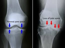
Osteoarthritis of the knee (knee OA) is the inflammation and wearing away of the cartilage on the bones that form the knee joint (osteo=bone, arthro=joint, itis=inflammation). The diagnosis of knee OA is based on 2 primary findings: radiographic evidence of changes in bone health (through medical images such as X-ray and magnetic resonance imaging [MRI]), and an individual’s symptoms (how you feel). Approximately 14 million people in the United States have symptomatic knee OA. Although more common in older adults, 2 million of the 14 million people with symptomatic knee OA were younger than 45 when diagnosed, and more than half were younger than 65.

download.jpg

What is Osteoarthritis of Knee?

Knee osteoarthritis (knee OA) is a progressive disease caused by inflammation and degeneration of the knee joint that worsens over time. It affects the entire joint, including bone, cartilage, ligaments, and muscles. Its progression is influenced by age, body mass index (BMI), bone structure, genetics, muscular strength, and activity level. Knee OA also may develop as a secondary condition following a traumatic knee injury. Depending on the stage of the disease and whether there are associated injuries or conditions, knee OA can be managed with physical therapy. More severe or advanced cases may require surgery.


How Does it Feel?

Individuals who develop knee OA may experience a wide range of symptoms and limitations based on the progression of the disease. Pain occurs when the cartilage covering the bones of the knee joint wears down. Areas where the cartilage is worn down or damaged exposes the underlying bone. The exposure of the bone allows increased stress and compression to the cartilage, and at times bone-on-bone contact during movement, which can cause pain. Because the knee is a weight-bearing joint, your activity level, and the type and duration of your activities usually have a direct impact on your symptoms. Symptoms may be worse with weight-bearing activity, such as walking while carrying a heavy object.

Symptoms of knee OA may include:

  • Worsening pain during or following activity, particularly with walking, climbing, or descending stairs, or moving from a sitting to standing position

  • Pain or stiffness after sitting with the knee bent or straight for a prolonged period of time

  • A feeling of popping, cracking, or grinding when moving the knee

  • Swelling following activity

  • Tenderness to touch along the knee joint

Typically these symptoms do not occur suddenly or all at once, but instead develop gradually over time. Sometimes individuals do not recognize they have osteoarthritis because they cannot remember a specific time or injury that caused their symptoms. If you have had worsening knee pain for several months that is not responding to rest or a change in activity, it is best to seek the advice of a medical provider.


How Is It Diagnosed?

Knee OA is diagnosed by 2 primary processes. The first is based on your report of your symptoms and a clinical examination. Your physical therapist will ask you questions about your medical history and activity. The therapist will perform a physical exam to measure your knee's movement (range of motion), strength, mobility, and flexibility. You might also be asked to perform various movements to see if they increase or decrease the pain you are experiencing.

The second tool used to diagnose knee OA is diagnostic imaging. Your physical therapist may refer you to a physician, who will order X-rays of the knee in a variety of positions to check for damage to the bone and cartilage of your knee joint. If more severe joint damage is suspected, an MRI may be ordered to look more closely at the overall status of the joint and surrounding tissues. Blood tests also may be ordered to help rule out other conditions that can cause symptoms similar to knee OA.


How Can a Physical Therapist Help?

Once you have received a diagnosis of knee OA, your physical therapist will design an individualized treatment program specific to the exact nature of your condition and your goals. Your treatment program may include:

Range-of-motion exercises. Abnormal motion of the knee joint can lead to a worsening of OA symptoms when there is additional stress on the joint. Your physical therapist will assess your knee’s range of motion compared with expected normal motion and the motion of the knee on your uninvolved leg. Your range-of-motion exercises will focus on improving your ability to bend and straighten your knee, as well as improve your flexibility to allow for increased motion.

Muscle strengthening. Strengthening the muscles around your knee will be an essential part of your rehabilitation program. Individuals with knee OA who adhere to strengthening programs have been shown to have less pain and an improved overall quality of life. There are several factors that influence the health of a joint: the quality of the cartilage that lines the bones, the tissue within and around the joints, and the associated muscles. Due to the wear and tear on cartilage associated with knee OA, maintaining strength in the muscles near the joint is crucial to preserve joint health. For example, as the muscles along the front and back of your thigh (quadriceps and hamstrings) cross the knee joint, they help control the motion and forces that are applied to the bones.

Strengthening the hip and core muscles also can help balance the amount of force on the knee joint, particularly during walking or running. The “core” refers to the muscles of the abdomen, low back, and pelvis. A strong core will increase stability throughout your body as you move your arms and legs. Your physical therapist will assess these different muscle groups, compare the strength in each limb, and prescribe specific exercises to target your areas of weakness.

Manual therapy. Physical therapists are trained in manual (hands-on) therapy. Your physical therapist will gently move your muscles and joints to improve their motion, flexibility, and strength. These techniques can target areas that are difficult to treat on your own. The addition of manual therapy techniques to exercise plans has been shown to decrease pain and increase function in people with knee OA.

Bracing. Compressive sleeves placed around the knee may help reduce pain and swelling. Devices such as realignment and off-loading braces are used to modify the forces placed on the knee. These braces can help "unload" certain areas of your knee and move contact to less painful areas of the joint during weight-bearing activities. Depending on your symptoms and impairments, your physical therapist will help determine which brace may be best for you.

Activity recommendations. Physical therapists are trained to understand how to prescribe exercises to individuals with injuries or pain. Since knee OA is a progressive disease, it is important to develop a specific plan to perform enough activity to address the problem, while avoiding excessive stress on the knee joint. Activity must be prescribed and monitored based on the type, frequency, duration, and intensity of your condition, with adequate time allotted for rest and recovery. Research has shown that individuals with knee OA who walked more steps per day were less likely to develop functional problems in the future. Your physical therapist will consider the stage and extent of your knee OA and prescribe an individualized exercise program to address your needs and maximize the function of your knee.

Modalities. Your physical therapist may recommend therapeutic modalities, such as ice and heat, to aid in pain management.

If Surgery Is Required

The meniscus (the shock absorber of the knee) may be involved in some cases of knee OA. In the past, surgery (arthroscopy) to repair or remove parts or all of this cartilage was common. Current research, however, has shown that—in a group of patients who were deemed surgical candidates for knee OA with involvement of the meniscus—60% to 70% of those who first participated in a physical therapy program did not go on to have surgery. One year later, those results were unchanged. This study suggests that physical therapy may be an effective alternative for people with knee OA, who would prefer to avoid surgery.

Sometimes, however, surgical intervention, such as arthroscopy or a total knee replacement, may be recommended. There are many factors to consider when determining the appropriate surgical treatment, including the nature of your condition, your age, activity level, and overall health. Your physical therapist will refer you to an orthopedic surgeon to discuss your surgical options.

Should you choose to have surgery, your physical therapist can assist you prior to and following your surgery. The treatment you require following surgery will depend on a variety of factors such as the type of surgery performed, your level of function, and fitness prior to surgery. Contrary to popular belief, surgery is not the easy choice; you will still require treatment following your surgery to maximize your level of function.


Can this Injury or Condition be Prevented?

Many conditions, including knee OA, can be prevented with the right fitness and exercise program. Physical therapists are experts in movement. Some ways that a physical therapist can help you prevent knee OA include:

  • Developing an appropriate exercise program. Inactivity is a significant contributor to many problems that affect individuals, including knee OA. Strengthening the muscles around the knee, as well as surrounding joints, can help decrease stress to the knee joint. Exercises to improve flexibility can help you maintain motion in the knee joint, which helps keep the cartilage healthy. Your physical therapist can design an individualized treatment program to boost your strength and flexibility, based on your specific condition.

  • Weight loss. Excessive weight can increase stress to the knee joint, which in turn can contribute to the wearing away of the protective cartilage, leading to knee OA. Your physical therapist can assess your weight, perform testing to determine your fitness level, establish an exercise program, and recommend lifestyle changes. The therapist also may refer you to another health care provider, such as a dietician, for further guidance.

  • Activity modification. Individuals often move or perform activities in a way that is unhealthy or inefficient, or that places excessive stress on the body, including the knee joint. Your physical therapist can teach you better ways to move in order to ease stress on your body and your knees.

  • Taking a “whole body” approach to movement. Lack of strength, mobility, and flexibility in surrounding areas of the body such as the ankle, hip, and spine also can affect the knee. Taking these body regions into consideration is important to help prevent knee OA. Your physical therapist will work with you to help ensure your whole body is moving correctly, as you perform your daily activities.


Real Life Experiences

Luke is a 50-year-old businessman who has just moved his family to the city so he can start a new job. For the last 2 months, Luke has been working hard to fix up his family’s new home, carrying heavy boxes and moving furniture up and down stairs. He also has worked late into the night installing appliances.

After starting his new job last week, sitting through numerous orientation sessions and meetings, Luke notices that his right knee is really hurting. He is used to occasional knee discomfort, but this is the worst it has felt in a long time. During his junior year at college, Luke suffered a significant knee injury while playing basketball, which required surgery.

These days, Luke coaches his son’s Little League team, exercises several times each week, and plays pickup basketball with his friends. But occasionally, particularly after long road trips, his knee pain flares up, and he has to resort to medication, icing, and rest. These bouts are starting to occur more regularly. Luke decides it's time to seek a consultation with a physical therapist.

During Luke’s first appointment, his physical therapist asks him questions regarding his medical history, prior injuries, current symptoms and complaints, and goals for physical therapy. She examines his knee motion, strength, balance, and walking mechanics. She also uses special tests and measures to determine the nature of Luke’s pain, ruling out any other possible conditions.

Based on her findings, Luke's physical therapist determines that his current knee pain is a result of posttraumatic osteoarthritis. She diagnoses knee OA. She explains that his history of significant knee injury in college put him at risk of developing knee OA at a young age. The recent increased demand on his knee joint during his move is likely responsible for the current flare-up of pain and swelling.

Over the next 6 weeks, Luke works with his physical therapist to decrease his joint pain and improve his knee motion and full-body flexibility. She uses manual therapy techniques to improve the mobility of his knee joint. She prescribes a progressive exercise program to strengthen the muscles of his hip, knee, and core. She tailors this program so that Luke can complete it daily, based on the equipment available at his office gym facility.

Six weeks later, Luke is able to climb and descend stairs, squat, and jog without pain. He can sit through a full day of meetings without noticing stiffness or swelling in his knee. On his last day of therapy, Luke’s physical therapist provides him with a detailed home-exercise program and suggestions for maintaining the improvements he has made. With the summer approaching, he's preparing to coach his son's baseball tournaments—and take his family to the beach!

This story was based on a real-life case. Your case may be different. Your physical therapist will tailor a treatment program to your specific case.


What Kind of Physical Therapist Do I Need?

All physical therapists are prepared through education and clinical experience to treat a variety of conditions or injuries. You may want to consider:

  • A physical therapist who is experienced in treating people with knee osteoarthritis and after knee replacement surgery. Some physical therapists have a practice with an orthopedic focus.

  • A physical therapist who is a board-certified orthopedic clinical specialist. This physical therapist will have advanced knowledge, experience, and skills that may apply to your condition.

You can find physical therapists who have these and other credentials by using Find a PT, the online tool built by the American Physical Therapy Association to help you search for physical therapists with specific clinical expertise in your geographic area.

General tips when you're looking for a physical therapist (or any other health care provider):

  • Get recommendations from family and friends or from other health care providers.

  • When you contact a physical therapy clinic for an appointment, ask about the physical therapists' experience in helping people with arthritis.

  • During your first visit with the physical therapist, be prepared to describe your symptoms in as much detail as possible, and report activities that make your symptoms worse.


Further Reading

The American Physical Therapy Association (APTA) believes that consumers should have access to information that could help them make health care decisions and also prepare them for their visit with their health care provider.

The following articles provide some of the best scientific evidence related to physical therapy treatment of arthritis. The articles report recent research and give an overview of the standards of practice both in the United States and internationally. The article titles are linked either to a PubMed* abstract of the article or to free full-text, so that you can read it or print out a copy to bring with you to your health care provider.

Brosseau L, Taki J, Desjardins B, et al. The Ottawa panel clinical practice guidelines for the management of knee osteoarthritis; part two: strengthening exercise programs. Clin Rehabil. 2017;31:596–611. Article Summary in PubMed.

Deshpande BR, Katz JN, Solomon DH, et al. Number of persons with symptomatic knee osteoarthritis in the US: impact of race and ethnicity, age, sex, and obesity. Arthritis Care Res (Hoboken). 2016;68:1743–1750. Article Summary in PubMed.

Ackerman IN, Bucknill A, Page RS, et al. The substantial personal burden experienced by younger people with hip or knee osteoarthritis. Osteoarthritis Cartilage. 2015;23:1276–1284. Article Summary in PubMed.

Katz JN, Brophy RH, Chaisson CE, et al. Surgery versus physical therapy for a meniscal tear and osteoarthritis [published correction appears in: N Engl J Med. 2013;369:683]. N Engl J Med. 2013;368:1675–1684. Free Article.

Segal NA. Bracing and orthoses: a review of efficacy and mechanical effects for tibiofemoral osteoarthritis. PM R. 2012;4(5 Suppl):S89–S96. Article Summary on PubMed.

Jansen MJ, Viechtbauer W, Lenssen AF, et al. Strength training alone, exercise therapy alone, and exercise therapy with passive manual mobilisation each reduce pain and disability in people with knee osteoarthritis: a systematic review. J Physiother. 2011;57:11–20. Free Article.

Bennell KL, Hinman RS. A review of the clinical evidence for exercise in osteoarthritis of the hip and knee. J Sci Med Sport. 2011;14:4–9. Article Summary on PubMed.

Lawrence RC, Felson DT, Helmick CG, et al. Estimates of the prevalence of arthritis and other rheumatic conditions in the United States, part II. Arthritis Rheum. 2008;58:26–35. Free Article.

* PubMed is a free online resource developed by the National Center for Biotechnology Information (NCBI). PubMed contains millions of citations to biomedical literature, including citations in the National Library of Medicine’s MEDLINE database.

Authored by Laura Stanley, PT, DPT, Board-Certified Clinical Specialist in Sports Physical Therapy. Reviewed by the MoveForwardPT.com editorial board.


Meniscus Tear

Meniscal tears are common injuries to the cartilage of the knee that can affect athletes and nonathletes alike. These tears can be either “acute,” meaning they happen as a result of a particular movement, or “degenerative,” meaning they happen over time. Your physical therapist can help you heal a meniscal tear and restore your strength and movement. If surgery is required, your physical therapist can help you prepare for the procedure and recover following surgery.

Unknown.jpeg


What is a Meniscal Tear?

The meniscus is a cartilage disc that cushions your knee. Each of your knees has 2 menisci (plural of meniscus); one on the inner (medial) part of the knee, and the other on the outer (lateral) part of the knee. Together, they act to absorb shock and stabilize the knee joint.

Meniscal tears can be classified in 2 ways: acute or degenerative. An acute meniscal tear typically is caused by twisting or turning quickly on a bent knee, often with the foot planted on the ground. This mechanism of injury often produces related injuries, such as an ACL tear. Degenerative meniscal tears occur over time, due to repetitive stress being put on the knee, such as in a job or sport that requires a lot of squatting.


How Does it Feel?

When you tear the meniscus, you might experience:

  • A sharp, intense pain in the knee area

  • A "pop" or a tearing sensation in the knee area (acute injury)

  • Swelling within the first 24 hours of injury

  • Difficulty walking or going up or down stairs because of pain or a "catching or locking" sensation in the knee

  • Difficulty straightening or bending the knee fully


How Is It Diagnosed?

Your physical therapist will:

  • Conduct a thorough evaluation that includes a detailed review of your injury, symptoms, and health history.

  • Perform special tests to measure the range of motion (amount of movement) in your knee and determine which specific movements and positions increase your symptoms.

  • Use a series of tests that apply pressure to the meniscus to determine whether it appears to be damaged.

The results of these tests may indicate the need for further diagnostic tests—such as magnetic resonance imaging (MRI)—or a referral to an orthopedic surgeon for consultation.


How Can a Physical Therapist Help?

Meniscal tears can often be managed without surgery. A short course of treatment provided by a physical therapist can help determine whether your knee will recover without surgery. Your physical therapist can help control pain and swelling in the knee area and work with you to restore full strength and mobility to your knee. Your treatment may include:

Manual therapy. Your physical therapist may apply manual therapy—hands-on treatment that may include massage, stretching, or joint mobilization—to help reduce swelling and stiffness, and restore muscle function around the knee.

Icing. Your physical therapist will apply ice packs to the knee to help control any pain and swelling, and may instruct you to apply icing at home. Swelling is an important "guide" during your rehabilitation and can indicate your level of ability and recovery. If you experience an increase in swelling, your physical therapist will modify your treatment program or activity level to ensure your safest, most effective recovery.

Compression. Your physical therapist may recommend the use of compression bandages, stockings, or pumps to assist in the reduction of or prevent further accumulation of edema (swelling). Your physical therapist may include them as part of your regular treatments and teach you and your family how to use them at home.

NMES. Your physical therapist may use a treatment called neuromuscular electrical stimulation (NMES). NMES uses electrical current to gently stimulate/contract the muscles around your knee to help improve their strength.

Assistive devices. It may be necessary to use assistive devices such as crutches, a cane, or a walker in the short term. Your physical therapist will make recommendations about which device is best for you and will instruct you in how to use it properly.

Strengthening exercises. Your physical therapist will design exercises to build and maintain your strength during recovery and help restore full movement to the knee. You will be given a home program of exercises that are specific to your condition. Strengthening the muscles around the knee and throughout the leg helps ease pressure on the healing knee tissue.

Fitness counseling. As you recover, your physical therapist will advise you on ways to improve and maintain your fitness and activity levels, and will help you decide when you are ready to return to full activity.

If Surgery Is Required

Patients with more serious meniscal tears, or those who don’t respond to a course of physical therapy, may need surgery to repair the injured knee. Surgically removing the torn cartilage (a procedure called a meniscectomy) usually is a simple procedure that requires a course of physical therapy treatment following surgery. Many people are able to return to their previous level of activity, including sports, after approximately 4 months of treatment.

Meniscus removal. Following a simple meniscectomy, your rehabilitation will likely be similar to that for nonsurgical meniscal injuries. Your physical therapist may use ice and compression to control pain and swelling and will show you how to use these treatments at home. The focus of your treatment will be on helping you get back your strength and movement through a series of exercises performed in the clinic and at home, as well as with hands-on treatment (manual therapy). Initially, it is likely that you will need to use crutches or a cane for walking. Your physical therapist will help guide you in gradually placing your weight on the knee to stand or walk, to allow the meniscus and other tissue in the knee joint to slowly adjust to increased pressure.

Meniscus repair. Sometimes your surgeon will decide that the torn meniscus can be repaired instead of removed. Research studies show that if a meniscal repair is possible, it can reduce the risk of arthritis developing later in life. Rehabilitation following a meniscal repair is slower and more extensive than with meniscal removal because the repaired tissue must be protected while it is healing. The type of surgical technique performed, the extent of your injury, and the preferences of the surgeon often determine how quickly you will be able to put weight on your leg, stop using crutches, and return to your previous activities.

Following surgery for meniscal repair, your physical therapist will:

  • Help you control pain and swelling

  • Guide you through progressive reloading of weight to the knee to allow the cartilage to slowly adjust to increased compressive stress and pressure

  • Help restore your knee and leg range of motion

  • Teach you exercises to help restore your muscle strength

Return to Activity

Whether your torn meniscus recovered on its own or required surgery, your physical therapist will play an important role in helping you return to your previous activities. Your physical therapist will help you learn to walk without favoring the leg and perform activities like going up and down stairs with ease.

Return to work. If you have a physically demanding job or lifestyle, your physical therapist can help you return to these activities and improve how you perform them.

Return to sport. If you are an athlete, you may need a more extensive course of rehabilitation. Your physical therapist will help you fully restore your strength, endurance, flexibility, and coordination to help maximize your return to sports and prevent reinjury. Return to sport varies greatly from one person to the next and depends on the extent of the injury, the specific surgical procedure, the preference of the surgeon, and the type of sport. Your physical therapist will consider these factors when designing and adjusting your rehabilitation program, and will work closely with your surgeon to help decide when it is safe for you to return to sports and other activities.

.

Can this Injury or Condition be Prevented?

There is little research at this time to support exercise or other interventions such as bracing for prevention of meniscal injuries. However, you can make choices to help improve your overall fitness and keep your knees as strong and as healthy as possible. Practices that can help keep your knees strong include:

  • Regular exercise to help strengthen the muscles that support your knees

  • Staying physically active to prepare your body for the demands of a sport or strenuous activity

  • Avoiding twisting or turning quickly while your foot is planted on the ground, to help prevent stress to the knee that can cause a meniscal tear

If you already have knee problems, your physical therapist can help you develop a fitness program that takes your knees into account. Some exercises are better than others for those with a history of knee pain. Many exercises can be modified to fit your specific needs.


Real Life Experiences

Beau is a college sophomore who plays on his intramural lacrosse team. During a recent practice game, Beau twisted his left knee while performing a sharp turn to make a catch. He immediately heard a “pop” and felt pain in his knee joint. He was helped off the field by teammates and led back to his room, where he applied ice and rested for the rest of the evening.

The next morning, Beau felt pain when he put weight on his leg to get out of bed, and found he had difficulty walking; he also noticed some swelling on the inside of his knee. His roommate is in the physical therapy program at his university; he suggested Beau see a physical therapist.

Beau’s physical therapist gets his medical history and asks him to describe what happened in the game to get a sense of what might have happened to his knee. She then performs some tests that include movements that selectively stress the tissues of the knee to see if the symptoms can be provoked. She tells Beau that his symptoms may indicate a meniscal tear. She recommends that he consult with an orthopedic surgeon, who orders magnetic resonance imaging (MRI). The surgeon confirms a diagnosis of a medial meniscal tear. After consultation with the surgeon, Beau chooses to have the tear “cleaned up,” and have a small piece of the meniscus removed—a procedure called a meniscectomy.

Prior to surgery, Beau works with his physical therapist, who prescribes exercises and manual therapy to reduce the swelling, improve the knee range of motion, and restore muscle function around the knee—treatments that have been shown to improve surgical outcomes.

Following his surgery, Beau’s physical therapist controls the swelling around the knee joint with ice, and shows Beau how to ice his knee at home. She applies electrical stimulation to speed the recovery of the quadriceps muscle. She teaches him range-of-motion exercises and tells him how often he should be doing them at home. She teaches him how to use crutches to avoid putting pressure and weight on the knee while its tissues heal.

After 1 week, Beau is able to walk without his crutches, but has difficulty bending his knee fully or straightening it when he walks. His physical therapist works with him on improving his walking pattern, and continues to address his knee range of motion, strength, swelling, and pain. His exercises are adjusted as he heals to continue to challenge him and move his recovery forward.

After 3 weeks, Beau has full range of motion and increased strength in his involved leg. He has good balance and no pain while walking. His physical therapist guides him as he returns to jogging and then running. She gives Beau feedback on how much he should be running, how running should feel, and what to do to ensure a symmetrical running pattern to keep his knee and his other joints safe.

At 4 weeks, Beau’s treatment progresses to sports-related rehabilitation activities, which include moving quickly from side to side and learning how to safely turn to field a catch. His physical therapist provides guidance and training to help Beau avoid reinjury. Beau reports his leg muscles have gained strength, and he feels more stable as he executes his movements.

After 6 weeks of treatment, Beau rejoins his team for a playoff game and, with newfound confidence, sets up his teammate for a winning goal!

This story was based on a real-life case. Your case may be different. Your physical therapist will tailor a treatment program to your specific case.


What Kind of Physical Therapist Do I Need?

All physical therapists are prepared through education and experience to treat a variety of conditions or injuries. You may want to consider:

  • A physical therapist who is experienced in treating people with orthopedic (musculoskeletal) problems or sports injuries.

  • A physical therapist who is a board-certified orthopaedic clinical specialist or who completed a residency or fellowship in orthopedic physical therapy or sports physical therapy has advanced knowledge, experience, and skills that may apply to your condition.

You can find physical therapists who have these and other credentials by using Find a PT, the online tool built by the American Physical Therapy Association to help you search for physical therapists with specific clinical expertise in your geographic area.

General tips when you're looking for a physical therapist:

  • Get recommendations from family and friends or from other health care providers.

  • When you contact a physical therapy clinic for an appointment, ask about the physical therapist's experience in helping people with a meniscal tear.

During your first visit with the physical therapist, be prepared to describe your symptoms in as much detail as possible, and say what makes your symptoms worse.


Further Reading

The American Physical Therapy Association (APTA) believes that consumers should have access to information that could help them make health care decisions and also prepare them for their visit with their health care provider.

The following articles provide some of the best scientific evidence related to physical therapy treatment of meniscal injuries. The articles report recent research and give an overview of the standards of practice for treatment both in the United States and internationally. The article titles are listed by year and are linked either to a PubMed* abstract of the article or to free access of the full article, so that you can read it or print out a copy to bring with you to your health care provider.

Beaufils P, Pujol N. Management of traumatic meniscal tear and degenerative meniscal lesions: save the meniscus. Orthop Tramatol Surg Res. 2017 September 2 [Epub ahead of print]. doi: 10.1016/j.otsr.2017.08.003. Article Summary in PubMed.

Moses MJ, Wang DE, Weinberg M, Strauss EJ. Clinical outcomes following surgically repaired bucket-handle meniscus tears. Phys Sportsmed. 2017 May 23 [Epub ahead of print]. doi: 10.1080/00913847.2017.1331688. Article Summary in PubMed.

Skou ST, Lind M, Holmich P, et al. Study protocol for a randomised controlled trial of meniscal surgery compared with exercise and patient education for treatment of meniscal tears in young adults. BMJ Open. 2017;7(8):e017436. Free Article.

Hare KB, Stefan Lohmander L, Kise NJ, et al. Middle-aged patients with an MRI-verified medial meniscal tear report symptoms commonly associated with knee osteoarthritis. Acta Orthop. 2017 August 8 [Epub ahead of print]. doi: 10.1080/17453674.2017.1360985. Free Article.

Katz JN, Brophy RH, Chaisson CE, et al. Surgery versus physical therapy for a meniscal tear and osteoarthritis. N Engl J Med. 2013;368:1675-1684. Article Summary in PubMed.

Logerstedt DS, Snyder-Mackler L, Ritter RC, Axe MJ. Knee pain and mobility impairments: meniscal and articular cartilage lesions. J Orthop Sports Phys Ther. 2010;40(6):A1-A35. Free Article.

Heckmann TP, Barber-Westin SD, Noyes FR. Meniscal repair and transplantation: indications, techniques, rehabilitation, and clinical outcome. J Orthop Sports Phys Ther. 2006;36:795-814. Article Summary in PubMed.

Lowery DJ, Farley TD, Wing DW, et al. A clinical composite score accurately detects meniscal pathology. Arthroscopy.2006;22:1174-1179.  Article Summary in PubMed.

Fitzgerald GK, Piva SR, Irrgang JJ. A modified neuromuscular electrical stimulation protocol for quadriceps strength training following anterior cruciate ligament reconstruction. J Orthop Sports Phys Ther. 2003;33:492-501. Article Summary in PubMed.

*PubMed is a free online resource developed by the National Center for Biotechnology Information (NCBI).  PubMed contains millions of citations to biomedical literature, including citations in the National Library of Medicine’s MEDLINE database.

 Authored by Christopher Bise, PT, DPT, MS. Reviewed by the MoveForwardPT.com editorial board.


Hip Impingement (Femoroacetabular Impingement)

Hip impingement involves a change in the shape of the surface of the hip joint that predisposes it to damage, resulting in stiffness and pain. Hip impingement is a process that may precede hip osteoarthritis. It most often occurs in young, active people. A recent study found that 87% of teens and adults with hip pain showed evidence of hip impingement on diagnostic images taken of their hip joints. To treat hip impingement, physical therapists prescribe stretches and strengthening exercises to better balance the muscles around the hip to protect it, and use manual therapies to help restore range of motion and increase comfort.

What is Hip Impingement?

There are 2 types of hip impingement; they may occur alone or together.

Pincer-Type Impingement

  • In pincer-type impingement, the hip socket (acetabulum), which is usually angled forward, may be angled toward the back, or protruding bone may be present on the pelvis side of the hip joint making the socket a deeper recess that covers more of the ball or head of the femur bone.

  • The overgrown bone or incorrect angle of the socket causes the labrum, a rim of connective tissue around the edge of the hip socket, to be pinched. Over time, this extra pressure to the labrum when flexing (moving the leg forward) leads to wear and tear that can cause inflammation and could result in a tear. If this condition persists, eventually the cartilage that lines the hip joint can become worn and form holes.

  • This condition affects men and women equally; symptoms often begin early, appearing at any time between 15 to 50 years of age.


Cam-Type Impingement

  • In cam-type impingement, the shape of the bone around the head of the femur—the ball at the top of the bone in the thigh—is misshapen. It can vary from the normal round ball shape, or have overgrown bone formed at the top and front. The nickname “pistol grip” deformity is given to the appearance of the bony overgrowth on x-rays.

  • The overgrown or misshapen bone contacts the cartilage that lines the hip socket, and can cause it to peel away from the bone in the socket. The labrum can become worn, frayed, or torn as well.

  • This condition affects men to women at a ratio of 3 to 1; symptoms often manifest during the teen years and 20s.

HipImpingement-SM.jpg

Signs and Symptoms

Hip impingement may cause you to experience:

  • Stiffness or deep aching pain in the front or side of the hip or front of the upper thigh while resting.

  • Sharp, stabbing pain when standing up from a chair, squatting, rising from a squat, running, "cutting," jumping, twisting, pivoting, or making lateral motions.

  • Hip pain described in a specific location by making a "C" with the thumb and hand and placing it on the fold at the front and side of the hip, known as the "C-sign."

  • Pain that most often starts gradually, but can also remain after another injury resolves.

  • Pain that increases with prolonged sitting or forward leaning.

  • Feeling less flexible at the hips, including a decreased ability to turn your thigh inward on the painful side.

How Is It Diagnosed?

Your physical therapist will evaluate the range of motion (movement) of the hip and surrounding joints, and test the strength of the muscles in that area. Your therapist will feel the hip joint and surrounding muscles to evaluate their condition. The examination will include observing how you move, standing from a sitting position, walking, running, or squatting, as appropriate. Your physical therapist may perform special tests to help determine whether the hip is the source of your symptoms. For instance, the therapist may gently roll your leg in and out (the “log roll” test), or bend your hip up and in while turning the lower leg out to the side (the "FADDIR" test) to assess your condition.

If further diagnosis is needed, your doctor may order diagnostic tests to help identify any joint changes, including x-rays, magnetic resonance imaging (MRI), or diagnostic injections. Hip impingement can occur at the same time as low back, buttock, or pelvic pain, or from conditions such as bursitis or groin strain. The final diagnosis of hip impingement may take some time, especially when other conditions are present.

How Can a Physical Therapist Help?

Without Surgery

When an active person develops hip pain, but does not have severe symptoms or joint damage, the recommended treatment is physical therapy. The following interventions can help decrease pain, improve movement, and avoid the progression of hip impingement and the need for surgery:

  • Improving the strength of your hips and trunk. Strengthening of the hips and trunk can reduce abnormal forces on the already injured joint and help with strategies to compensate.

  • Improving hip muscle flexibility and joint mobility. Stretching tight muscles can reduce abnormal forces that cause pain with motion. Joint mobilization may help ease pain from the hip joint; however, these treatments do not always help range of motion, especially if the shape of the bone at the hip joint has changed.

  • Improving tolerance of daily activities. Your physical therapist can consider your job and recreational activities and offer advice regarding maintaining postures that are healthier for your hip and activity modification. Often this involves limiting the amount of bending at the hip to avoid further hip damage.


Following Surgery

Surgery for hip impingement is performed with arthroscopy. This is a minimally invasive type of surgery, where the surgeon makes small incisions in the skin and inserts pencil-sized instruments into the joint to repair damage. The surgeon may perform 1 or several techniques during your procedure as needed. The surgeon may remove or reshape the bone on the pelvis or femur side of the joint, and repair or remove the damaged labrum or cartilage of the hip joint.

Postsurgical physical therapy varies based on the procedure performed. It may include:

  • Ensuring your safety as you heal. Your physical therapist may recommend that you limit the amount of weight you put on the operated leg if there was a repair of the labrum. You may wear a brace to help limit the amount of bending at the hip. You might also use crutches to avoid overloading the leg if the bone on the femur was reshaped.

  • Improving your range of motion, strength, and balance. Your physical therapist will guide you through safe range-of-motion, strengthening, and balance activities to improve your movement as quickly as possible while allowing the surgical site to heal properly.

  • Instructions on returning to an active lifestyle. Most people return to normal daily activities about 3 months after surgery, and to high-level activities and sports 4 to 6 months after surgery. Your physical therapist will recommend a gradual return to activity based on your condition—research shows that 60% to 90% of athletes return to their previous playing ability depending on the surgical procedure performed and the sport.

Can this Injury or Condition be Prevented?

Currently there are no recommendations to prevent hip impingement. Despite a major increase in research to learn more about hip impingement, there is a great deal that is unknown. For instance, many active young people whose x-rays show hips as being abnormal do not have pain despite continuing to live active lives and participate in sports.

However, there is evidence that physical therapy interventions along with anti-inflammatory drugs can decrease pain, slow joint damage, and improve function. This is particularly important in those with mild hip impingement, those who are attempting to avoid surgery, and those who are not candidates for surgery.

Real Life Experiences

Lindsay is an active high school senior who plays shortstop for her school's softball team. Over the last several months, she has had progressively worsening pain on the front and side of her left hip. It started as an occasional sharp pain when she fielded ground balls at practice, and it eventually developed into aching and stiffness of the hip while resting. Lindsay occasionally develops hip pain while sitting in class or at the movies. In the past couple of weeks, she has found it hard to lean forward to tie her shoes. Her mom has been worrying about her pain and takes Lindsay to her physical therapist.

At the evaluation, the physical therapist finds that Lindsay has weakness around her hip and trunk muscles, decreased hip mobility, pain when flexing the hip, pain returning to a standing position after squatting, and decreased balance when standing on her affected leg. Her physical therapist diagnoses mild hip impingement in her left hip. Lindsay sees her physical therapist 1-2 times a week for the next 6 weeks.

Her treatments focus on developing a home program for strengthening her hips and trunk, and the therapist uses manual therapy for the hip to improve her comfort and allow her to perform more activities. The therapist works with Lindsay to change how she moves when standing from a seated position, and also to modify how she moves when playing the infield in softball. Lindsay also spends less time in the positions that bother her hip in the weight room and on the practice field, following recommendations from her physical therapist. After 3 weeks, the majority of her pain has subsided, and by 6 weeks, she is playing in games pain-free.

Lindsay meets her goal of finishing her senior year with the softball team. However, she is considering other ways to stay active after she graduates that don’t involve bending forward as much.

This story was based on a real-life case. Your case may be different. Your physical therapist will tailor a treatment program to your specific case.

What Kind of Physical Therapist Do I Need?

All physical therapists are prepared through education and experience to treat patients who have hip impingement. You may want to consider:

  • A physical therapist who is experienced in treating people with musculoskeletal problems. Some physical therapists have a practice with a sports or orthopaedic focus.

  • A physical therapist who is a board-certified clinical specialist or who completed a residency or fellowship in sports or orthopaedic physical therapy. This therapist has advanced knowledge, experience, and skills that may apply to your condition.

You can find physical therapists who have these and other credentials by using Find a PT, the online tool built by the American Physical Therapy Association to help you search for physical therapists with specific clinical expertise in your geographic area.

General tips when you're looking for a physical therapist (or any other health care provider):

  • Get recommendations from family and friends or from other health care providers.

  • When you contact a physical therapy clinic for an appointment, ask about the physical therapists' experience in helping people with hip impingement.

  • During your first visit with the physical therapist, be prepared to describe your symptoms in as much detail as possible, and say what makes your symptoms worse.

Further Reading

The American Physical Therapy Association (APTA) believes that consumers should have access to information that could help them make health care decisions and also prepare them for their visit with their health care provider.

The following articles provide some of the best scientific evidence related to physical therapy treatment of problems related to hip impingement. The articles report recent research and give an overview of the standards of practice for treatment both in the United States and internationally. The article titles are linked either to a PubMed* abstract of the article or to free full text, so that you can read it or print out a copy to bring with you to your health care provider.

Byrd JW. Femoroacetabular impingement in athletes, part I: cause and assessment. Sports Health. 2010;2:321-333. Free Article.

Byrd JW. Femoroacetabular impingement in athletes, part II: treatment and outcomes. Sports Health. 2010;2:403-409. Free Article.

Enseki KR, Martin RL, Draovitch P, et al. The hip joint: arthroscopic procedures and postoperative rehabilitation. J Orthop Sports Phys Ther. 2006:36:516-525. Article Summary in PubMed.

*PubMed is a free online resource developed by the National Center for Biotechnology Information (NCBI). PubMed contains millions of citations to biomedical literature, including citations in the National Library of Medicine’s MEDLINE database.

Authored by Jennifer Miller, PT, MPT, SCS. Reviewed by the MoveForwardPT.com editorial board.

Greater Trochanteric Bursitis

Greater trochanteric bursitis (GTB) is an irritation of the bursa, a fluid-filled sac that sits on top of the greater trochanter, a bony prominence on the outside of the hip bone (femur). The bursa acts as a cushion to decrease friction between the outside of the hip bone and muscles attaching to the bone; bursitis results when the bursa on the outside of the hip bone becomes irritated. Greater trochanteric pain syndrome is the term used when the condition also includes irritation to the tendons of the gluteal muscles that sit beneath the bursa. 

Most often, GTB is the result of repetitive friction to the bursa due to a combination of muscle weakness and tightness affecting the outside of the hip. The condition is most often treated with physical therapy to restore normal function.

GTB may result from a combination of several different variables, including:

  • Gluteal muscle weakness

  • Iliotibial (IT) band (a thick band of tissue that runs along the outside of the leg from the pelvis to the knee) tightness

  • Hip muscle tightness

  • Abnormal hip or knee structure

  • Abnormal hip or knee mechanics (movement)

  • Improper movement technique with repetitive activities

  • Change in an exercise routine or sport activity

  • Improper footwear


images-1.jpeg


How Does it Feel?

People with GTB may experience:

  • Tenderness to touch on the outside of the hip

  • Pain that can vary from sharp to dull, and can radiate to the buttock, groin, thigh, or knee

  • Pain that is intermittent and symptomatic for a prolonged period

  • Pain when lying on the involved side

  • Pain and stiffness with prolonged sitting, walking (worst with the first few steps), negotiating stairs, or squatting

  • Pain that may increase during prolonged activity


How Is It Diagnosed?

The goals of the initial examination are to assess the degree of the injury, and determine the cause and contributing factors to it. GTB is a condition that develops as a consequence of repetitive irritation in the hip; it seldom results from a single injury. Your physical therapist will begin by gathering information about your condition, including your health history and your current symptoms. Your therapist will then examine your hip and thigh region to determine the presence of GTB. Your physical therapist may ask you questions about:

  • Your health history

  • Your current symptoms and how they may affect your typical day

  • The location and intensity of your pain, and how it may vary during the day

  • How the pain affects your activity level, and what you do to reduce the pain

  • How any injury may have occurred prior to your symptoms developing

  • How you have sought treatment, such as seeing other health care practitioners or having imaging or other tests done

Your physical examination will focus on the region of your symptoms, but also include other areas that may have been affected as your body has adjusted to pain. Your physical therapist may watch you walk, step onto a stair, squat, or balance on one leg. Following the interview and physical examination, your physical therapist will assess the results and develop an individualized treatment program to address your specific condition and goals. 

Imaging techniques, such as X-ray or MRI, are typically not needed to diagnose GTB.


How Can a Physical Therapist Help?

You and your physical therapist will work together to develop a plan to help achieve your specific goals. To do so, your physical therapist will select treatment strategies in any or all of the following areas:

  • Patient education. Your physical therapist will work with you to identify and change any external factors causing your pain, such as the type and amount of exercises you perform, your athletic activities, or your footwear. Your therapist will recommend improvements in your daily activities, and develop a personalized exercise program to help ensure a pain-free return to your desired activity level.

  • Pain management. Your physical therapist will design a program to address your pain that includes applying ice to the affected area as well as a trial of heat, such as a hot shower or heating pad. The exercises discussed below also can have a pain-reducing component. Your physical therapist also may recommend decreasing some activities that cause pain. Physical therapists are experts in prescribing pain-management techniques that reduce or eliminate the need for medication.

  • Range-of-motion exercise. Your low back, hip, or knee joint may be moving improperly, causing increased tension at the greater trochanter. Your physical therapist may teach you self-stretching techniques to decrease tension and help restore normal motion in the back, hip, and knee.

  • Manual therapy. Your physical therapist may apply “hands-on” treatments to gently move your muscles and joints, most likely in your low back, hip, or thigh. These techniques help improve motion and strength, and often address areas that are difficult to treat on your own.

  • Muscle strength. Muscle weaknesses or imbalances can result in excessive strain at the greater trochanter. Based on your specific condition, your physical therapist will design a safe, individualized, progressive resistance program for you, likely including your core (midsection) and lower extremity. You may begin by performing strengthening exercises lying on a table or at home on the bed or floor (eg, lifting your leg up while lying in different positions). You then may advance to exercises in a standing position (eg, standing squats). Your physical therapist will choose what exercises are right for you based on your age and physical condition.

  • Functional training. Once your pain, strength, and motion improve you will need to safely transition back into more demanding activities. To minimize the tension on the hip and your risk of repeated injury, it is important to teach your body safe, controlled movements. Based on your own unique movement assessment and goals, your physical therapist will create a series of activities to help you learn how to use and move your body correctly and safely.

Physical therapy promotes recovery from GTB by addressing issues, such as pain in the body structure, that is under stress from any lack of strength, flexibility, or body control. Your physical therapist may also recommend a period of relative rest, then help you slowly resume activities and carefully guide your progression. When GTB remains untreated, however, your pain will persist and result in long-term difficulty performing your desired activities.


Can this Injury or Condition be Prevented?

GTB may be the result of changes in the body’s shape, such as one leg being longer or shorter on the involved side. This condition can occur from an injury to the lower extremity or subtle differences that occur in the body’s growth and development.

Maintaining appropriate lower-extremity mobility and muscle strength, and paying particular attention to your exercise routine—especially changes in an exercise activity, the volume of exercises performed, and your footwear—are the best methods for preventing GTB.

Your physical therapist will help guide you through a process that will progressively reintegrate more demanding activities into your routine without overstraining your hip. Keep in mind that returning to activities too soon after injury can cause the condition to be more difficult to fix, and often leads to persistent pain.


Real Life Experiences

Karen is a 47-year-old teacher who is training for her first 5K road race. She runs 3 to 4 days each week, then walks the other days. Over the past 2 weeks, she has begun to experience pain in the outside of her right hip. Her pain is worse while running and lying on her right side; she experiences hip pain and stiffness when taking her first steps in the morning and walking up stairs, and also notes a dull ache with prolonged sitting and standing. She typically performs stretches for 5 minutes before her runs. Karen had not run consistently before she began training for the 5K.

Karen is concerned about the sharp hip pain she feels when running and her inability to complete her training due to pain. She is worried about her ability to perform daily activities and train for her upcoming race. She decides to seek the help of a physical therapist.

Karen's physical therapist takes a full history of her condition. Karen describes her typical daily running routine, including distance, pace, and running surface; her stretching routine; and her footwear. Her physical therapist then assesses Karen’s motion, strength, balance, movement, and running mechanics. He skillfully palpates (gently presses on) the front, side, and back of her hip to determine the precise location of her pain. Based on these findings, he diagnoses greater trochanteric bursitis.

Karen and her physical therapist work together to establish short- and long-term goals and identify immediate treatment priorities, including icing and stretching to decrease her pain, as well as gentle hip-strengthening exercises. They also discuss temporary alternative methods for Karen to maintain her fitness without continuing to aggravate her injury and prolong her recovery, such as swimming or biking. She is also prescribed a home-exercise program consisting of a series of activities to perform daily to help speed her recovery.

Together, they outline a 4-week rehabilitation program. Karen sees her physical therapist 1 to 2 times each week. He assesses her progress, performs manual therapy techniques, and advances her exercise program as appropriate. He advises her as to when she can begin to carefully resume running, and establishes a day-by-day plan to help her safely build back up to her desired mileage. Karen performs an independent daily exercise routine at home, including stretching and strengthening activities, which her physical therapist modifies as she regains strength and movement.

After 4 weeks of patient work, Karen no longer experiences pain or stiffness in her hip, and resumes her desired training program in preparation for her upcoming 5K race.

On the day of the race, Karen runs pain free and crosses the finish line in a personal best time!


What Kind of Physical Therapist Do I Need?

All physical therapists are prepared through education and experience to treat greater trochanteric bursitis. However, you may want to consider:

  • A physical therapist who is experienced in treating people with greater trochanteric bursitis. Some physical therapists have a practice with an orthopedic or musculoskeletal focus.

  • A physical therapist who is a board-certified clinical specialist or who completed a residency or fellowship in orthopedic or sports physical therapy. This physical therapist has advanced knowledge, experience, and skills that may apply to your condition.

You can find physical therapists who have these and other credentials by using Find a PT, the online tool built by the American Physical Therapy Association to help you search for physical therapists with specific clinical expertise in your geographic area.

General tips when you're looking for a physical therapist (or any other health care provider):

  • Get recommendations from family and friends or from other health care providers.

  • When you contact a physical therapy clinic for an appointment, ask about the physical therapists' experience in helping people who have greater trochanteric bursitis.

  • During your first visit with the physical therapist, be prepared to describe your symptoms in as much detail as possible, and describe what makes your symptoms worse.


Further Reading

The American Physical Therapy Association (APTA) believes that consumers should have access to information that could help them make health care decisions and also prepare them for a visit with their health care provider.

The following articles provide some of the best scientific evidence related to physical therapy treatment of greater trochanteric bursitis. The articles report recent research and give an overview of the standards of practice both in the United States and internationally. The article titles are linked either to a PubMed* abstract of the article or to free full text, so that you can read it or print out a copy to bring with you to your health care provider.

Tan LA, Benkli B, Tuchman A, et al. High prevalence of greater trochanteric pain syndrome among patients presenting to spine clinic for evaluation of degenerative lumbar pathologies. J Clin Neurosci. 2018;53:89–91. Article Summary in PubMed.

Mulligan EP, Middleton EF, Brunette M. Evaluation and management of greater trochanter pain syndrome. Phys Ther Sport. 2015;16(3):205–214. Article Summary in PubMed.

Grumet RC, Frank RM, Slabaugh MA, et al. Lateral hip pain in an athletic population: differential diagnosis and treatment options. Sports Health. 2010;2(3):191–196. Free Article.

* PubMed is a free online resource developed by the National Center for Biotechnology Information (NCBI). PubMed contains millions of citations to biomedical literature, including citations in the National Library of Medicine’s MEDLINE database.

Authored by Allison Mumbleau, PT, DPT, SCS. Reviewed by the MoveForwardPT.com editorial board. Revsied by Caleb Pagliero, PT, of APTA's Academy of Orthopaedic Physical Therapy. Reviewed by APTA Section liaison.  


Shin Splints (Medial Tibial Stress Syndrome)

Medial tibial stress syndrome (MTSS) develops when too much stress is placed on the tibia (main shin bone). The muscles that attach to the tibia can cause an overload of stress on the bone, and strain themselves at their insertion onto the bone as well. These muscles include the posterior tibialis muscle, the soleus muscle, and the flexor digitorum longus muscle.

The most common risk factors of MTSS include:

  • Flattening of the arch of the foot (overpronation) while standing and walking/running

  • Participation in a sport that requires repetitive jumping and/or running

  • Excessive hip motion (moving the hip through a greater range than is typical)

  • A high body mass index (>20.2 BMI)

  • A previous running injury

shin splint.jpg

How Does it Feel?

If you have developed MTSS, you may feel pain in the middle or bottom third of the inside of the shin. The pain may be sharp when you touch the tender area, or occur as an ache during or after exercise. Generally, however, the pain is initially provoked with activity and lessens with rest. When MTSS is developing, the pain may be present during the beginning of exercise and less noticeable as exercise progresses. Over time, the condition can worsen and pain may be felt throughout any exercise regimen and continue after exercise.

How Is It Diagnosed?

Your physical therapist will perform a thorough examination that will include taking a full health history and observing you as you walk and perform the activity that causes your symptoms, such as running or jumping.

Your physical therapist will further perform a series of tests and measures of your musculoskeletal system that assess your strength, mobility, flexibility, and pain response. The most reliable symptom of MTSS is pain felt when pressure is applied to specific locations on the shin.

If the results of the examination suggest MTSS, your physical therapist will discuss with you the goals of treatment and develop a specialized rehabilitation program for you. If a more serious condition could be contributing to your pain, you may be referred to a physician for further tests.

How Can a Physical Therapist Help?

Your physical therapist will determine what risk factors have caused your MTSS and will teach you how to address those causes. A treatment plan will be developed that is specific to you and what your body needs to recover and to prevent reinjury.

To relieve pain, your physical therapist may prescribe:

  • Rest from the aggravating activity or exercise

  • Icing the tender area for 5 to 10 minutes, 1 to 3 times a day

  • Exercises to gently stretch the muscles around the shin

  • Taping the arch of the foot or the affected leg muscles

  • Hands-on massage of the injured tissue

To help strengthen weak muscles, your physical therapist may teach you:

  • Exercises that increase the strength of hip rotation, hip abduction (lifting the leg away from the other leg), and hip extension (lifting the leg behind your body) to decrease stress to the lower leg

  • Exercises that increase your arch and shin muscle strength to decrease the overpronation (flattening out) of the arch of the foot

Your treatment also may include:

  • Calf and foot muscle stretches

  • Single-leg exercises including squats, reaching exercises, or heel raises

  • Modified take-off and landing techniques for jumping athletes

  • Modified leg and foot control during walking and running

  • Suggestions for footwear that provides better support when walking or exercising

Your physical therapist also may prescribe orthotics or shoe inserts that support the arch of the foot if your feet flatten out too much, or if your foot muscles are weak.

Physical therapists help athletes with MTSS alter their training schedules to safely return to sport, and offer specific guidance for reducing the possibility of reinjury. 

Can this Injury or Condition be Prevented?

To prevent MTSS, physical therapists recommend that you:

  • Get an annual functional fitness examination, including strength, flexibility, mobility, and sport-specific analyses.

  • Perform dynamic stretches before exercising and static stretches after exercising.

  • Perform strength and endurance exercises for the foot, hip, and pelvic muscles.

  • Perform balance exercises on each leg.

  • Follow a recommended training program when starting or increasing an exercise program. MTSS is commonly seen in the early part of a person’s training or the beginning of an athlete’s season. All exercise programs should begin gently and progress slowly.

  • Choose appropriate footwear for the activity that is being performed.

  • Choose appropriate cross-training activities to condition the core and leg muscles.

  • Exercise on a softer, more pliable surface whenever possible.

Your physical therapist can teach you exercises to ensure maximum strength and health, and prevent MTSS.

Real Life Experiences

John is a 35-year-old high school teacher who is training for his third half marathon. Recently, John began to feel shin pain in both legs during the first mile of his runs, which went away during the remaining miles. Over the next few days, the pain lasted longer during his run. John had been to physical therapy before for treatment of a knee problem. Concerned that he might be causing a new injury, John contacted his physical therapist.

John’s physical therapist begins his session with a detailed interview concerning his shin pain. She also asks John about his general health to rule out any other condition that could be a contributing factor to his pain.

John's physical therapist conducts a thorough examination to assess his pelvic, trunk, hip, leg, foot, and ankle strength. She asks him to try to hold test positions as she applies pressure to his legs and hips. John can't hold his position when she applies pressure to the hip area. During further tests, John demonstrates excessive flattening of each of his feet, and his knees show weakness. John’s physical therapist applies pressure to the muscles surrounding the shins and reproduces pain over the muscles on the lower one-third of the inside of the shin on each leg. She diagnoses MTSS in both legs.

John’s physical therapist begins his treatment by applying gentle massage to the painful area in both shins. She shows John how to apply ice to the painful areas for 5 to 10 minutes, 1 to 3 times a day. She teaches him gentle strengthening exercises for the hip and foot muscles. She also recommends that he temporarily modify his training program to run fewer overall miles, and to stop when his symptoms reappear. She offers John suggestions for specific footwear that will provide better support and cushioning, and educates him about choosing safer surfaces to run on when he resumes his full marathon training.

Because John sought help as soon as his symptoms began, after 2 weeks of treatment, his pain is much less. He slowly rebuilds his marathon training program with the advice of his physical therapist. He continues his prescribed exercise regimen and his physical therapy treatments for a few more weeks.

The following month, feeling stronger than he has in years, John competes pain free in the half marathon, and is proud to report a personal-best finishing time!

What Kind of Physical Therapist Do I Need?

All physical therapists are prepared through education and experience to treat MTSS. However, you may want to consider:

  • A physical therapist who is experienced in treating people with MTSS, or who has experience treating patients who participate in your sport.

  • A physical therapist who is a board-certified clinical specialist or who has completed a residency or fellowship in orthopaedic or sports physical therapy. This physical therapist has advanced knowledge, experience, and skills that may apply to your condition.

You can find physical therapists who have these and other credentials by using Find a PT, the online tool built by the American Physical Therapy Association to help you search for physical therapists with specific clinical expertise in your geographic area.

General tips when you’re looking for a physical therapist (or any other health care provider):

  • Get recommendations from family and friends or from other health care providers.

  • When you contact a physical therapy clinic for an appointment, ask about the physical therapists’ experience helping people who have MTSS.

  • During your first visit with the physical therapist, be prepared to describe your symptoms in as much detail as possible, and say what makes your symptoms worse.

Further Reading

The American Physical Therapy Association (APTA) believes that consumers should have access to information that could help them make health care decisions and also prepare them for a visit with their health care provider.

The following articles provide some of the best scientific evidence related to physical therapy treatment of MTSS. The articles report recent research and give an overview of the standards of practice for treatment of it both in the United States and internationally. The article titles are linked either to a PubMed* abstract of the article or to free full text, so that you can read it or print out a copy to bring with you to your health care provider.

References

Newman P, Witchalls J, Waddington G, Adams R. Risk factors associated with medial tibial stress syndrome in runners: a systematic review and meta-analysis. Open Access J Sports Med. 2013;4:229–241. Free Article.

Moen MH, Holtslag L, Bakker E, et al. The treatment of medial tibial stress syndrome in athletes: a randomized clinical trial. Sports Med Arthrosc Rehabil Ther Technol. 2012;4:12. Free Article.

Moen MH, Bongers T, Bakker EW, et al. Risk factors and prognostic indicators for medial tibial stress syndrome. Scand J Med Sci Sports. 2012;22(1):34–39. Article Summary on PubMed.

Moen MH, Tol JL, Weir A, et al. Medial tibial stress syndrome: a critical review. Sports Med. 2009;39(7):523-546. Article Summary on PubMed.

* PubMed is a free online resource developed by the National Center for Biotechnology Information (NCBI). PubMed contains millions of citations to biomedical literature, including citations in the National Library of Medicine’s MEDLINE database.

Revised by Stephen Reischl, PT, DPT.  He is a board-certified orthopaedic clinical specialist. Authored by Kari Brown Budde, PT, DPT. She is a board-certified sports clinical specialist. Reviewed by the MoveForwardPT.com editorial board.


Guide to Osgood-Schlatter Disease

What is Osgood-Schlatter Disease?

Osgood-Schlatter disease occurs when there is irritation to the top, front portion of the shin bone (tibia) where the tendon attached to the kneecap (patella) meets the shin bone. It occurs when there is an increased amount of stress placed upon the bones where the tendons attach. This is most often the result of increased activity levels by an adolescent athlete.

Our musculoskeletal system is made up of bones and surrounding soft tissue structures, including muscles, ligaments (which connect bone to bone), and tendons (which connect muscle to bone). These structures all play a role in helping us move.

During adolescence our bodies grow at a rapid rate. As our bodies develop, our bones are growing longer. Throughout this phase, our growth plates (epiphyseal plates) are susceptible to injury. A growth plate is the site at the end of a bone where new bone tissue is made and bone growth occurs. Females typically experience the most rapid growth between approximately 11 to 12 years of age, and males typically experience this growth surge between approximately 13 and 14 years of age. Males experience OS more frequently than females, likely due to an increased rate of sports participation.

Structures in our body might become irritated if they are asked to do more than they are capable of doing. Injuries can occur in an isolated event, but OS disease is most likely the cumulative effect of repeated trauma. OS is most frequently experienced in adolescents who regularly participate in running, jumping, and "cutting" (rapid changes in direction) activities.

When too much stress is present (ie, from rapid growth) and when the body is overworked (ie, either too much overall volume of exercise, or too much repetition), the top of the shin can become painful and swollen. As this condition progresses, the body’s response to bone stress can be an increase in bone production; an adolescent may begin to develop a boney growth that feels like a bump on the front of the upper shin.

OS can start as mild soreness, but can progress to long-lasting pain and limited function, if not addressed early and appropriately.

Back to Top

How Does it Feel?

With Osgood-Schlatter, you may experience:

  • Gradually worsening pain below your knee, at the top of the shin bone.

  • Pain that worsens with exercise.

  • Swelling and tenderness at the top of the shin.

  • A boney growth at the top of the shin.

  • Loss of strength in the quadriceps muscle (connecting the hip to the knee).

  • Increased tightness in the quadriceps muscle.

  • Loss of knee motion.

  • Discomfort with daily activities that use your knee, like kneeling, squatting, or walking up and down stairs.

Back to Top

How Is It Diagnosed?

Diagnosis of OS begins with a thorough medical history, including specific questions regarding athletic participation (sports played, frequency of practices/games, positions). Your physical therapist will assess different measures, such as sensation, motion, strength, flexibility, tenderness, and swelling. Your physical therapist will perform several tests specific to the knee joint, and may ask you to briefly demonstrate the activities or positions that cause your pain, such as walking, squatting, and stepping up or down stairs.

Because the knee and hip are both involved in these aggravating activities, your physical therapist will likely examine your hip as well. Other nearby areas, such as your feet and core, will also be examined to determine whether they, too, might be contributing to your knee condition.

If your physical therapist suspects there may be a more involved injury than increased stress-related irritation (ie, if there is a recent significant loss of motion or strength, or severe pain when the knee is moved), your therapist will likely recommend a referral to an orthopedic physician for diagnostic imaging, such as ultrasound, x-ray, or MRI.

Back to Top

How Can a Physical Therapist Help?

Once other conditions have been ruled out and OS is diagnosed, your physical therapist will work with you to develop an individualized treatment plan tailored to your specific knee condition and your goals. The goal of physical therapy is to accelerate your recovery and return to pain-free activity. There are many physical therapy treatments that have been shown to be effective in treating OS, and among them are:

Range of Motion Therapy. Your physical therapist will assess the motion of your knee and its surrounding structures, and design gentle exercises to help you work through any stiffness and swelling to return to a normal range of motion.

Strength Training. Your physical therapist will teach you exercises to strengthen the muscles around the knee so that each muscle is able to properly perform its job, and stresses are eased so the knee joint is properly protected.

Manual Therapy.Physical therapists are trained in manual (hands-on) therapy. If needed, your physical therapist will gently move your kneecap or patellar tendon and surrounding muscles as needed to improve their motion, flexibility, and strength. These techniques can target areas that are difficult to treat on your own. 

Pain Management.Your physical therapist may recommend therapeutic modalities, such as ice and heat, or a brace to aid in pain management.

Functional Training.Physical therapists are experts at training athletes to function at their best. Your physical therapist will assess your movements and teach you to adjust them to relieve any extra stress on the front of your knee.

Education. The first step to addressing your knee pain is rest. Your physical therapist will explain why this is important and develop a plan for your complete rehabilitation.

Back to Top

Can this Injury or Condition be Prevented?

Fortunately, there is much that can be done to prevent the cascade of events that lead to OS. Physical therapists focus on:

  • Educating coaches, parents, and athletes on guidelines for sports participation, explaining common causes of overuse injuries, and providing strategies for prevention.

  • Educating athletes on the risks of playing through pain.

  • Scheduling adequate rest time to recover between athletic events.

  • Tracking a young athlete’s growth curves (height, weight, BMI) to identify periods of increased injury risk.

  • Developing an athlete-specific flexibility and strengthening routine to be followed throughout the athletic season.

  • Encouraging consultation with a physical therapist whenever symptoms appear.

Back to Top

Real Life Experiences

Caleb is a 13-year-old boy who has been playing basketball since he was in the first grade. When younger, Caleb played only during the winter season. Over the course of the last year, however, he has attended 2 basketball camps during the summer, played on his middle-school team during the winter, and is now playing AAU basketball in the spring.

Caleb also chose to join the track team this spring, competing in the high jump and sprint events to improve his basketball skills. Over the past 3 months, Caleb has grown 2 inches, and both he and his basketball coaches are excited about his recent growth.

Recently, Caleb has been busy playing in weekly AAU tournaments with 1 to 2 track meets during the week. But when he got home from track practice on Monday, he told his dad that his leg was hurting. He said that it had begun getting sore while playing basketball over the weekend, but he didn’t want to tell his coach because he wanted to continue to play. Now he feels like the top of his shin is tender to touch, and he is unable to fully bend his knee without increased pain.

His dad realizes that this is more than the expected postactivity soreness; he immediately calls their local physical therapist.

Caleb's physical therapist takes his health history and performs an extensive examination. It becomes clear that Caleb has not scheduled appropriate rest times between his athletic activities, and that he is experiencing a growth spurt. The physical examination reveals that the top of Caleb’s shin is very tender, the area around his knee is swollen, and he has lost knee motion and strength. OS is diagnosed.

Together, Caleb and his physical therapist, father, and basketball and track coaches develop a treatment plan to help him return to pain-free sport participation. It begins with a 2-week period of rest where Caleb performs only minimal running exercise, and works regularly with his physical therapist on stretching, strengthening, balance, and coordination exercises, and on improving his squatting movements.

While at basketball practice, Caleb continues to work on his ball handling and free-throw shooting, activities that won't increase his knee pain.

After 2 weeks, when his knee is less tender, Caleb, his physical therapist, and his coaches develop a plan for his gradual return to full participation in track and basketball. They help Caleb understand how important it is to be honest about his knee pain, and to communicate with his coach if it starts to bother him again.

A month later, Caleb is back participating in all his track and basketball activities. He has changed his routine to allow for adequate warm-up time before and after each practice, and sufficient rest periods between activities. He makes sure his dad or coaches are keeping track of how much time he spends at practice and at rest.

Caleb decides to spend more time during the week working on his stretching, so he can reduce any risk of pain as he continues to grow. At the end of the season, Caleb’s AAU team wins the state championship—and he sets a new personal-best record in both the high jump and the 100-meter sprint!

Back to Top

What Kind of Physical Therapist Do I Need?

All physical therapists are prepared through education and clinical experience to treat a variety of conditions or injuries. You may want to consider:

  • A physical therapist who is experienced in treating people with orthopedic or musculoskeletal injuries.

  • A physical therapist who is a board-certified specialist or has completed a residency in orthopedic or sports physical therapy, as the therapist will have advanced knowledge, experience, and skills that apply to an athletic population.

You can find physical therapists that have these and other credentials by using Find a PT, the online tool built by the American Physical Therapy Association to help you search for physical therapists with specific clinical expertise in your geographic area.

General tips when you're looking for a physical therapist (or any other health care provider):

  • Get recommendations from family and friends or from other health care providers.

  • When you contact a physical therapy clinic for an appointment, ask about the physical therapists' experience in helping young athletes with knee pain.

  • During your first visit with the physical therapist, be prepared to describe your symptoms in as much detail as possible, and report activities that make your symptoms worse.

Back to Top

Further Reading

The American Physical Therapy Association (APTA) believes that consumers should have access to information that could help them make health care decisions, and also prepare them for a visit with their health care provider.

The following articles provide some of the best scientific evidence related to physical therapy treatment of athletic injuries. The articles report recent research and give an overview of the standards of practice both in the United States and internationally. The article titles are linked either to a PubMed* abstract of the article or to free full text, so that you can read it or print out a copy to bring with you to your health care provider.

Whitmore A. Osgood-Schlatter disease. JAAPA. 2013;26(10):51–52. Article Summary on PubMed.

Maffulli N, Longo UG, Spiezia F, Denaro V. Aetiology and prevention of injuries in elite young athletes. Med Sport Sci. 2011;56:187–200. Article Summary on PubMed.

Stein CJ, Micheli LJ. Overuse injuries in youth sports. Phys Sportsmed. 2010;38(2):102–108. Article Summary on PubMed.

* PubMed is a free online resource developed by the National Center for Biotechnology Information (NCBI). PubMed contains millions of citations to biomedical literature, including citations in the National Library of Medicine’s MEDLINE database.

 Authored by Allison Mumbleau, PT, DPT, SCS. Reviewed by the MoveForwardPT.com editorial board.


Avoid Chronic Disease With Regular Physical Activity

Most Americans don’t move enough despite proven benefits, such as reduced risk of cancer and chronic diseases, and improved bone health, cognitive function, weight control, and overall quality of life.

The second edition of the Physical Activity Guidelines for Americans, reports that approximately 80% of US adults and adolescents are insufficiently active. As a result, many Americans currently have or are likely to experience chronic diseases, including heart disease, diabetesobesity, and depression.

The good news is that regular physical activity can prevent and improve many chronic conditions. America, it's time to get moving!

How Much Physical Activity Should I Do?

According to the guidelines, the following is recommended:

  1. Preschool-aged children (ages 3 through 5 years) should be physically active at least for 3 hours, if not more. Adult caregivers should encourage active play that includes a variety of activity types and limits sitting-around time, such as screen time.

  1. Children and adolescents (ages 6 through 17 years) need at least 60 minutes or more of activity a day. This includes activities to strengthen bones, build muscles, and get the heart beating faster.

  1. Adults should do at least 150 minutes of moderate-intensity aerobic activity a week, and at least 2 days for muscle-strengthening activities. Adding more time provides further benefits.

  1. Older adults (ages 65 and older) should do at least 150 minutes of aerobic activity a week and include muscle-strengthening activities 2 days a week. You should also add components, such as balance training as well. If you have limitations due to preexisting conditions, consult with a health care provider and be as physically active as your abilities allow.

  1. Pregnant and postpartum women who were physically active before pregnancy can continue these activities during pregnancy and in the postpartum period, but they should consult their health care provider about any necessary adjustments.

  1. Adults with chronic health conditions and disabilities, who are able, should do at least 150 minutes of moderate-intensity activity a week. You should consult with a health care provider about the types and amounts that are appropriate for you.

How a Physical Therapist Can Help

Physical therapists are movement experts who optimize quality of life through prescribed exercise, hands-on care, and patient education.

After making a diagnosis, physical therapists create personalized treatment plans that help their patients improve mobility, manage pain and other chronic conditions, recover from injury, and prevent future injury and chronic disease.

Physical therapists empower people to be active participants in their own treatment, and they work collaboratively with other health professionals to ensure patients receive comprehensive care.

Resources

US Department of Health and Human Services. Physical Activity Guidelines for Americans. 2nd ed. Washington, DC: US Dept of Health and Human Services; 2018. Accessed November 14, 2018.

US Department of Health and Human Services. Are My Kids Getting Enough Physical Activity. Move Your Way. Washington, DC: US Dept of Health and Human Services. Accessed November 14, 2018

US Department of Health and Human Services. 60 A Day! Move Your Way. Washington, DC: Dept of Health and Human Services. Accessed November 14, 2018.

US Department of Health and Human Services. What’s your move? Move Your Way. Washington, DC: Dept of Health and Human Services. Accessed November 14, 2018.


Physical Therapist's Guide to Osteoporosis

Osteoporosis is a common disease that causes a thinning and weakening of the bones. It can affect people of any age. Women have the greatest risk of developing the disease, although it also occurs in men. Osteoporosis affects 55% of Americans aged 50 or older; one-half of women and a quarter of men will fracture a bone as a result of low bone density (osteopenia) or osteoporosis. Thin bones are the cause of 1.5 million fractures per year in the United States; hip fractures alone result in 300,000 hospitalizations. It is important to diagnosis low bone density or osteoporosis early so that steps can be taken to rebuild bone strength and lessen the risk of fracture.

What is Osteoporosis?

Osteoporosis is a bone disease characterized by low bone density (thickness of the bone), decreased bone strength, and a change in the bone structure, which can lead to an increased risk of fracture. The normal bone structure becomes thinned out and porous with poor nutrition, aging, or when osteoporosis develops, lessening the ability of the bone to withstand the typical forces that are applied in everyday living. Fractures from low bone density and osteoporosis can be serious, causing pain and affecting quality of life.

Bone is living tissue. Normally, one type of cell removes bone and another type of cell adds bone in a balanced, ongoing process. In osteoporosis, bones weaken when not enough new bone is formed and/or too much bone is lost. This imbalance commonly begins in women during the first 5 years of menopause. However, it can also occur in men and in children, often due to diseases that affect bone development, such as celiac disease, inflammatory bowel disease, rheumatoid arthritis, spina bifida, cystic fibrosis, or kidney disease. Some medicines, such as steroids, may increase the risk of developing osteoporosis. Athletes who are underweight during the time of peak bone development are also susceptible.

There are many factors that can cause a person to be at risk for developing osteoporosis. It is important to know your risks so that you can be diagnosed and proactive in your treatment.

Risk Factors for Osteoporosis

Noncontrollable risks

  • Female gender

  • Small frame

  • Advanced age

  • Hormone levels

  • Genetics

  • Predisposing medical conditions

Controllable risks

  • Cigarette smoking

  • Excessive alcohol intake

  • Inactive lifestyle

  • Excessive caffeine intake

  • Lack of weight-bearing exercise

  • Drugs (eg, steroids, heparin)

  • Poor health

  • Low weight

  • Calcium-poor diet

  • Low vitamin D levels


How Does it Feel?

Osteoporosis is a disease that can be "silent." There may be no outward symptoms until a fracture occurs. If you are middle-aged or older, you may notice a loss of height or the appearance of a humpback. You may also begin to experience pain between your shoulder blades or above the crest of the pelvis.

People with low bone density may experience fractures in everyday situations that would not occur in persons with healthy bones, such as breaking a hip or a wrist with a fall from a standing height, breaking a rib when opening a window or when receiving a hug, or breaking an ankle after stepping off a curb. These are called fragility fractures and are a red flag for bone disease. Spinal compression fractures, particularly those in the upper back or thoracic spine (area between the neck and the lower back), are the most common fractures, followed by hip and wrist fractures.

How Is It Diagnosed?

If you are seeing a physical therapist for back pain or other rehabilitation issues, the therapist will review your medical, family, medication, exercise, dietary, and hormonal history, conduct a complete physical examination, and determine your risk factors for osteoporosis. The assessment may lead the physical therapist to recommend further testing.

Osteoporosis is best diagnosed through a quick and painless specialized X-ray called the DXA, which measures bone density. The results are reported using T-scores and Z-scores.

  • The T-score compares your score to that of healthy 30-year-old adults. If you have a T-score of -1 or less, you have a greater risk of having a fracture.

  • If the T-score is -2.5 or less you will receive the diagnosis of osteoporosis.

  • The Z-score compares your bone mineral density to those of the same sex, weight, and age. It is used for those whose bone mass has not yet peaked, premenopausal women, and men older than 50.

Other methods of measuring bone density include X-ray, ultrasound, and CT scan. 

Back to Top

How Can a Physical Therapist Help?

Your physical therapist can develop a specific program based on your individual needs to help improve your overall bone health, keep your bones healthy, and help you avoid fracture. Your physical therapist may teach you:

  • Specific exercises to build bone or decrease the amount of bone loss

  • Proper posture to protect your spine from fracture

  • Proper alignment during activities of daily living

  • How to improve your balance so as to reduce your risk of falling

  • How to adjust your environment to protect your bone health

Healthy bone is built and maintained through a healthy lifestyle. Your physical therapist will teach you specific exercises to meet your particular needs.

The exercise component for bone building or slowing bone loss is very specific and similar for all ages. Bone grows when it is sufficiently and properly stressed, just as muscle grows when challenged by more than usual weight. Two types of exercise are optimal for bone health: weight-bearing and resistance.

It is best for a physical therapist to provide your individual bone-building prescription to ensure that you are neither overexercising nor underexercising. Typically, exercises are performed 2 to 3 times a week as part of an overall fitness program.

Weight-bearing exercises

  • Dancing

  • Jogging (if your bone density is higher than -3.0)

  • Racquet sports

  • Heel drops

  • Stomping

Resistance exercises

  • Weight lifting in proper spine and lower-extremity alignment

  • Use of exercise bands

  • Gravity resistance (eg, push-ups, prone trunk extension with cushion to protect lowest ribs, single-leg heel raises, squats, lunges, sustained standing yoga poses in neutral spine position)

  • Exercises that reduce or stabilize kyphosis (hunchback)

  • Balance exercises

If you are diagnosed with osteoporosis or low bone density, your physical therapist will work with you to:

  • Build bone or lessen the amount of bone loss at areas most vulnerable to fracture through exercise—hip, spine, shoulder, arms.

  • Improve your dynamic balance to avoid falls.

  • Improve your posture.

  • Adjust your work and living environments to limit risk.

  • Help you avoid exercises and movements that may contribute to spinal fracture, including any type of sit-up or crunch, and excessive spinal or hip twisting.

Conservative treatment of a fracture includes bed rest and appropriate pain treatment. Your physical therapist will work with you to:

  • Decrease your pain through positioning and other pain-relieving modalities. Individualized physical therapist regimens can help reduce pain without the need for medications, such as opioids.

  • Provide appropriate external devices, such as bracing, to promote healing and improve posture.

  • Decrease your risk of a fall, strengthen your muscles, and improve your postural alignment.

  • Avoid exercises that involve too much forward or side bending or twisting.

  • Avoid water or endurance exercises, as they have been shown to negatively affect bone density.

If your pain lasts longer than 6 weeks following a spinal fracture, you can discuss surgical options, such as vertebroplasty or kyphoplasty, with your physical therapist, primary care physician, and surgeon.

Children and adolescents. Physical therapists can educate families and youth groups on proper exercise and posture, and about the need to move daily to build bone strength and prevent bone loss. Children with health issues such as spina bifida, diabetes, Crohn's disease, and cerebral palsy are at a greater risk for bone disease and can particularly benefit from the guidance of a physical therapist. Proper physical conditioning is crucial for children and adolescents: the majority of bone is built during adolescence and peaks by the third decade of life.

Middle-aged and older adults. As people age, they may begin to notice postural, balance, and strength changes. Physical therapists work with middle-aged and older adults to:

  • Develop individualized exercise programs to promote bone growth or lessen bone loss

  • Improve dynamic balance to avoid falls

  • Improve posture

  • Improve the strength of back muscles

  • Improve hip strength and mobility

Can this Injury or Condition be Prevented?

Osteoporosis can be prevented by building adequate bone density through childhood, adolescence, and early adulthood. Building strong bones requires an adequate intake of calcium and vitamin D, and regular exercise.

There are steps to take to improve bone health at any age. An active lifestyle that includes resistance and weight-bearing exercise is important to maintain healthy bone. It is also important to avoid habits that promote bone loss, such as smoking, excessive alcohol consumption, and an inadequate intake of calcium in your diet. Maintaining good body mechanics and posture also contribute to good bone health. We have no control over the genetic tendencies we have inherited, but we can choose to manage osteoporosis through proper medication, diet, and appropriate exercise.

As with any health issue, an overall healthy lifestyle is important for staying well.

Real Life Experiences

Anna is a 69-year-old retired legal secretary. She has enjoyed her early years of retirement, taking long walks in beautiful settings across the United States. Two years into her retirement, however, she began having knee pain during some of her walks, which gradually grew worse. Last year, she had a total knee replacement due to arthritis. She now walks with a cane because of chronic knee and ankle pain, and has experienced a loss of balance. She also has developed a rounded upper back, and low back pain. She seeks the help of a physical therapist.

Anna's physical therapist performs an assessment that includes a medical review for osteoporosis risk factors and for other health issues. He evaluates her range of motion and strength, testing her arms, legs, and trunk—especially her upper back. He tests the flexibility of her spine and her balance, her walking ability, and her risk of falling. Anna's walking style is uneven and she leans heavily on her cane. A DXA scan reveals that Anna has lost bone density in her spine and both hips. A vertebral fracture assessment X-ray shows that she has painless compression fractures of her spine. Her physical therapist diagnoses osteoporosis of the spine.

Anna first works with her physical therapist to improve her posture and knee function through flexibility and strengthening exercises, so she can walk more normally while working on her balance to lower her fall risk. She tells him her main goal is to be able to take walks in the park again.

Anna’s physical therapist teaches her safe trunk movement to avoid spinal fracture. Anna agrees to wear a dynamic trunk brace 2 hours a day to help make her posture more upright. She practices weight-bearing exercises with considerations for her arthritis, and learns resistive strengthening exercises for her spine and hip. Anna's physical therapist designs a gentle home-exercise program for her as well.

By her last visit, the flexibility and strength of Anna’s trunk and legs and her tolerance of physical activity have improved. The quality of her walking and dynamic balance are measurably improved, and her risk of falling has decreased. Anna feels much more confident about managing her condition.

Just this past week, Anna joined a therapeutic senior walking group that meets at the local botanic garden twice a week. She is thrilled to be enjoying gentle walks in nature again, and looks forward to coordinating other activities with her new group of friends!

This story was based on a real-life case. Your case may be different. Your physical therapist will tailor a treatment program to your specific case.

What Kind of Physical Therapist Do I Need?

All physical therapists are prepared through education and experience to treat those with osteoporosis. However, if you have a diagnosis of osteoporosis or low bone density, you may want to consider:

  • A physical therapist who is a board-certified clinical specialist or who completed a residency or fellowship in orthopedic physical therapy or geriatric physical therapy. This physical therapist has advanced knowledge, experience, and skills that may apply to your condition.

  • A physical therapist who specializes in the treatment of osteoporosis.

You can find physical therapists with these and other credentials by using Find a PT, the online tool by the American Physical Therapy Association that can help you search for physical therapists with specific clinical expertise in your geographic area.

General tips when you're looking for a physical therapist (or any other health care provider):

  • Get recommendations from family and friends or from other health care providers.

  • When you contact a physical therapy clinic for an appointment, ask about the physical therapists' experience in helping people who have osteoporosis.

Further Reading

The American Physical Therapy Association (APTA) believes that consumers should have access to information that can help them make health care decisions and also prepare them for a visit with their health care provider.

The following websites are important and reputable resources to obtain more information about improving your bone health:

National Osteoporosis Foundation. Accessed March 28, 2018.   

American Bone Health. Accessed March 28, 2018.

American Bone Health. FORE fracture risk calculator. Accessed March 28, 2018.

Osteoporosis Canada. Accessed March 28, 2018.

Osteoporosis Canada. Too fit to fracture series. Accessed March 28, 2018.

National Bone Health Alliance. Accessed March 28, 2018.

Own the Bone. Accessed March 28, 2018.

National Osteoporosis Foundation and Pilates Anytime. Safe movement video series. Accessed March 28, 2018.

MedBridge. Osteoporosis education courses for physical therapists. Accessed March 28, 2018.

Office of the US Surgeon General. The 2004 Surgeon General’s report on bone health and osteoporosis. Accessed March 28, 2018. 

Physical Activity Guidelines Advisory Committee, US Dept of Health and Human Services. Physical Activity Guidelines Advisory Committee Report, 2008Washington, DC: US Department of Health and Human Services. Published June 2008. Accessed March 28, 2018. 

The following articles provide some of the best scientific evidence related to physical therapy treatment of osteoporosis and fracture prevention. The articles report recent research and give an overview of the standards of practice both in the United States and internationally. The article titles are linked either to a Pub Med* abstract of the article or to free full text, so that you can read it or print out a copy to bring with you to your health care provider.

Watson SL, Weks BK, Weis LJ, Harding AT, Horan SA, Beck BR. High-intensity resistance and impact training improves bone mineral density and physical function in postmenopausal women with osteopenia and osteoporosis: the LIFTMOR randomized controlled trial. J Bone Miner Res. 2018;33(2):211–220. Article Summary in PubMed.

Beck BR, Daly RM, Singh MA, Taaffe DR. Exercise and Sports Science Australia (ESSA) position statement on exercise prescription for the prevention and management of osteoporosis. J Sci Med Sport. 2017;20(5):438–445. Article Summary in PubMed.

Sözen T, Özışık L, Başaran NÇ. An overview and management of osteoporosis. Eur J Rheumatol. 2017;4(1):46–56. Free Article.

Giangregorio LM, McGill S, Wark JD, et al. Too fit to fracture: outcomes of a Delphi consensus process on physical activity and exercise recommendations for adults with osteoporosis with or without vertebral fractures. Osteoporos Int. 2015;26(3):891–910. Free Article.

Bansal S, Katzman WB, Giangregorio LM. Exercise for improving age-related hyperkyphotic posture: a systematic review. Arch Phys Med Rehabil. 2014;95(1):129–140. Free Article.

Clark EM, Carter L, Gould VC, Morrison L, Tobias JH. Vertebral fracture assessment (VFA) by lateral DXA scanning may be cost-effective when used as part of fracture liaison services or primary care screening. Osteoporos Int. 2014;25(3):953–964. Article Summary in PubMed.

Siris ES, Adler R, Bilezikian J, et al. The clinical diagnosis of osteoporosis: a position statement from the National Bone Health Alliance Working Group. Osteoporos Int. 2014;25(5):1439–1443. Free Article.

Silva BC, Boutroy S, Zhang C, et al. Trabecular bone score (TBS): a novel method to evaluate bone microarchitectural texture in patients with primary hyperparathyroidism. J Clin Endocrinol Metab. 2013;98(5):1963–1970. Free Article.

Cheung AM, Giangregorio L. Mechanical stimuli and bone health: what is the evidence? Curr Opin Rheumatol. 2012;24:561–566. Article Summary in PubMed.

Pfeifer M, Kohlwey L, Begerow B, Minne HW. Effects of two newly developed spinal orthoses on trunk muscle strength, posture, and quality-of-life in women with postmenopausal osteoporosis: a randomized trial. Am J Phys Med Rehabil. 2011;90:805–815. Article Summary on PubMed.

Kasukawa Y, Miyakoshi N, Hongo M, et al. Relationships between falls, spinal curvature, spinal mobility and back extensor strength in elderly people. J Bone Miner Metab. 2010;28:82–87. Article Summary in PubMed.

Nikander R, Kannus P, Dastidar M, et al. Targeted exercises against hip fragility. Osteoporos Int. 2009;20:1321–1328. Article Summary in PubMed.

Hongo M, Itoi E, Sinaki M, et al. Effect of low-intensity back exercise on quality of life and back extensor strength in patients with osteoporosis: a randomized controlled trial. Osteoporos Int. 2007;18:1389–1395. Article Summary in PubMed.

Vainionpaa A, Korpelainen R, Leppaluoto J, Jamsa T. Effects of high-impact exercise on bone mineral density: a randomized controlled trial in premenopausal women. Osteoporos Int. 2005;16:191–197. Article Summary in PubMed.

*PubMed is a free online resource developed by the National Center for Biotechnology Information (NCBI). PubMed contains millions of citations to biomedical literature, including citations in the National Library of Medicine's MEDLINE database.

Authored by Mary Saloka Morrison, PT, DScPT, MHS. Reviewed by the MoveForwardPT.com editorial board.


Patellofemoral Knee Pain

Patellofemoral pain syndrome (PFPS) refers to pain at the front of the knee, in and around the kneecap (patella). PFPS is one of the most common types of knee pain experienced in the United States, particularly among athletes, active teenagers, older adults, and people who perform physical labor. Patellofemoral pain affects more women than men and accounts for 20% to 25% of all reported knee pain. Physical therapists design exercise and treatment programs for people experiencing PFPS to help them reduce their pain, restore normal movement, and avoid future injury.

Current research indicates that PFPS is an "overuse syndrome," which means that it may result from repetitive or excessive use of the knee. Other contributing factors may include:

  • Weakness, tightness, or stiffness in the muscles around the knee and hip

  • An abnormality in the way the lower leg lines up with the hip, knee, and foot

  • Improper tracking of the kneecap

These conditions can interfere with the ability of the kneecap to glide smoothly on the femur (the bone that connects the knee to the thigh) in the femoral groove (situated along the thigh bone) during movement. The friction between the undersurface of the kneecap and the femur causes the pain and irritation commonly seen in PFPS. The kneecap also may fail to track properly in the femoral groove when the quadriceps muscle on the inside front of the thigh is weak, and the hip muscles on the outside of the thigh are tight. The kneecap gets pulled in the direction of the tight hip muscles and can track or tilt to the side, which irritates the tissues around the kneecap.

PFPS often occurs in people who are physically active or who have suddenly increased their level of activity, especially when that activity involves repeated knee motion, such as running, stair climbing, squatting, or repeated carrying of heavy loads. Older adults may experience age-related changes that cause the cartilage on the undersurface of the kneecap to wear out, resulting in pain and difficulty completing daily tasks without pain.

PatellofemoralPain_SM.jpg


How Does it Feel?

People with PFPS may experience:

  • Pain when walking up or down stairs or hills

  • Pain when walking on uneven surfaces

  • Pain that increases with activity and improves with rest

  • Pain that develops after sitting for long periods of time with the knee bent

  • A "crack" or "pop" when bending or straightening the knee

How Is It Diagnosed?

Your physical therapist will review your health history, perform a thorough examination, and conduct a series of tests to evaluate the knee. Your therapist may observe the alignment of your feet, analyze your walking and running patterns, and test the strength of your hip and thigh muscles to find out whether there is a weakness or imbalance that might be contributing to your pain. Your physical therapist also will check the flexibility of the muscles in your leg, paying close attention to those that attach at the knee.

Generally, X-rays are not needed to diagnose PFPS. Your physical therapist may consult with an orthopedic physician who may order an X-ray to rule out other conditions.

How Can a Physical Therapist Help?

After a comprehensive evaluation, your physical therapist will analyze the findings and, if PFPS is present, your therapist will prescribe an exercise and rehabilitation program just for you. Your program may include:

Strengthening exercises. Your physical therapist will teach you exercises targeted at the hip (specifically, the muscles of the buttock and thigh), the knee (specifically, the quadriceps muscle located on the front of your thigh that straightens your knee), and the ankle. Strengthening these muscles will help relieve pressure on the knee, as you perform your daily activities.

Stretching exercises. Your physical therapist also will choose exercises to gently stretch the muscles of the hip, knee, and ankle. Increasing the flexibility of these muscles will help reduce any abnormal forces on the knee and kneecap.

Positional training. Based on your activity level, your physical therapist may teach you proper form and positioning when performing activities, such as rising from a chair to a standing position, stair climbing, squatting, or lunging, to minimize excessive forces on the kneecap. This type of training is particularly effective for athletes.

Cross-training guidance. PFPS is often caused by overuse and repetitive activities. Athletes and active individuals can benefit from a physical therapist’s guidance about proper cross-training techniques to minimize stress on the knees.

Taping or bracing. Your physical therapist may choose to tape the kneecap to reduce your pain and retrain your muscles to work efficiently. There are many forms of knee taping, including some types of tape that help align the kneecap and some that just provide mild support to irritated tissues around it. In some cases, a brace may be required to hold the knee in the best position to ensure proper healing.

Electrical stimulation. Your physical therapist may prescribe treatments with gentle electrical stimulation to reduce pain and support the healing process.

Activity-based exercises. If you are having difficulty performing specific daily activities, or are an athlete who wants to return to a specific sport, your physical therapist will design individualized exercises to rebuild your strength and performance levels.

Fitting for an orthosis. If the alignment and position of your foot and arch appear to be contributing to your knee pain, your physical therapist may fit you with a special shoe insert called an orthosis. The orthosis can decrease the stress to your knee caused by low or high arches.

Can this Injury or Condition be Prevented?

PFPS is much easier to treat if it is caught early. Timely treatment by a physical therapist may help stop any underlying problems before they become worse. If you are experiencing knee pain, contact a physical therapist immediately. 

Your physical therapist can show you how to adjust your daily activities to safeguard your knees, and teach you exercises to do at home to strengthen your muscles and bones—and help prevent PFPS.

Physical therapists can assess athletic footwear and recommend proper choices for runners and daily walkers alike. Wearing the correct type of shoes for your activity and changing them when they are no longer supportive is essential to injury prevention.

Real Life Experiences

Amelia is a 25-year-old office assistant who loves to start her day with a 5-mile run. Over the past 6 months, she has been training for her first marathon. She began by training on very flat ground and has just moved to a hilly area.

Last week, Amelia began feeling pain in the front of her left knee when running downhill. Today, she had to stop running after 3 miles because of her knee pain. She called her physical therapist.

Amelia's physical therapist completes a comprehensive evaluation, including a screening for other possible conditions that might be causing her pain. He uses special tests to measure her strength and finds that she has weak hip muscles and tenderness around the kneecap. He determines that she has developed PFPS. Amelia is shocked to learn that she also has flat feet, and she’s not wearing the right supportive running shoes.

To begin her treatments, Amelia’s physical therapist applies special tape to the front of her knee to help reduce her pain, and instructs her in the use of ice to decrease her symptoms. He performs gentle movements of her kneecap and the surrounding tissues to help increase mobility and decrease pain. He teaches her special exercises to gently strengthen the weak muscles that support the knee.

He also designs a specific home-exercise program for Amelia to perform between sessions. He provides information about proper shoe choices for her foot and body type, and advises her to purchase shoes that will give her feet the right type of support. He also recommends that she try deep-water running or swimming for a week instead of her regular running program, until her condition improves.

After her first week of physical therapy, Amelia notices a decrease in her pain and an increased ability to walk up and down stairs without pain. Her physical therapist approves her new footwear, and adds more challenging exercises to her session and her home program. He gives her the go-ahead to race-walk. She applies ice only when she has pain.

After 2 weeks, Amelia reports she is feeling even less pain. Her physical therapist continues to increase the intensity of her exercises, and she starts to run again—but only on flat surfaces and short distances combined with longer walk intervals.

After a few more weeks of therapy, Amelia occasionally feels only slight twinges of pain and gradually resumes her prior level of training. Her physical therapist recommends continuation of her stretching and strengthening exercises, and discharges her from physical therapy.

A few months later, Amelia completes her first marathon pain free. She is thrilled to learn that her time was a personal best!

This story was based on a real-life case. Your case may be different. Your physical therapist will tailor a treatment program to your specific case.

What Kind of Physical Therapist Do I Need?

All physical therapists are prepared through education and experience to treat a variety of conditions or injuries. You may want to consider:

  • A physical therapist who is experienced in treating people with orthopedic, or musculoskeletal, problems.

  • A physical therapist who is a board-certified clinical specialist or who has completed a residency or fellowship in orthopedic physical therapy and has advanced knowledge, experience, and skills that may apply to your condition.

You can find physical therapists who have these and other credentials by using Find a PT, the online tool built by the American Physical Therapy Association to help you search for physical therapists with specific clinical expertise in your geographic area.

General tips when you're looking for a physical therapist:

  • Get recommendations from family and friends or from other health care providers.

  • When you contact a physical therapy clinic for an appointment, ask about the physical therapist's experience in helping people with patellofemoral pain syndrome.

During your first visit with the physical therapist, be prepared to describe your symptoms in as much detail as possible, and say what makes your symptoms worse.

Further Reading

The American Physical Therapy Association (APTA) believes that consumers should have access to information that could help them make health care decisions and also prepare them for their visit with their health care provider.

The following articles provide some of the best scientific evidence related to physical therapy treatment of patellofemoral pain syndrome. The articles report recent research and give an overview of the standards of practice both in the United States and internationally. The article titles are linked either to a PubMed* abstract of the article or to free access of the full article, so that you can read it or print out a copy to bring with you to your health care provider.

Tevhen DS, Robertson J. Knee pain: strengthen my hips? But it's my knees that hurt! J Orthop Sports Phys Ther. 2011-41-571. Article Summary on PubMed.

Davis IS, Powers CM. Patellofemoral pain syndrome: proximal, distal and local factors, an international retreat, April 30-May 2, 2009, Fells Point, Baltimore, MD. J Orthop Sports Phys Ther. 2010;40:A1–A16. Article Summary on PubMed.

Fukuda TY, Rossetto FM, Magalhaes E, et al. Short-term effects of hip abductors and lateral rotators strengthening in females with patellofemoral pain syndrome: a randomized controlled clinical trial. J Orthop Sports Phys Ther. 2010;40:736–742. Article Summary on PubMed.

Dixit S, DiFiori JP, Burton M, Mines B. Management of patellofemoral pain syndrome. Am Fam Physician. 2007;75:194–202. Free Article.

Powers CM, Ward SR, Chan LD, et al. The effect of bracing on patella alignment and patellofemoral joint contact area. Med Sci Sports Exerc. 2004;36:1226-1232. Article Summary on PubMed.

Bizzini M, Childs JD, Piva SR, Delitto A. Systematic review of the quality of randomized controlled trials for patellofemoral pain syndrome. J Ortho Sports Phys Ther. 2003;33:4–20. Article Summary on PubMed.

Crossley K, Bennell K, Green S, et al. Physical therapy for patellofemoral pain: a randomized, double-blinded, placebo-controlled trial. Am J Sports Med. 2002;30:857–865. Article Summary on PubMed.


* PubMed is a free online resource developed by the National Center for Biotechnology Information (NCBI).  PubMed contains millions of citations to biomedical literature, including citations in the National Library of Medicine’s MEDLINE database.

 Authored by Christopher Bise, PT, MS, DPT. Revised by Julie Mulcahy, PT. Reviewed by the MoveForwardPT.com editorial board.


Ulnar Collateral Ligament Injury

Ulnar collateral ligament (UCL) injuries generally occur when repetitive stress damages the inside of the elbow, compromising stability. UCL injuries are most common in athletes who play "overhead" sports, such as volleyball and baseball, which require using the arms in an overhead position. These injuries are occurring in greater frequency with the rise of sport specialization. They are often referred to as "Tommy John" injuries, named after the famous baseball pitcher who underwent the first surgery for a UCL injury in 1974. A physical therapist can help improve your arm's strength and range of motion, and your body's overall stability and balance following a UCL injury.

What Are Ulnar Collateral Ligament Injuries?

The ulnar collateral ligament is a band of tissue that connects the inside of your upper arm (humerus) to the inside of your forearm (ulna). This ligament helps to support and stabilize your arm when you perform a motion, such as throwing a ball. A UCL injury may at first cause pain and tightness in the area. However, over time and with repetitive stress or trauma, the UCL can become stretched and even tear. Surgery is not always necessary to heal a UCL injury, but it may be performed if pain persists or the elbow feels unstable upon a return to sport or other activities.

Signs and Symptoms

With a UCL injury, you may experience:

  • Soreness or tightness along the inside of your elbow

  • Minor swelling and possible bruising along the inside of your arm

  • Possible numbness and tingling in your arm

  • Instability at your elbow joint (a feeling like your elbow might “give out” when you move it through certain motions)

  • Pain when using your arm in an overhead position (eg, throwing/pitching a ball, swinging a racquet)

  • Difficulty warming up for a sport, or needing a longer time to warm up

  • Poorer performance (eg, a decrease in pitching speed)

How Is It Diagnosed?

Your physical therapist will conduct a thorough evaluation that includes taking your health and activity history. Your physical therapist may ask you questions including:

  • When and how did this injury occur? (Sudden or gradual?)

  • How long have you had pain?

  • Have you had any numbness and tingling in your arm?

  • Did you feel a "pop" near your elbow when throwing or performing an overhead activity?

  • Have you experienced any instability (eg, a feeling of your arm “giving out”) when performing an overhead activity?

  • Have you experienced a decrease in job or sport performance?

  • What other sports or activities do you participate in?

  • Have you had to stop playing your sport, or performing your job, because of the injury to your elbow?

Your physical therapist may gently touch the area around your elbow joint to locate the specific area of pain. Your physical therapist may slightly bend your arm while applying pressure along the outside of your elbow joint, or ask you to mimic a throwing motion as the therapist resists against it.

To provide a definitive diagnosis, your physical therapist may collaborate with an orthopedic surgeon. The surgeon may order further tests, such as magnetic resonance imaging (MRI) or magnetic resonance arthrogram (MRA), to confirm the diagnosis and to rule out other possible damage.

How Can a Physical Therapist Help?

Your physical therapist can help improve your arm's strength and range of motion following a UCL injury, and help restore your shoulder and core stability, coordination, and balance. Your therapist also will work with you before and after any necessary surgery, and can help identify other issues that may have contributed to your injury, such as range of motion and strength deficits, or improper throwing mechanics. Your physical therapist will help you:

Boost your healing process. Decreasing stress across the injured area is the best way to promote healing of a UCL injury. Your physical therapist will likely tell you to take some time off from your sport or other activity. Your therapist may educate you on the RICE (rest, ice, compression, elevation) principle and may implement "cross-friction massage" to help the body supply nutrients to the injured ligament.

Strengthen your muscles. After your injury your arm may feel weaker. Strengthening the muscles of your shoulder, upper back, and shoulder blades in addition to those of the forearm will help decrease the stress at the elbow joint. Addressing lower-body balance or any weakness through your hips and trunk also may help decrease stress across your elbow.

Improve your range of motion. After your injury you may notice more difficulty straightening or bending your arm. Your physical therapist will work with you to improve your arm's range of motion, including possibly stretching your shoulder to help decrease stress on your elbow when performing overhead movements.

Correct your movements. While every sport requires different arm positions, certain positions may put an athlete at greater risk for injury to the elbow. Examining and modifying the movements you perform may help you safely return to your sport. Your physical therapist will help design a specific program to allow a gradual full return to activity.

Prepare to return to sport. An important component of preparing for a return to sports after an UCL injury is preparing the arm to properly withstand the stress placed on it during throwing or other overhead motions. Your physical therapist will work with you to establish and implement a progressive program to prepare you for a return to practice and competition.

If Surgery Is Required

If surgery is necessary, your physical therapist may measure your arm strength and range of motion prior to surgery to define a baseline goal to achieve following the procedure.

Immediately following surgery, your arm will likely be placed in a splint, brace, or sling to protect your elbow. Physical therapy will begin within the first week to 10 days following surgery. Your physical therapist will:

  • Provide appropriate guidance. You will receive an individualized treatment program of gradual rehabilitation that will ensure you heal in the safest and most effective way possible. 

  • Protect the graft/repair site in the early postoperative period. You will be provided a brace that will likely need to be worn for 5 to 6 weeks, depending on your surgeon’s preference. Your physical therapist will show you how to ensure you don’t bend your arm too much or rotate your shoulder too far during this time.

  • Improve how far you can move your shoulder and elbow. When you are ready, your physical therapist will help you gently bend and straighten your arm through different exercises and stretching techniques. Your therapist also will gently stretch your shoulder to help decrease stress across the elbow.

  • Improve the strength of your arm. Through a series of exercises, your physical therapist will work with you to improve your arm strength. Your hand grip and forearm strength will likely be the first things you will work on following surgery. As you progress, the exercises will begin to focus more on your shoulder blade and upper back muscles.

  • Improve muscle strength and coordination. As you begin to heal and progress, your exercises will become more specific to your sport or other activity.

Resuming sport-specific activities. An athlete who has experienced a UCL injury can begin to return to throwing at approximately 6 months after surgery. The return is based on the surgeon and physical therapist providing clearance to do so.

Returning to full competition. An athlete generally can be cleared to return to game competition approximately 12 to 14 months after surgery.

Can this Injury or Condition be Prevented?

Certain factors may increase a person’s chances of injuring the UCL. For example, shoulder and elbow range of motion imbalances may play a role in creating too much stress at the elbow. Balance and coordination deficits also can lead to improper movement during sporting or other activities. Your physical therapist will design an individualized treatment program to address and correct these deficits.

Current evidence suggests the biggest factors for athletes developing this injury are pitch velocity, and the overall volume of throwing and other overhead activities performed in a specific sport. Throwing with high velocity (>83 mph), pitching too many pitches, pitching on short rest, pitching while fatigued, and introducing new pitches in excess are all factors related to exposing the UCL to force that it may not be able to withstand. Other factors such as age, type of sport, and position played also may affect overall arm fitness and health. It is important to keep up with regular arm care and exercises in order to reduce the likelihood of injury. 

Real Life Experiences

Jason is an 18-year-old college baseball player who is also on the Dean’s List at school. Last week, he “pulled an all-nighter” studying for an important test, and pitched an important game on exam day.

Jason pitched a great first inning, but noticed his right elbow began to feel tight in the second inning; he lost some control over his pitches in the third. By the fourth inning, he was pushing through pain and tightness because he didn’t want to let his team down. When throwing a fast ball to the second batter in the fifth inning, he felt a “pop” and a sharp pain in his right elbow. He then felt numbness and tingling on the inside of his right forearm and was unable to continue pitching.

The school’s athletic trainer examined Jason, applied ice to the arm, and put it in a sling. He referred Jason to an orthopedic surgeon who specialized in baseball injuries. The surgeon diagnosed a severe UCL injury. After talking with the surgeon and his family, Jason decided to have surgery to reconstruct the UCL on his right elbow.

Immediately after surgery, Jason was placed in a custom splint that held his elbow at a 90° angle with a sling around his shoulder to support his arm. He began his physical therapy 10 days after his surgery.

Jason’s physical therapist gently removed his splint and helped him begin to move his right elbow and shoulder. He gave Jason a series of exercises to perform at home, to work on his posture, shoulder blade strength, and the overall range of motion of his elbow and shoulder.

Over the next few weeks, Jason teamed with his physical therapist to work on his shoulder and elbow range of motion, single-leg balance exercises, core strengthening, and posture and shoulder-blade exercises. As he regained strength and motion, Jason learned new exercises to strengthen the muscles of his shoulder. His physical therapist measured his range of motion to ensure he was on track, and introduced more intense exercises at the shoulder and elbow.

Jason then began a throwing program that gradually increased the stresses across his elbow as he moved from shorter- to longer-distance throws. His physical therapist and pitching coach instructed him to focus on his mechanics and be aware of the position of his arm, trunk, and legs when he threw.

When the new baseball season began, Jason was able to return to the starting lineup! With careful attention to the instructions of his physical therapist on adequate warm ups, safe throwing motions, maintaining shoulder and arm strength and overall balance, and not throwing too much, he was able to pitch a complete season.

Jason called his physical therapist after his last postseason game, proud to report that he had set a personal record for number of wins and earned run average!

This story was based on a real-life case. Your case may be different. Your physical therapist will tailor a treatment program to your specific case.

What Kind of Physical Therapist Do I Need?

Although all physical therapists are prepared through education and experience to treat UCL injuries, you may want to consider:

  • A physical therapist who is experienced in treating people with UCL injuries. Some physical therapists have a specialized practice with a focus on sports and orthopedics, and more specifically, the upper extremity.

  • A physical therapist who is a board-certified clinical specialist or who has completed a residency or fellowship in sports or orthopaedic physical therapy. This physical therapist has advanced knowledge, experience, and skills that may apply to your condition.

You can find physical therapists who have these and other credentials by using Find a PT, the online tool developed by the American Physical Therapy Association to help you search for physical therapists with specific clinical expertise in your geographic area.

General tips when you're looking for a physical therapist:

  • Get recommendations from family and friends or from other health care providers.

  • When you contact a physical therapy clinic for an appointment, ask about the physical therapists' experience in helping people who have UCL injuries.

  • During your first visit with the physical therapist, be prepared to describe your symptoms in as much detail as possible, and say what makes your symptoms worse.

Further Reading

The American Physical Therapy Association (APTA) believes that consumers should have access to information that could help them make health care decisions and also prepare them for a visit with their health care provider.

The following articles provide some of the best scientific evidence related to physical therapy treatment of UCL injuries. The articles report recent research and give an overview of the standards of practice for treatment both in the United States and internationally. The article titles are listed by year and are linked either to a PubMed* abstract of the article or to free access of the full article, so that you can read it or print out a copy to bring with you to your health care provider.

Whiteside D, Martini DN, Lepley AS, Zernicke RF, Goulet GC. Predictors of ulnar collateral ligament reconstruction in Major League Baseball pitchers. Am J Sports Med. 2016;44(9):2202–2209. Article Summary in PubMed.

Bruce JR, Andrews JR. Ulnar collateral ligament injuries in the throwing athlete. J Am Acad Orthop Surg. 2014;22(5):315–325. Article Summary in PubMed.

Garrison JC, Cole MA, Conway JE, et al. Shoulder range of motion deficits in baseball players with an ulnar collateral ligament tear. Am J Sports Med. 2012;40(11):2597–2603. Article Summary on PubMed.

Shanley E, Rauh MJ, Michener LA, et al. Shoulder range of motion measures as risk factors for shoulder and elbow injuries in high school softball and baseball players. Am J Sports Med. 2011;39(9):1997–2006. Article Summary on PubMed.

Wilk KE, Macrina LC, Fleisig GS, et al. Correlation of glenohumeral internal rotation deficit and total rotational motion to shoulder injuries in professional baseball pitchers. Am J Sports Med. 2011;39(2):329–335. Article Summary on PubMed.

Fleisig GS, Andrews JR, Cutter GR, et al. Risk of serious injury for young baseball pitchers: a 10-year prospective study. Am J Sports Med. 2011;39(2):253–257. Article Summary on PubMed.

Hariri S, Safran MR. Ulnar collateral ligament injury in the overhead athlete. Clin Sports Med. 2010;29(4):619–644. Article Summary on PubMed.

Lin YC, Thompson A, Kung JT, et al. Functional isokinetic strength ratios in baseball players with injured elbows. J Sport Rehabil. 2010;19(1):21–29. Article Summary on PubMed.

Dines JS, Frank JB, Akerman M, Yocum LA. Glenohumeral internal rotation deficits in baseball players with ulnar collateral ligament insufficiency. Am J Sports Med. 2009;37(3):566–570. Article Summary on PubMed.

Reinold MM, Wilk KE, Macrina LC, et al. Changes in shoulder and elbow passive range of motion after pitching in professional baseball players. Am J Sports Med. 2008;36(3):523–527. Article Summary on PubMed.

Kibler WB, Sciascia AD, Uhl TL, et al. Electromyographic analysis of specific exercises for scapular control in early phases of shoulder rehabilitation. Am J Sports Med. 2008;36(9):1789–1798. Article Summary on PubMed.

Petty DH, Andrews JR, Fleisig GS, Cain EL. Ulnar collateral ligament reconstruction in high school baseball players. Am J Sports Med. 2004;32(5):1158–1164. Article Summary in PubMed.

*PubMed is a free online resource developed by the National Center for Biotechnology Information (NCBI). PubMed contains millions of citations to biomedical literature, including citations in the National Library of Medicine’s MEDLINE database.

Revised by David Colvin, PT. Authored by Craig Garrison, PT, PhD, ATC, and Joseph Hannon, PT, DPT. Reviewed by the MoveForwardPT.com editorial board.


Tarsal Tunnel Syndrome

Tarsal tunnel syndrome (TTS) is a condition that develops when a nerve within the tarsal tunnel of the inner ankle is compressed. TTS can lead to alterations in sensation and movement of the foot, ankle, and lower leg, and/or pain. It is often associated with conditions causing increased compression or swelling in the lower leg. Physical therapists help people experiencing TTS to relieve their pain and restore their normal function.

What is Tarsal Tunnel Syndrome?

Often described as the carpal tunnel syndrome of the lower extremity, tarsal tunnel syndrome is a condition that results from the compression of the posterior tibial nerve as it runs through the tarsal tunnel (a structure made up of bone and tissue (retinaculum) on the inside of the ankle). As it passes through the tarsal tunnel, the tibial nerve divides into 3 branches that provide sensation for the heel and bottom of the foot, and aid in the foot's function. When this structure becomes compressed, symptoms, such as pain, numbness, and/or tingling may occur and radiate into the lower leg, foot, and toes. Individuals may also experience muscle weakness in the area.

How Does it Feel?

The most common symptoms of TTS result from irritation of the tibial nerve and its branches. People with TTS may experience:

  • Pain, numbness, or tingling in the foot or ankle, which may radiate into the lower leg, foot, and toes

  • Weakness in the muscles of the lower leg and foot

  • Weakness of the big toe

  • Foot swelling

  • Symptoms that increase with prolonged standing or walking

  • Symptoms that decrease with rest

  • Altered temperatures of the foot and ankle

  • Pain that disrupts sleep

How Is It Diagnosed?

There are several tests that can help a clinician determine if TTS is present. Your physical therapist and/or physician will first take a comprehensive health history, and inquire about your current symptoms. Then your physical therapist may conduct tests, such as:

  • Gently tapping over the posterior tibial nerve in an attempt to reproduce your symptoms.

  • Tensing of the posterior tibial nerve, a maneuver that looks and feels like a "stretch," in an attempt to reproduce your symptoms.

  • Conducting a nerve condition study—a diagnostic test to determine the speed at which a nerve conducts information.

  • Ruling out other conditions, such as plantarfasciitis (inflammation of the tissue that runs along the bottom of the foot).

How Can a Physical Therapist Help?

Physical therapists play a vital role in helping people experiencing TTS to improve and maintain their daily function and activities. Your physical therapist will work with you to develop a treatment plan to help address your specific needs and goals.  

Because the signs and symptoms of TTS can vary, the approach to care will also vary. Your physical therapist may provide the following recommendations and care:

Nerve Gliding Activities. Gentle exercises that move and "glide" the nerves may help reduce symptoms and improve function.

Muscle Strengthening Exercises. Strengthening activities for any muscles affected by TTS, such as the tibialis posterior muscle in the back of your lower leg.

Balance and Coordination Activities. Work to improve your balance and coordination, which are often affected by TTS.

Orthotics/Taping/Bracing. Apply ankle taping, a custom orthotic, or bracing to position the foot to decrease stress on the posterior tibial nerve.

As with many conditions, education is key. Understanding the underlying mechanisms of TTS, and learning to recognize early signs and symptoms of stress may help you better manage the condition.

Can this Injury or Condition be Prevented?

Although there are no proven strategies for preventing TTS, there are ways to minimize stress to the foot and ankle, such as choosing appropriate footwear, wearing custom orthotics, minimizing the amount of time spent standing on hard surfaces, and improving and maintaining strength in the muscles of your legs, ankles, and feet. These strategies can be discussed further with your physical therapist.

In addition, early detection of the signs and symptoms of TTS will help you and your medical providers begin appropriate management of the condition, which may enhance your long-term well-being.

Real Life Experiences

Kim is a 46-year-old woman who works on an automobile assembly line. Her job involves standing on hard surfaces for prolonged periods of time. Kim recently noticed an onset of pain in her inner ankle after working a few hours, as well as an occasional shooting pain in her big toe. Now, her pain gets progressively worse throughout the day, and often interrupts her sleep. She is afraid of losing her job if she mentions her symptoms to her boss, and really can't afford to miss work. Kim decides to call her physical therapist.

Kim's physical therapist asks about her medical history, and learns that Kim has been diagnosed with high blood pressure and high cholesterol. They discuss Kim’s current symptoms.

He examines Kim’s ankle motion and strength, and gently performs procedures to provoke her symptoms. He also observes how she walks, and assesses her balance. Based on these signs and symptoms, he diagnoses tarsal tunnel syndrome.

Over the next several weeks, Kim works with her physical therapist to reduce her pain and improve her function. Her treatments include:

  • Application of a custom orthotic to better support her foot/ankle.

  • Nerve-gliding activities to improve the mobility of the posterior tibial nerve.

  • Balance exercises.

  • Manual therapy to ease her pain and improve her ankle mobility.

  • Strengthening exercises for her affected muscles.

  • Education about modifying her work positioning and activities.

After 4 weeks of physical therapy, Kim reports a significant reduction in her symptoms. She says she no longer fears going to work, and believes that she has taken control of her current situation. She continues to perform the home-based exercises her physical therapist taught her, and is amazed at how comfortable her feet feel throughout the workday—in her new shoes with custom orthotic inserts.

This story highlights an individualized experience of TTS. Your case may be different. Your physical therapist will tailor a treatment program to your specific needs.

What Kind of Physical Therapist Do I Need?

All physical therapists are prepared through education and experience to treat conditions, such as TTS. However, when seeking a provider, you may want to consider:

  • A physical therapist who is a board-certified clinical specialist, or who has completed a residency or fellowship in physical therapy. This therapist has advanced knowledge, experience, and skills that may apply to your condition.

  • A physical therapist who is well-versed in the treatment of TTS or other neuropathic disorders.

You can find physical therapists who have these and other credentials by using Find a PT, the online tool built by the American Physical Therapy Association to help you search for physical therapists with specific clinical expertise in your geographic area.

General tips when you're looking for a physical therapist (or any other health care provider):

  • Get recommendations from family and friends or from other health care providers.

  • When you contact a physical therapy clinic for an appointment, ask about the physical therapists' experience in helping people with painful conditions

  • During your first visit with the physical therapist, be prepared to describe your symptoms in as much detail as possible. Keeping a journal highlighting when you experience pain will help the physical therapist identify the best way of approaching care.


Further Reading

The American Physical Therapy Association (APTA) believes that consumers should have access to information that could help them make health care decisions, and also prepare them for a visit with their health care provider.

The following articles provide some of the best scientific evidence for the treatment of Tarsal Tunnel Syndrome. The articles report recent research and give an overview of the standards of practice both in the United States and internationally. The article titles are linked either to a PubMed* abstract of the article or to free full text, so that you can read it or print out a copy to bring with you to your health care provider.

Alshama AM, Souvlis T, Coppieters MW. A review of plantar heel pain of neural origin: differential diagnosis and management. Man Ther. 2008;13(2):103–111. Article Summary on PubMed.

Kavlak Y, Uygur F. Effects of nerve mobilization exercise as an adjunct to the conservative treatment for patients with tarsal tunnel syndrome. J Manipulative Physiol Ther. 2011;34(7):441–448. Article Summary on PubMed.

*PubMed is a free online resource developed by the National Center for Biotechnology Information (NCBI). PubMed contains millions of citations to biomedical literature, including citations in the National Library of Medicine’s MEDLINE database.

Acknowledgements: Joseph Brence, PT, DPT, FAAOMPT, COMT, DAC. Reviewed by the MoveForwardPT.com editorial board.


Total Shoulder Replacement (Arthroplasty)

Total shoulder arthroplasty (TSA), often called a total shoulder replacement, is a surgical procedure in which part or all of the shoulder joint is replaced. It is estimated that 53,000 people in the United States have shoulder replacement surgery each year, according to the Agency for Healthcare Research and Quality. That number compares to the more than 900,000 Americans a year who have knee and hip replacement surgery. Physical therapists can help patients who undergo a TSA return to their previous levels of physical activity, including fitness training, or participation in sports like swimming or golf.

What is Total Shoulder Arthoplasty?

Total shoulder arthroplasty is a surgical procedure in which part or all of the shoulder joint is replaced. It is performed on the shoulder when medical interventions, such as other conservative surgeries, medication, and physical therapy no longer provide pain relief. The decision to have a TSA is made following consultation with your orthopedic surgeon and your physical therapist.

A shoulder replacement may be needed if you have any of the following conditions affecting the shoulder, causing severe shoulder pain and limiting your ability to use the affected shoulder:

A TSA involves removing the ends of the bone at the shoulder joint, and replacing them with artificial parts. The upper part of the arm bone (humerus) is shaped like a ball; it is called the "head" of the humerus. During a TSA, the head of the humerus is replaced by a metal ball. The socket that the head of the humerus sits in is called the glenoid fossa. During a TSA, the socket is replaced by a plastic cup.

Due to various physical limitations, your orthopedic surgeon may decide that you are a candidate for another form of TSA, such as:

  • Shoulder hemiarthroplasty, where only the head of the humerus is replaced with a metal ball.

  • Reverse TSA, where the metal ball and plastic socket are reversed. This procedure is recommended when the rotator cuff muscles of the shoulder are damaged. The plastic socket is attached to the top of the humerus, and the metal ball is attached to the socket. This procedure allows another shoulder muscle, called the deltoid, to take over for the damaged rotator cuff muscles, improving functional range of motion, strength, and stability of the shoulder

ShoulderReplacement-SM.jpg

How Can a Physical Therapist Help?

Physical therapy plays a vital role in ensuring a safe recovery by improving shoulder function, and limiting pain following a TSA. Your physical therapist will work with you prior to and following your surgery, to help you safely return to your previous levels of activity, including performing household chores, job duties, and recreational activities.


Before Surgery

The better physical condition your shoulder is in prior to surgery, the better your recovery will be. Your physical therapist will teach you exercises to build shoulder strength, and improve your shoulder and upper back movement to keep the shoulder as strong and mobile as possible up until the time of surgery.

After Surgery

Your physical therapist will educate you about precautions to take after surgery, such as wearing a sling to perform all activities, and gradually beginning to safely move your arm. If you are a smoker, quitting smoking will improve your healing process.

After your TSA, you will likely stay in the hospital for 2 to 3 days. If you have other medical conditions, such as diabetes or heart disease, your hospital stay may be a few days longer. Your shoulder will be placed in a sling for the next 2 to 6 weeks; you will be advised to not move your shoulder on your own.

Your physical therapy will begin within a day or two of your surgery. A hospital physical therapist will visit your room to teach you how to perform simple tasks like brushing your teeth, and tell you what movements (such as pushing, pulling, or reaching with the affected arm) you simply cannot perform. Your physical therapist will teach you how to get in and out of bed safely, how to get the sling on and off, and how to get dressed while keeping your shoulder in a safe position. You will also learn how to minimize pain and swelling in the area by applying an ice pack, and elevating the upper arm.

You may need some help from friends or family members with daily activities for the first few days or weeks after your surgery. You will not be able to drive for the first few weeks after surgery.


As You Recover

When you are discharged from the hospital, continuation of physical therapy is essential. Your surgeon and physical therapist will work as a team to ensure your safe recovery. Your physical therapist will teach you exercises that may include:

Range-of-Motion Exercises. It is important to not move your shoulder suddenly or with any force for the first 2 to 6 weeks following surgery, to allow proper healing. Your physical therapist will passively move your shoulder in different directions to allow you to safely begin regaining movement. Your physical therapist will also teach you gentle exercises to perform at home. You will also learn range-of-motion exercises for the elbow and hand, so these joints do not get stiff from being held in a sling. Squeezing a ball or putty will help keep your grip strong, while your shoulder recovers. You will use ice packs on the shoulder and elevate your arm on pillows to allow gravity to help reduce the swelling in the shoulder, as instructed by your physical therapist.

Strengthening Exercises. As your shoulder mobility returns within a few weeks or months, your physical therapist will guide you through a shoulder strengthening program. You may use resistive bands and weights to perform gentle strengthening exercises.

Functional Training. Your physical therapist will help you regain everyday shoulder movements, such as reaching into a cupboard, reaching behind your body to tuck in your shirt, or reaching across your body to fasten a seat belt.

Job and Sport-Specific Training. Your physical therapist will design a personalized program to enable you to resume your job tasks without pain. These may include reaching, pushing, or carrying movements. You will also receive sport-specific training if you are planning to return to a sport. Your physical therapist will create a specialized home or fitness-center exercise program based on your individual needs, to be continued long after formal physical therapy has been completed.

Can this Injury or Condition be Prevented?

If you begin noticing your shoulder is painful and you are losing the ability to move your shoulder, a physical therapist can help. A properly designed exercise program can delay or even help you avoid surgery. A physical therapist will teach you specific, safe exercises to improve your shoulder flexibility and strength, and teach you how to manage your pain. Proper nutrition and physical activity will keep all of your joints healthy. Avoiding smoking is essential for proper healing and overall recovery from any injury.

Real Life Experiences

Charles is a 59-year-old golfer and swimmer with a history of osteoarthritis that began when he was 45 years old. Recently, Charles began to notice an increase in pain and difficulty when he reached overhead with his right arm. He also noticed that he couldn't throw a ball like he used to, and his shoulder was hurting during his golf swing and swim stroke. Just this month, Charles began to have difficulty shifting gears while driving, and realized that he could no longer lift his arm to reach into the cupboard to get his coffee cup. He called his doctor.

Charles's doctor took his medical history and thoroughly examined his shoulder. He diagnosed severe shoulder arthritis. He referred Charles to an orthopedic surgeon, who scheduled Charles for a TSA. Charles had a presurgery consultation with his physical therapist to learn what to expect from his recovery after surgery. His physical therapist explained how to wear and use a sling, and how to manage any pain or swelling. He also showed Charles the exercises that he would be performing.

The first day after his surgery, Charles' hospital physical therapist visited his room to teach him some deep-breathing exercises to keep his lungs inflated and reduce any risk of developing complications, such as pnemonia. She taught him how to properly use his sling, and guided him through a few gentle elbow and hand exercises. She also showed him how to safely get in and out of bed and a chair, without putting pressure on his right shoulder.

The second day after surgery, Charles' physical therapist taught him how to remove the sling safely to perform gentle pendulum exercises that helps to keep the shoulder from getting stiff. He learned how to avoid using his right shoulder at all other times, and to keep it in the sling, except when doing the pendulum exercises, and gentle elbow and hand exercises. He learned safe techniques for washing and other activities of daily living, including putting on a shirt.

The third day after surgery was Charles' last day in the hospital. His physical therapist helped him make arrangements for outpatient physical therapy.

Charles began his outpatient physical therapy just days after his TSA. His physical therapist performed passive movements with his right shoulder to ensure that it regained full mobility. She designed a home-exercise program for him, continuing the pendulum exercises and active elbow and hand exercises, as well as conservative shoulder blade squeezes.

As his shoulder strength and movement began to be restored, Charles' physical therapist added "active assisted" exercises (movement patterns assisted by a pulley or by the opposite shoulder) to gently increase his right shoulder mobility. She taught Charles how to squeeze a tennis ball a few times a day to improve his grip strength. Charles also learned how to apply an ice pack, and elevate his right shoulder at home and after each physical therapy session.

Eight weeks following his TSA, Charles was able to reach his right arm farther overhead than he was able to before his surgery!

After 12 weeks, under the guidance of his physical therapist, Charles has more shoulder motion and much less pain than he had prior to his TSA. He is able to slowly return to golf and swimming by performing his guided exercises, which target specific muscles needed to safely return to these activities. He began with gently swinging a golf club, and now he is able to perform a full golf swing.

Now, 4 months after his TSA, Charles reaches into his cupboard each Saturday morning for his coffee cup, and enjoys a healthy breakfast before heading out to the golf course for a pain-free round of golf. His scores are better than in many recent years, and he plans to lead his team to a league championship!

What Kind of Physical Therapist Do I Need?

All physical therapists are prepared through education and experience to treat a total shoulder arthroplasty condition/injury. However, you may want to consider:

  • A physical therapist who is experienced in treating people with an orthopedic condition/injury. Some physical therapists have a practice with an orthopedic, manual therapy, and sports medicine focus.

  • A physical therapist who is a board-certified clinical specialist, or who completed a residency or fellowship in orthopedics physical therapy. This therapist has advanced knowledge, experience, and skills that may apply to your condition.

You can find physical therapists who have these and other credentials by using Find a PT, the online tool built by the American Physical Therapy Association to help you search for physical therapists with specific clinical expertise in your geographic area.

General tips when you're looking for a physical therapist (or any other health care provider):

  • Get recommendations from family and friends or from other health care providers.

  • When you contact a physical therapy clinic for an appointment, ask about the physical therapists' experience in helping people who have underlying shoulder or orthopedic conditions.

  • During your first visit with the physical therapist, be prepared to describe your symptoms in as much detail as possible, and say what makes your symptoms worse.

Further Reading

The American Physical Therapy Association (APTA) believes that consumers should have access to information that could help them make health care decisions, and also prepare them for their visit with their health care provider.

The following articles provide some of the best scientific evidence related to physical therapy treatment ofcervical radiculopathy. The articles report recent research and give an overview of the standards of practice both in the United States and internationally. The article titles are linked either to a PubMed* abstract of the article or to free full text, so that you can read it or print out a copy to bring with you to your health care provider.

ScienceDaily.  Published July 13, 2009. Accessed February 11, 2015

Golant A, Christoforou D, Zuckerman JD, Kwon YW. Return to sports after shoulder arthroplasty: a survey of surgeons' preferences. J Shoulder Elbow Surg. 2012;21(4):554–560. Article Summary in PubMed.

Schumann K, Flury MP, Schwyzer HK, Simmen BR, Drerup S, Goldhahn J. Sports activity after anatomical total shoulder arthroplasty. Am J Sports Med. 2010;38(10):2097–2105. Article Summary in PubMed.

Boardman ND III, Cofield RH, Bengtson KA, Little R, Jones MC, Rowland CM. Rehabilitation after total shoulder arthroplasty. J Arthroplasty. 2001;16(4):483–486. Article Summary in PubMed.

Wirth MA, Rockwood CA Jr. Complications of total shoulder-replacement arthroplasty. J Bone Joint Surg Am. 1996;78(4):603–616. Article Summary in PubMed.

*PubMed is a free online resource developed by the National Center for Biotechnology Information (NCBI). PubMed contains millions of citations to biomedical literature, including citations in the National Library of Medicine’s MEDLINE database.

Authored by Julie A. Mulcahy, PT, MPT. Reviewed by the MoveForwardPT.com editorial board.


Total Knee Replacement (Arthroplasty)

The knee is the most commonly replaced joint in the body. The decision to have knee replacement surgery is one that you should make in consultation with your orthopedic surgeon and your physical therapist. Usually, total knee replacement surgery is performed when people have:

  • Knee joint damage due to osteoarthritisrheumatoid arthritis, other bone diseases, or fracture that has not responded to more conservative treatment options

  • Knee pain or alignment problems in the leg that cause difficulty with walking or performing daily activities, which have not responded to more conservative treatment options

What is a Total Knee Replacement (TKR)?

A total knee replacement (TKR), also known as total knee arthroplasty, involves removing the arthritic parts of the bones at the knee joint (the tibia, sometimes called the shin bone; the femur, or thigh bone; and the patella, or kneecap) and replacing them with artificial parts. These parts consist of a metal cap at the end of the femur and a cemented piece of metal in the tibia with a plastic cap on it to allow the surfaces to move smoothly. When appropriate, the back part of the kneecap also may be replaced with a smooth plastic surface.

KneeReplacement-SM.jpg

How Can a Physical Therapist Help?

The physical therapist is an integral part of the team of health care professionals who help people receiving a total knee replacement regain movement and function, and return to daily activities. Your physical therapist can help you prepare for and recover from surgery, and develop an individualized treatment program to get you moving again in the safest and most effective way possible.

Before Surgery

The better physical shape you are in before TKR surgery, the better your results will be (especially in the short term). A recent study has shown that even 1 visit with a physical therapist prior to surgery can help reduce the need for short-term care after surgery, such as a short stay at a skilled nursing facility, or a home health physical therapy program.

Before surgery, your physical therapist may:

  • Teach you exercises to improve the strength and flexibility of the knee joint and surrounding muscles.

  • Demonstrate how you will walk with assistance after your operation, and prepare you for the use of an assistive device, such as a walker.

  • Discuss precautions and home adaptations with you, such as removing loose accent rugs that could cause you to “catch” your leg on them when maneuvering with an assistive device, or strategically placing a chair so that you can sit instead of squatting to get something out of a low cabinet. It is always easier to make these modifications before you have TKR surgery.

Longer-term adjustments that are recommended prior to surgery include:

  • Stopping smoking. Seek assistance or advice from your physician on stopping smoking, as you schedule and plan for your surgery. Being tobacco-free will improve your healing process following surgery.

  • Losing weight. Losing excess body weight may help you recover more quickly, and help improve your function and overall results following surgery.

Immediately Following Surgery

You may stay in the hospital for a few days following surgery, or you may even go home on the same day, depending on your condition. If you have other medical conditions, such as diabetes or heart disease, you might need to stay in the hospital or go to a skilled nursing facility for a few days before returning home. While you are in the hospital, a physical therapist will:

  • Educate you on applying ice, elevating your leg, and using compression wraps or stockings to control swelling in the knee area and help the incision heal.

  • Teach you breathing exercises to help you relax, and show you how to safely get in and out of bed and a chair.

  • Show you how to walk with a walker or crutches, and get in and out of a car.

  • Help you continue to do the flexibility and strengthening exercises that you learned before your surgery.

As You Begin to Recover

The goal of the first 2 weeks of recovery is to manage pain, decrease swelling, heal the incision, restore normal walking, and initiate exercise. Following those 2 weeks, your physical therapist will tailor your range-of-motion exercises, progressive muscle-strengthening exercises, body awareness and balance training, functional training, and activity-specific training to address your specific goals and get you back to the activities you love!

Range-of-motion exercises. Swelling and pain can make you move your knee less. Your physical therapist can teach you safe and effective exercises to restore movement (range of motion) to your knee, so that you can perform your daily activities.

Strengthening exercises. Weakness of the muscles of the thigh and lower leg could make you need to still use a cane when walking, even after you no longer need a walker or crutches. Your physical therapist can determine which strengthening exercises are right for you.

Body awareness and balance training. Specialized training exercises help your muscles "learn" to respond to changes in your world, such as uneven sidewalks or rocky ground. When you are able to put your full weight on your knee without pain, your physical therapist may add agility exercises (such as turning and changing direction when walking, or making quick stops and starts) and activities using a balance board that challenge your balance and knee control. Your program will be based on the physical therapist’s examination of your knee, on your goals, and on your activity level and general health.

Functional training. When you can walk freely without pain, your physical therapist may begin to add activities that you were doing before your knee pain started to limit you. These might include community-based actions, such as crossing a busy street or getting on and off an escalator. Your program will be based on the physical therapist's examination of your knee, on your goals, and on your activity level and general health.

The timeline for returning to leisure or sports activities varies from person-to-person; your physical therapist will be able to estimate your unique timeline based on your specific condition.

Activity-specific training. Depending on the requirements of your job or the type of sports you play, you might need additional rehabilitation that is tailored to your job activities (such as climbing a ladder) or sport activities (such as swinging a golf club) and the demands that they place on your knee. Your physical therapist can develop an individualized rehabilitation program for you that takes all of these demands into account.

Can this Injury or Condition be Prevented?

If you have knee pain, you may be able to delay the need for surgery by working with a physical therapist to improve the strength and flexibility of the muscles that support and move the knee. This training could even help you avoid surgery altogether. Participating in an exercise program designed by a physical therapist can be one of your best protections against knee injury. And staying physically active in moderately intense physical activities and controlling your weight through proper diet might help reduce the risk of osteoarthritis of the knee getting worse.

Real Life Experiences

Carmella is a 67-year-old grandmother of 3 who has had osteoarthritis in her right knee for a few years. She used to take care of her grandchildren after school each day before her daughter got home from work. Then Carmella's knee became so painful that she could no longer walk up and down stairs or stand for long periods of time. She also had a lot of difficulty getting up from a chair. She had to tell her daughter that she couldn't take care of her grandchildren anymore. She decided to see a physical therapist.

Carmella’s physical therapist began her first session by asking detailed questions about her knee, such as what other treatments Carmella had tried and the outcomes of those treatments. Carmella said she had seen an orthopedic surgeon who had suggested injections, which helped reduce her pain for a period of time. Her physical therapist then asked her how her current knee pain affected her ability to do the things she wanted to do. Carmella said it made her unable to care for her grandchildren, participate in a regular walking program for fitness, or do the things she enjoyed for recreation.

Her physical therapist then took some measurements of her knee range of motion and strength and conducted tests to get a better idea of what was generating her pain. He suggested that she consult with an orthopedic surgeon. After carefully reviewing her condition and learning about her previous treatments and current activity limitations, the surgeon suggested it was time for a total knee replacement. Carmella agreed. The surgeon scheduled the procedure for 1 month later.

To prepare for surgery, Carmella’s physical therapist taught her strengthening and stretching exercises, showed her how to use crutches following surgery, and advised her on preparing her home environment to make it safe post surgery.

The first day after her surgery, a hospital-based physical therapist came to Carmella's room to begin a gentle recovery program. She showed Carmella how to bend and straighten her knee and how to tense and then relax and release her knee, calf, and hip muscles to strengthen them. She then helped Carmella practice sitting at the edge of her hospital bed and standing up using crutches.

The second day after surgery, Carmella started walking with crutches with the physical therapist’s assistance, putting a little weight on her right leg. The physical therapist also instructed her in some gentle leg-strengthening exercises.

On the third day after surgery, Carmella was able to walk using her crutches, monitored by the physical therapist but without her help, in the hospital hallways and up and down a few stairs. Her physical therapist designed an at-home exercise program just for her, and taught it to her. Carmella was discharged home with a pair of crutches.

Once Carmella returned home, a home-care physical therapist regularly visited her at her house to continue her rehabilitation. As she improved, he prescribed more challenging exercises for her that added weights for strengthening. Carmella also began to practice walking with a cane instead of her crutches.

Two weeks after her surgery, Carmella began going to outpatient physical therapy. Her pain progressively decreased and she had noticeable improvements in her knee range of motion and the strength of her lower body. She and her physical therapist developed a plan that would help allow her to get back to her recreational activities as well as allow her to care for her grandchildren.

A few weeks laterCarmella felt hardly any pain in her knee. She could walk without using a cane, but still needed to use a handrail when going up or down stairs. At times, her knee felt "shaky." She told her physical therapist she was still not comfortable taking care of her grandchildren because of these remaining challenges.

Carmella's physical therapist instructed her in more aggressive strengthening and movement exercises for her hips, knees, and ankles. She also worked with her on improving her stair climbing, balance, and agility. Carmella began to feel more confident walking up and down stairs, getting in and out of her car and driving, and performing other daily activities. She felt that her new knee was much more stable.

A few weeks later, Carmella was able to take care of her grandchildren again! She also joined a health club that offered exercise programs for older adults, so she could maintain the benefits she had gained from her physical therapy.

This story was based on a real-life case. Your case may be different. Your physical therapist will tailor a treatment program to your specific case.

What Kind of Physical Therapist Do I Need?

Although all physical therapists are prepared through education and experience to treat people who have a TKR, you may want to consider:

  • A physical therapist who is experienced in treating people with orthopedic, or musculoskeletal, problems

  • A physical therapist who is a board-certified clinical specialist or who has completed a residency or fellowship in orthopedic physical therapy, giving the physical therapist advanced knowledge, experience, and skills that may apply to your condition

You can find physical therapists who have these and other credentials by using Find a PT, the online tool built by the American Physical Therapy Association to help you search for physical therapists with specific clinical expertise in your geographic area.

General tips when you're looking for a physical therapist:

  • Get recommendations from family and friends or from other health care providers.

  • When you contact a physical therapy clinic for an appointment, ask about the physical therapist's experience in helping people with TKR.

During your first visit with the physical therapist, be prepared to describe your symptoms in as much detail as possible, and say what makes your symptoms worse.

Further Reading

The American Physical Therapy Association (APTA) believes that consumers should have access to information that could help them make health care decisions and also prepare them for their visit with their health care provider.

The following articles provide some of the best scientific evidence about physical therapist treatment of TKR. The articles report recent research and give an overview of the standards of practice for treatment both in the United States and internationally. The article titles are linked either to a PubMed abstract (summary) of the article or to free access of the entire article, so that you can read it or print out a copy to bring with you when you see your health care provider.

Harmelink KE, Zeegers AV, Hullegie W, et al. Are there prognostic factors for one-year outcome after total knee arthroplasty: a systematic review. J Arthroplasty. 2017 August 1 [Epub ahead of print]. doi: 10.1016/j.arth.2017.07.011. Article Summary in PubMed.

Pua YH, Seah FJ, Poon CL, et al. Age- and sex-based recovery curves to track functional outcomes in older adults with total knee arthroplasty. Age Ageing. 2017 August 30 [Epub ahead of print]. doi: 10.1093/ageing/afx148. Article Summary in PubMed.

Sobh AH, Siljander MP, Mells AJ, et al. Cost analysis, complications, and discharge disposition associated with simultaneous vs staged bilateral total knee arthroplasty. J Arthroplasty. 2017 September 13 [Epub ahead of print]. doi: 10.1016/j.arth.2017.09.004. Article Summary in PubMed.

Bistolfi A, Zanovello J, Ferracini R, et al. Evaluation of the effectiveness of neuromuscular electrical stimulation after total knee arthroplasty: a meta-analysis. Am J Phys Med Rehabil. 2017 October 7 [Epub ahead of print]. Article Summary in PubMed.

Otero-López A, Beaton-Comulada D. Clinical considerations for the use lower extremity arthroplasty in the elderly. Phys Med Rehabil Clin N Am. 2017;28(4):795–810. Article Summary in PubMed.

Loyd BJ, Jennings JM, Judd DL, et al. Influence of hip abductor strength on functional outcomes before and after total knee arthroplasty: post hoc analysis of a randomized controlled trial. Phys Ther. 2017;97(9):896–903. Article Summary in PubMed.

Piva SR, Teixeira PE, Almeida GJ, et al. Contribution of hip abductor strength to physical function in patients with total knee arthroplasty. Phys Ther. 2011;91:225–233. Free Article.

Dowsey MM, Liew D, Choong PF. The economic burden of obesity in primary total knee arthroplasty. Arthritis Care Res(Hoboken). 2011;63(10):1375–1381. Article Summary on PubMed.

Piva SR, Gil AB, Almeida GJ, et al. A balance exercise program appears to improve function for patients with total knee arthroplasty: a randomized clinical trial. Phys Ther. 2010;90:880–894. Free Article.

Bade MJ, Kohrt WM, Stevens-Lapsley JE. Outcomes before and after total knee arthroplasty compared to healthy adults. J Ortho Sports Phys Ther. 2010;40:559–567. Free Article.

Walls RJ, McHugh G, O’Gorman DJ, et al. Effects of preoperative neuromuscular electrical stimulation on quadriceps strength and functional recovery in total knee arthroplasty: a pilot study. BMC Musculoskelet Disord. 2010;11:119. Free Article.

Topp R, Swank AM, Quesada PM, et al. The effect of prehabilitation exercise on strength and functioning after total knee arthroplasty. PM R. 2009;1:729–735. Article Summary on PubMed.

Kirkley A, Birmingham TB, Litchfield RB, et al. A randomized trial of arthroscopic surgery for osteoarthritis of the knee [published correction appears in: N Engl J Med. 2009;361:2004]. N Engl J Med. 2008;359:1097–1107. Free Article.

Minns Lowe CJ, Barker KL, Dewey M, Sackley CM. Effectiveness of physiotherapy exercise after knee arthroplasty for osteoarthritis: systematic review and meta-analysis of randomised controlled trials. BMJ. 2007;335:812. Free Article.

Moffet H, Collet JP, Shapiro SH, et al. Effectiveness of intensive rehabilitation on functional ability and quality of life after first total knee arthroplasty: a single-blind randomized controlled trial. Arch Phys Med Rehabil. 2004;85:546–556. Free Article.

Deyle GD, Henderson NE, Matekel RL, et al. Effectiveness of manual physical therapy and exercise in osteoarthritis of the knee: a randomized, controlled trial. Ann Intern Med. 2000;132:173–181. Free Article.

 *PubMed is a free online resource developed by the National Center for Biotechnology Information (NCBI). PubMed contains millions of citations to biomedical literature, including citations in the National Library of Medicine’s MEDLINE database.

Authored by Anne Reicherter, PT, DPT, PhDThe author is a board-certified clinical specialist in orthopaedic physical therapyReviewed by the MoveForwardPT.com editorial board.


De Quervain’s Tendinitis

De Quervain's (dih-kwer-VAINS) tendinitis is a condition that causes pain and tenderness at the thumb side of the wrist, at the base of the thumb and forearm. Pain is worsened with grasping or extending the thumb (pulling it back like "thumbing a ride"). People of all ages can develop this condition, which usually happens when the tendons are strained by prolonged or repetitive use of the hand, rapid or forceful hand use, or use of the hand or arm in an awkward position. Tendons at the wrist become irritated and thickened, resulting in pain when moving the thumb and grasping objects. Common forms of treatment for De Quervain’s include splinting and range-of-motion exercises. Injection for cortisone by a doctor is common treatment. Persistent cases may require surgery.

What is De Quervain’s Tendinitis?

De Quervain's tendinitis is a common condition involving tendons of the wrist. Tendons are tough cords or bands of connective tissue that attach muscles to a bone. The thumb and wrist extensor tendons (Abductor Pollicus Longus and Extensor Pollicus Brevis) are encased in a "sheath" or a tunnel at the wrist, which keeps the tendons in place. De Quervain's tendinitis can occur gradually or suddenly, when the tendons become inflamed or thickened from overload or repetitive use, and have difficulty sliding through the extensor tunnel.

Risk factors for developing De Quervain's tendinitis include:

  • Chronic overuse of the hand.

  • Excessive use of the thumb from texting and gaming.

  • Being female (women are 8 to 10 times more likely to develop this condition than men).

  • Pregnancy.

  • Using the hand or arm in a position that feels awkward.

  • Participation in sports that stress the hand and wrist, such as golf and tennis.

  • Age greater than 40 years.

  • Race; members of the black community may be more likely to experience this problem.

How Does it Feel?

A person who has De Quervain's tendinitis may:

  • Feel localized tenderness, pain, and, swelling at the wrist near where the thumb is attached to the forearm.

  • Have difficulty pinching or grasping with the thumb or hand.

  • Feel pain when moving the wrist from side to side or twisting it.

  • Experience limited motion and feeling of weakness in the thumb.

  • Have difficulty flexing the thumb.

  • Notice a "catching" or "snapping" sensation with movement of the thumb (a less common symptom).

Active use of the hand with activities, such as writing, opening jars, lifting a child, hammering, sports, and any workplace or home activity that involves pinching or grasping with the thumb, can provoke the symptoms of pain, stiffness, and weakness.

Note: Other conditions of the wrist and hand can cause symptoms similar to those stated here. Your physical therapist will help to identify any underlying problems of your joints, tissues, or nerves that may be causing similar symptoms.

How Is It Diagnosed?

Your physical therapist will ask you how and when you first experienced symptoms, and what it feels like at the present time. Your therapist will perform a physical exam that will include feeling for tender spots, measuring the flexibility and range of motion of the thumb and wrist, and testing the strength of the thumb muscles and grip. Your physical therapist will also perform a Finkelstein test, which gently stretches the tendons on the thumb side of the wrist through the extensor tunnel. Pain during this test is common with De Quervain’s tendinitis.

Your physical therapist will also perform other tests to rule out any underlying conditions that may mimic De Quervain's symptoms.

How Can a Physical Therapist Help?

If your physical therapist confirms De Quervain’s Tendinitis from an evaluation, they will work with you to develop an individualized plan with you for this condition.

Your physical therapist will review and evaluate how you use your hand and wrist for functional activity. The review will include your daily activities, work, and sports activities. The physical therapist will try to help you identify what activities or positions that you use that may contribute to the problem. They will instruct you how to make changes in your function to help healing and reduce risk of the problem in the future.

Specific instructions may include avoiding repetitive thumb and/or wrist movements, avoid flexing the thumb, and avoid moving the hand toward the pinkie finger as much as possible. You should also avoid forceful hand movements, and any movements or activities that increase pain.

The therapist may provide a wrist splint to position your wrist and thumb for rest, and to provide compression to help pain and swelling.

Your therapist may also work with you to reduce pain and inflammation.

Ultrasound therapy may be applied to improve pain. This treatment uses ultrasonic sound waves applied over the involved area to improve circulation, reduce swelling, and aide healing of the tissues and tendons.

Iontophoresis is another option to reduce swelling and pain. Iontophoresis is a type of electrical stimulation that is used to administer medication to the problem area through your skin.

Ice or heat may be recommended for short term pain relief. Your therapist will advise you for what is best for your condition.

Exercise is prescribed to improve range of motion and prevent stiffness. Early on, exercise is restricted to avoid aggravating the condition. As the condition improves, exercises will be progressed to improve strength for functional activity, as well as improve active range of motion of the thumb and wrist.

If your symptoms do not respond to conservative care, your physical therapist will refer you to a physician who will determine if you need medication, injection, or surgical care for further recovery.

How Can a Physical Therapist Help Before & After Surgery?

If your De Quervain's problem does require surgery, your physical therapist may fit a splint to your hand and wrist after the procedure. Your physical therapist will help you to control any swelling, maintain and improve your hand and wrist flexibility, build your strength, and improve your range of motion, allowing you to safely return to your preinjury activity levels.

Can this Injury or Condition be Prevented?

It may be possible to prevent De Quervain's tendinitis. Some risk factors cannot be controlled, such as gender, race, or age; however, physical therapists recommend that you:

  • Avoid chronic overuse of the hand.

  • Avoid or restrict overly forceful use of the wrist.

  • Avoid excessive use of the thumbs for texting and gaming.

  • Avoid putting the wrist and hand in awkward positions while using the hand or arm.

  • Train and condition in sports, such as golf and tennis to minimize wrist and thumb strain.

Your physical therapist can teach you correct and safe hand and wrist positions to maintain during your daily home, work, and sport activities.

Real Life Experiences

Janet is a mother of a 2-year-old boy, and is expecting her second child in 3 months. Her part-time job as a secretary requires her to spend 90% of her work day typing on a keyboard. Recently, Janet noticed her right wrist near her thumb was starting to feel sore after work. When she got home, she had trouble picking up her child because of the pain she felt on the thumb side of her wrist. On a recent weekend, Janet spent a lot of time painting to get her new nursery ready. On Monday morning she felt a constant pain in her wrist and forearm that was worse with grasping. She could hardly pick up her coffee cup. She was unable to work. She called her physical therapist.

Janet's physical therapist performed an evaluation of her wrist and hand. He found swelling and tenderness of the thumb extensor tendons. Gently bending the wrist to the "pinkie side" and flexing the thumb increased her pain. The Finkelstein test confirmed De Quervain’s tendinitis. He also noticed her hand and wrist movements were limited.

Janet's physical therapist treated the area with ultrasound to reduce the pain and swelling, and fitted her with a wrist-thumb spica splint to limit use of the affected tendons, and to provide compression. He showed her how to perform gentle movements of the thumb for stiffness in a way that did not increase her symptoms. He cautioned her to avoid lifting and typing as much as possible for 2 weeks. He also recommended ice to the area for 10 minutes, 2 to 3 times a day, to reduce pain and swelling.

Janet felt improvement after her first treatment. She returned for treatment 2 times a week for ultrasound, soft-tissue massage, and modified exercises.

Two weeks later, Janet reported that her pain was no longer constant, and when present, it felt 50% better. She still experienced pain when lifting her child and using the keyboard for longer than 30 minutes. At her physical therapist's suggestion, Janet modified her work habits; for example, she began using a voice dictation program to reduce the amount of time she spent typing. She was happy to note that holding her coffee cup, and similar activities of daily living were no longer painful.

Janet kept using her splint daily with activity throughout her rehabilitation, and continued to restrict any activities that created or increased her pain symptoms. She also made changes to her workspace as recommended by her physical therapist, and changed the position of her hands to reduce joint stress at her wrist and hand during work.

Four weeks after her initial visit with the physical therapist, Janet's pain was minimal and only occurred with movements that stressed the thumb side of the wrist, such as holding a gallon of milk. She continues her home exercise program for strengthening her wrist, thumb, and grip. She continues to use her splint intermittently with activity. She feels stronger and more confident lifting her child, and is gradually returning to her full activity levels.

What Kind of Physical Therapist Do I Need?

All physical therapists are prepared through education and experience to treat De Quervain's tendinitis. However, you may want to consider:

  • A physical therapist who is experienced in treating people with hand conditions, sports injuries, or repetitive stress injuries of the upper limb.

  • Physical therapists who have a practice with a focus on hand rehabilitation.

  • A physical therapist who is a board-certified clinical specialist, or who has completed a residency or fellowship in Orthopedic or hand therapy. This therapist has advanced knowledge, experience, and skills that may apply to your condition.

You can find physical therapists who have these and other credentials by using Find a PT, the online tool built by the American Physical Therapy Association to help you search for physical therapists with specific clinical expertise in your geographic area.

General tips when you're looking for a physical therapist (or any other health care provider):

  • Get recommendations from family and friends or from other health care providers.

  • When you contact a physical therapy clinic for an appointment, ask about the physical therapists' experience in helping people who have De Quervain's tendinitis.

  • During your first visit with the physical therapist, be prepared to describe your symptoms in as much detail as possible, and say what makes your symptoms worse.

Further Reading

The American Physical Therapy Association (APTA) believes that consumers should have access to information that could help them make health care decisions and also prepare them for their visit with their health care provider.

The following articles provide some of the best scientific evidence related to physical therapy treatment of De Quervain’s tendinitis. The articles report recent research and give an overview of the standards of practice both in the United States and internationally. The article titles are linked either to a PubMed* abstract of the article or to free full text, so that you can read it or print out a copy to bring with you to your health care provider.

Ashurst JV, Turco DA, Lieb BE. Tenosynovitis caused by texting: an emerging disease. J Am Osteopath Assoc. 2010;110(5):294–296. Free Article

Consensus on a Multidisciplinary Treatment Guideline for deQuervain Disease: Results From the European HANDGUIDE Study, Huisstede BMA, Coert JH, Friden J, Hoogvliet P. Physical Therapy 2014; 94:1095-1110.

Frontera WR, Silver JK, Rizzo TD Jr. Essentials of Physical Medicine and Rehabilitation: Musculoskeletal Disorders, Pain, and Rehabilitation. 2nd ed. Philadelphia, PA: Saunders Elsevier Publishers; 2008:129-132. 

De Quervain’s tendinitis: MedlinePlus Medical Encyclopedia. Accessed May 12, 2014.

*PubMed is a free online resource developed by the National Center for Biotechnology Information (NCBI). PubMed contains millions of citations to biomedical literature, including citations in the National Library of Medicine’s MEDLINE database.

Authored by Mary Kay Zane, PT, OCSReviewed by the MoveForwardPT.com editorial board.


3 Ways a Physical Therapist Can Help Manage Headaches

Headaches affect 47% of the global population and are described by the type and location of pain in the head. Many headaches are harmless and resolve gradually. However, more frequent moderate to severe headaches can impact your ability to do daily activities and quality of life. 

Different types of headaches include:

  • Tension

  • Cervicogenic or neck muscle-related

  • Migraine

  • Secondary headaches from an underlying condition, such as fever, infectious disease, sinus disorder, or in rare cases, a tumor or more serious illness

  • Unspecified headaches

A physical therapist will perform a clinical examination to diagnose the type of headache and develop an effective treatment plan. Physical therapy has been proven to:

  • Decrease or resolve the intensity, frequency, and duration of headache

  • Decrease medication use

  • Improve function and mobility

  • Improve ease of motion in neck

  • Improve quality of life

A physical therapist treatment plan may include:

  1. Manual therapy: Proven hands-on techniques are designed to alleviate joint and muscle stiffness, increase mobility of the head and neck, decrease muscle tension and spasms, and improve muscle performance.

  1. Exercise: Research has shown that various types of specific exercises will decrease pain, improve endurance, decrease inflammation, and promote overall healing. In addition to individualized prescribed exercises, customized home-exercise programs are an essential part of the treatment plan.

  1. Education: Educational strategies have been found helpful at lessening severity and/or frequency of headaches. These strategies include identifying highly individualized triggers (ie, dietary, sleep, movement/postural habits, stressors, hydration). Effective strategies to alleviate symptoms also include a wide variety of relaxation techniques.


Resources

Blanpied PR, Gross AR, Elliott JM, et al. Neck pain: revision 2017. J Orthop Sports Phys Ther. 2017;47(7):A1–A83. Free Article.

Ferracini G, Florencio LL, Dach F, et al. Myofascial trigger points and migraine-related disability in women with episodic and chronic migraine. Clin J Pain. 2017;33(2):109–115. Article Summary in PubMed.

Bevilaqua-Grossi D, Goncalves MC, Carvalho GF, et al. Additional effects of a physical therapy protocol on headache frequency, pressure pain threshold, and improvement perception in patients with migraine and associated neck pain: a randomized clinical trial. Arch Phys Med Rehabil. 2016;97(6):866–874. Free Article.

Fernandez-de-las-Penas C, Cuadrado ML Physical therapy for headaches. Cephalalgia. 2016;36(12):1134–1142. Article Summary in PubMed.

Irby MB, Bond DS, Lipton RB, Nicklas B, Houle TT, Penzien DB. Aerobic exercise for reducing migraine burden: mechanisms, markers, and models of change processes. Headache. 2016;56(2):357–369. Free Article.

Louw A, Puentedura EJ, Zimney K, Schmidt S. Know pain, know gain? A perspective on pain neuroscience education in physical therapy. J Orthop Sports Phys Ther. 2016;46(3):131–134. Free Article.

Fernandez-de-las-Penas C, Courtney CA. Clinical reasoning for manual therapy management of tension type and cervicogenic headache. J Man Manip Ther. 2014;22(1):45–50. Free Article.

Page P. Cervicgogenic headaches: an evidence-led approach to clinical management. Int J Sports Phys Ther. 2011;6(3):254–266. Free Article.

Zito G, Jull G, Story I. Clinical tests of musculoskeletal dysfunction in the diagnosis of cervicogenic headache. Man Ther. 2006;11(12):118–129. Article Summary in PubMed.

Jull G, Trott P, Potter H, et al. A randomized controlled trial of exercise and manipulative therapy for cervicogenic headaches. Spine (Phila Pa 1976). 2002;27(17):1835-1843. Article Summary in PubMed. 


Authored by Denise Schneider, PT.

Frozen Shoulder (Adhesive Capsulitis)

Often called a stiff or “frozen shoulder,” adhesive capsulitis occurs in about 2% to 5% of the American population. It affects women more than men and is typically diagnosed in people over the age of 45. Of the people who have had adhesive capsulitis in 1 shoulder, it is estimated that 20% to 30% will get it in the other shoulder as well. Physical therapists help people with adhesive capsulitis address pain and stiffness, and restore shoulder movement in the safest and most effective way possible.

What is Frozen Shoulder (Adhesive Capsulitis)?

Adhesive capsulitis is the stiffening of the shoulder due to scar tissue, which results in painful movement and loss of motion. The actual cause of adhesive capsulitis is a matter for debate. Some believe it is caused by inflammation, such as when the lining of a joint becomes inflamed (synovitis), or by autoimmune reactions, where the body launches an "attack" against its own substances and tissues. Other possible causes include:

  • Reactions after an injury or surgery

  • Pain from other conditions, such as arthritis, a rotator cuff tear, bursitis, or tendinitis, that has caused a person to stop moving the shoulder

  • Immobilization of the arm, such as in a sling, after surgery or fracture

Often, however, there is no clear reason why adhesive capsulitis develops.


FrozenShoulder_SM.jpg

How Does it Feel?

Most people with adhesive capsulitis have worsening pain and a loss of movement. Adhesive capsulitis can be broken down into 4 stages; your physical therapist can help determine what stage you are in.

Stage 1: "Prefreezing"

During stage 1 of its development, it may be difficult to identify your problem as adhesive capsulitis. You've had symptoms for 1 to 3 months, and they're getting worse. Movement of the shoulder causes pain. It usually aches when you're not using it, but the pain increases and becomes "sharp" with movement. You'll begin to limit shoulder motion during this period and protect the shoulder by using it less. The movement loss is most noticeable in "external rotation" (this is when you rotate your arm away from your body), but you might start to lose motion when you raise your arm or reach behind your back. Pain is the hallmark feature of this stage; you may experience pain during the day and at night.

Stage 2: "Freezing"

By this stage, you've had symptoms for 3 to 9 months, most likely with a progressive loss of shoulder movement and an increase in pain (especially at night). The shoulder still has some range of movement, but it is limited by both pain and stiffness.

Stage 3: "Frozen"

Your symptoms have persisted for 9 to 14 months, and you have a greatly decreased range of shoulder movement. During the early part of this stage, there is still a substantial amount of pain. Toward the end of this stage, however, pain decreases, with the pain usually occurring only when you move your shoulder as far you can move it.

Stage 4: "Thawing"

You've had symptoms for 12 to 15 months, and there is a big decrease in pain, especially at night. You still have a limited range of movement, but your ability to complete your daily activities involving overhead motion is improving at a rapid rate.

How Is It Diagnosed?

Often, physical therapists don't see patients with adhesive capsulitis until well into the freezing phase or early in the frozen phase. Sometimes, people are being treated for other shoulder conditions when their physical therapist notices the signs and symptoms of adhesive capsulitis. Your physical therapist will perform a thorough evaluation, including an extensive health history, to rule out other diagnoses. Your physical therapist will look for a specific pattern in your decreased range of motion called a "capsular pattern" that is typical with adhesive capsulitis. In addition, your physical therapist will consider other conditions you might have, such as diabetes, thyroid disorders, and autoimmune disorders, that are associated with adhesive capsulitis.

How Can a Physical Therapist Help?

Your physical therapist's overall goal is to restore your movement, so you can perform your daily activities. Once the evaluation process has identified the stage of your condition, your physical therapist will create an individualized exercise program tailored to your specific needs. Exercise has been found to be most effective for those who are in stage 2 or higher. Your treatment may include:

Stages 1 and 2

Exercises and manual therapy. Your physical therapist will help you maintain as much range of motion as possible and will help reduce your pain. Your therapist may use a combination of range-of-motion exercises and manual therapy (hands-on) techniques to maintain shoulder movement.

Modalities. Your physical therapist may use heat and ice treatments (modalities) to help relax the muscles prior to other forms of treatment.

Home-exercise program. Your physical therapist will give you a gentle home-exercise program designed to help reduce your loss of motion. Your therapist will warn you that being overly aggressive with stretching in this stage may make your shoulder pain worse.

Your physical therapist will match your treatment activities and intensity to your symptoms, and educate you on appropriate use of the affected arm. Your therapist will carefully monitor your progress to ensure a safe healing procedure is followed. 

Pain medication. Sometimes, conservative care cannot reduce the pain of adhesive capsulitis. In that case, your physical therapist may refer you for an injection of a safe anti-inflammatory and pain-relieving medication. Research has shown that although these injections don’t provide longer-term benefits for range of motion and don’t shorten the duration of the condition, they do offer short-term pain reduction.

Stage 3

The focus of treatment during phase 3 is on the return of motion. Treatment may include:

Stretching techniques. Your physical therapist may introduce more intense stretching techniques to encourage greater movement and flexibility.

Manual therapy. Your physical therapist may take your manual therapy to a higher level, encouraging the muscles and tissues to loosen up.

Strengthening exercises. You may begin strengthening exercises targeting the shoulder area as well as your core muscles. Your home-exercise program will change to include these exercises.

Stage 4

In the final stage, your physical therapist will focus on the return of "normal" shoulder body mechanics and your return to normal, everyday, pain-free activities. Your treatment may include:

Stretching techniques. The stretching techniques in this stage will be similar to previous ones you’ve learned, but will focus on the specific directions and positions that are limited for you. 

Manual therapy. Your physical therapist may perform manual therapy techniques in very specific positions and ranges that are problematic for you. They will focus on eliminating the last of your limitations.

Strength training. Your physical therapist will prescribe specific strengthening exercises related to any weakness that you may have to help you perform your work or recreational tasks. 

Return to work or sport. Your physical therapist will address movements and tasks that are required in your daily and recreational life.

Can this Injury or Condition be Prevented?

The cause of adhesive capsulitis is debatable, with no definitive cause. Therefore, to date, there is no known method of prevention. The onset of the condition is usually gradual, with the disease process needing to "run its course." However, the sooner you contact your physical therapist, the sooner you will receive appropriate information on how to most effectively address your symptoms. 

Real Life Experiences

Cheryl is 47-year-old office manager who swims and hikes on the weekends. A few months ago, Cheryl began having pain in her left shoulder when she reached up to file archived reports at work. At first she ignored it, but then noticed her shoulder was aching after work and sometimes at night. She began to limit her movement due to pain. Just this week, she chose not to visit her local pool for her regular swim. She decided to contact a physical therapist.

Cheryl’s physical therapist took her health history, and asked her to describe when the pain began, and how her current symptoms are affecting her. Cheryl reports no accident or trauma, and that the pain has slowly increased over the past few months. She notes that she has to make adjustments at work because she can’t lift her arm above shoulder level, and that the pain is now affecting her sleep. Her physical therapist conducts a thorough physical examination and diagnoses stage 2 frozen shoulder (adhesive capsulitis).

He begins Cheryl’s rehabilitation with heat treatments to relax her muscles, and designs an individualized home-exercise program to address her symptoms and help stall any loss of motion. He encourages Cheryl to perform her home exercises every day.

Cheryl’s treatments during this phase consist of gentle movements performed by her physical therapist (manual therapy), to help maintain the shoulder joint’s current range of motion. At this point, he focuses treatment not on increasing the shoulder’s range of motion, but on mobilizing the joint to reduce pain and reduce the amount of movement that is lost.

When Cheryl progresses into stage 3 ("frozen") adhesive capsulitis, her visits to the physical therapist are increased. He uses stretching and manual therapy techniques to improve her range of motion. He updates Cheryl’s home-exercise program to match her current limitations and function.

After a few more weeks of treatment, Cheryl reports minimal pain, and her range of motion is beginning to increase. Her treatment is reduced to weekly visits, and then to twice monthly visits. She begins to slowly return to swimming; her physical therapist prescribes a safe and appropriate program to follow, as she resumes her activities. 

After 2 more months of treatment, Cheryl’s range of motion is normal, and her pain has stopped. She has happily returned to her regular swimming schedule, and feels stronger than she has in years! Cheryl's physical therapist credits her excellent recovery to her full participation in her treatment and home-exercise programs.

This story was based on a real-life case. Your case may be different. Your physical therapist will tailor a treatment program to your specific case.

What Kind of Physical Therapist Do I Need?

All physical therapists are prepared through education and experience to treat people who have frozen shoulder, or adhesive capsulitis. You may want to consider:

  • A physical therapist who is experienced in treating people with orthopedic, or musculoskeletal, problems.

  • A physical therapist who is a board-certified clinical specialist or who completed a residency or fellowship in orthopaedic physical therapy, manual physical therapy, or specializes in the treatment of the upper extremity. This therapist has advanced knowledge, experience, and skills that may apply to your condition.

You can find physical therapists who have these and other credentials by using Find a PT, the online tool built by the American Physical Therapy Association to help you search for physical therapists with specific clinical expertise in your geographic area.

General tips when you're looking for a physical therapist:

  • Get recommendations from family and friends or from other health care providers.

  • When you contact a physical therapy clinic for an appointment, ask about the physical therapist's experience in helping people with frozen shoulder.

  • During your first visit with the physical therapist, be prepared to describe your symptoms in as much detail as possible, and say what makes your symptoms worse.

Further Reading

The American Physical Therapy Association (APTA) believes that consumers should have access to information that could help them make health care decisions and also prepare them for their visit with their health care provider.

The following articles provide some of the best scientific evidence related to physical therapy treatment of adhesive capsulitis. The articles report recent research and give an overview of the standards of practice for treatment of adhesive capsulitis both in the United States and internationally. The article titles are linked either to a PubMed abstract of the article or to free full text, so that you can read it or print out a copy to bring with you to your health care provider.

Jain TK, Sharma NK. The effectiveness of physiotherapeutic interventions in the treatment of frozen shoulder/adhesive capsulitis: a systematic review. J Back Musculoskelet Rehabil. 2014;27:247–273. Article Summary in PubMed.

Russell S, Jariwala A, Conlon R, et al. A blinded, randomized, controlled trial assessing conservative management strategies from frozen shoulder. J Shoulder Elbow Surg. 2014;23:500–507. Article Summary in PubMed.

Rill BK, Fleckenstein CM, Levy MS, et al. Predictors of outcome after nonoperative and operative treatment of adhesive capsulitis.Am J Sports Med. 2011;39:567–574. Article Summary in PubMed.

Neviaser AS, Hannafin JA. Adhesive capsulitis: a review of current treatment. Am J Sports Med. 2010;38:2346–2356. Article Summary in PubMed.

Jewell DV, Riddle DL, Thacker LR. Interventions associated with an increased or decreased likelihood of pain reduction and improved function in patients with adhesive capsulitis: a retrospective cohort study. Phys Ther. 2009;89:419–429. Free Article.

Kelley MJ, McClure PW, Leggin BG. Frozen shoulder: evidence and a proposed model guiding rehabilitation. J Ortho Sports Phys Ther. 2009;39:135-148. Article Summary in PubMed.

Levine WN, Kashyap CP, Bak SF, et al. Nonoperative management of idiopathic adhesive capsulitis. J Shoulder Elbow Surg. 2007;16:569–573. Article Summary in PubMed.

Sheridan MA, Hannafin JA. Upper extremity: emphasis on frozen shoulder. Orthop Clin North Am. 2006;37:531–539. Article Summary in PubMed.

Diercks RL, Stevens M. Gentle thawing of the frozen shoulder: a prospective study of supervised neglect versus intensive physical therapy in seventy-seven patients with frozen shoulder syndrome followed up for two years. J Shoulder Elbow Surg.2004:13:499–502. Article Summary in PubMed.

Reviewed by the MoveForwardPT.com editorial board.


Guide to Osteoarthritis

"Arthritis" is a term used to describe inflammation of the joints. Osteoarthritis (OA) is the most common form of arthritis and usually is caused by the deterioration of a joint. Typically, the weight-bearing joints are affected, with the knee and the hip being the most common.

An estimated 27 million Americans have some form of OA. According to the Centers for Disease Control and Prevention, 1 in 2 people in the United States (US) may develop knee OA by age 85, and 1 in 4 may develop hip OA in their lifetime. Until age 50, men and women are equally affected by OA; after age 50, women are affected more than men. Over their lifetimes, 21% of overweight and 31% of obese adults are diagnosed with arthritis.

OA affects daily activity and is the most common cause of disability in the US adult population. Although OA does not always require surgery, such as a joint replacement, it has been estimated that the use of total joint replacement in the US will increase 174% for hips and 673% for knees by the year 2030.

Physical therapists help patients understand OA and its complications, provide treatments to lessen pain and improve movement, and offer education about obesity and healthy lifestyle choices. Research has shown that light to moderate activities such as walking, biking, and swimming can provide more benefits than harm to your joints, and promote weight loss. One study showed that just an 11-pound weight loss reduced the risk of OA in the women studied.

More Information on Osteoarthritis:

Other Arthritis Resources:

What is Osteoarthritis?

Your bones are connected at joints such as the hip and knee. A rubbery substance called cartilage coats the bones at these joints and helps reduce friction when you move. A protective oily substance called synovial fluid is also contained within the joint, helping to ease movement. When these protective coverings break down, the bones begin to rub together during movement. This can cause pain, and the process itself can lead to more damage in the remaining cartilage and the bones themselves.

The cause of OA is unknown. Current research points to aging as the main cause. Factors that may increase your risk for OA include:

  • Age. Growing older increases your risk for developing OA because degeneration and aging of the cartilage and synovial fluid increases over time.

  • Genetics. Research indicates that some people's bodies have difficulty forming cartilage. Individuals can pass this problem on to their children.

  • Past injury. Individuals with prior injury to a specific joint, especially a weight-bearing joint (such as the hip or knee), are at increased risk for developing OA.

  • Occupation. Jobs that require repetitive squatting, bending, and twisting (eg, construction, landscaping, childcare) are risk factors for OA. People who perform jobs that require prolonged kneeling (eg, miners, flooring specialists) also are at high risk.

  • Sports. Athletes who repeatedly use a specific joint in extreme ways (eg, pitchers, football linemen, ballet dancers, runners) and those who engage in high-impact joint loading done in a repetitive manner (eg, running, jumping, landing on hard surfaces) may increase their risk for developing OA later in life.

  • Obesity. Being overweight causes increased stress to the weight-bearing joints (such as knees), increasing the risk for development of OA.

How Does it Feel?

Typically, OA causes pain and stiffness in the affected joint. Common symptoms include:

  • Stiffness in the joint, especially in the morning, which eases in less than 30 minutes

  • Stiffness in the joint after sitting or lying down for long periods

  • Pain during activity that is relieved by rest

  • Cracking, creaking, crunching, or other types of joint noise

  • Pain when you press on the joint

  • Increased bone growth around the joint that you may be able to feel

Caution: Swelling and warmth around the joint is not usually seen with OA and may indicate a different condition or signs of inflammation. Please consult a doctor if you have swelling, redness, and warmth in or around a joint.

How Is It Diagnosed?

Osteoarthritis is typically diagnosed by your doctor using an X-ray, but there are signs that may lead your physical therapist to suspect you have OA. Joint stiffness; difficulty moving; joint cracking, creaking, or crunching; and pain that is relieved with rest are typical symptoms. Physical therapists often use the American Academy of Rheumatology criteria for diagnosing knee OA, which lists the presence of pain, plus at least 3 of the following 5 criteria:

  • Age >50 years

  • Stiffness <30 minutes

  • Crepitus (a grinding/crunching sound emitted from the joint with movement)

  • Bone tenderness

  • Bony enlargement

How Can a Physical Therapist Help?

Physical therapist treatment has proven to be an effective treatment for OA, and may help you avoid surgery and use of prescription painkillers. Although the symptoms and progression of OA are different for each person, starting an individualized exercise program and addressing risk factors can help relieve your symptoms and slow the condition's advance.

Your physical therapist may:

  • Perform a thorough examination to determine your symptoms.

  • Observe what activities are difficult for you.

  • Design an individualized exercise program to address your specific needs and improve your movement.

  • Use manual (hands-on) physical therapy to improve movement of the affected joint.

  • Offer suggestions for adjusting your work area to lessen the strain on your joints.

  • Teach you aerobic and strengthening exercises to improve your movement and overall health.

  • Design and teach you a home-exercise program to improve your strength and movement.

  • Teach you an exercise program for safe weight loss, if you need to lose weight to ease pressure on your joints.

  • Recommend simple lifestyle changes that will help keep the weight off.

In cases of severe OA that are not helped by physical therapy alone, surgery, such as a knee or hip replacement, may be necessary. Your physical therapist will refer you to an orthopedic surgeon to discuss the possibility of surgery.

Can this Injury or Condition be Prevented?

The development of OA cannot be completely prevented. The best way to slow the onset or progression of OA is to choose a healthy lifestyle by avoiding obesity and participating in regular physical activity or exercise programs.

Workers or athletes who perform repetitive movements that put pressure on specific joints can seek guidance from a physical therapist on proper movement techniques and muscle strengthening to help lessen joint strain, and prolong joint health.

Workers and athletes, as well as older adults, also should learn proper balance and movement techniques to lower their risk of falling and causing injury to a joint.

Real Life Experiences

Estelle is a 65-year-old executive assistant who sits for long hours at work. In recent months, she has noticed stiffness and pain in her right knee when getting up from her desk. Over the previous few weeks, Estelle has felt more pain in her knee when performing everyday tasks like going up and down stairs, or bending to open a file drawer. Her walks to and from the bus on her commute home have become more challenging due to her knee pain. She now has an increasingly hard time getting up and moving in the morning, due to pain and stiffness. Her friend suggests that she see a physical therapist.

At her first visit, Estelle’s physical therapist reviews her overall medical history, and asks her to describe her current symptoms, when and how they started, and her current level of activities. She notes that Estelle is limited in a number of key daily living activities. She conducts a full physical examination, and notes stiffness and limited range of motion in the right knee, as well as bone tenderness. Considering Estelle’s age and her reported incidence of pain, her physical therapist diagnoses knee OA.

Based on Estelle’s current condition and her personal goals, her physical therapist develops a plan of care to help ease her pain and stiffness and get her moving again. She first works with Estelle to help her gain some lost range of motion in her knee area. She applies manual (hands-on) therapy to gently increase movement in the knee area, and teaches Estelle gentle exercises to strengthen the muscle around her knee without increasing her pain.

Because Estelle’s physical therapist knows the importance of cardiovascular fitness and weight management, she has her ride a stationary bike at her physical therapy sessions, and recommends that she begin a low-impact aerobic or aquatic program at her local gym. She also designs a home-exercise program for Estelle, which she adjusts as Estelle’s strength and movement improve.

Over the next couple of months, Estelle notices an increase in motion and flexibility in the knee area, and reports that she feels much less pain when performing her daily living tasks. She has grown more comfortable and consistent with exercising at her local gym, and with her home-exercise program.

At work, Estelle now finds that she can rise from a chair pain free, and perform her office tasks with much more flexibility. Following her physical therapist’s advice, she makes sure that she gets up from her desk regularly to move about and “stretch her legs.”

Just this week, Estelle decides she’ll walk to a bus stop 2 blocks farther away from work, just for the fun of the added exercise!

This story was based on a real-life case. Your case may be different. Your physical therapist will tailor a treatment program to your specific case.

What Kind of Physical Therapist Do I Need?

All physical therapists are prepared through education and experience to treat OA, regardless of the affected joint. However, you may want to consider:

  • A physical therapist who is a board-certified clinical specialist or who completed a residency or fellowship in orthopedic physical therapy. This physical therapist has advanced knowledge, experience, and skills that may apply to your condition.

  • A physical therapist who specializes in the treatment of osteoarthritis.

You can find physical therapists with these and other credentials by using Find a PT, the online tool by the American Physical Therapy Association that can help you search for physical therapists with specific clinical expertise in your geographic area.

General tips when you're looking for a physical therapist (or any other health care provider):

  • Get recommendations from family and friends or from other health care providers.

  • When you contact a physical therapy clinic for an appointment, ask about the physical therapists' experience in helping people who have osteoarthritis.

Further Reading

The American Physical Therapy Association (APTA) believes that consumers should have access to information that could help them make health care decisions and also prepare them for their visit with their health care provider.

The following articles provide some of the best scientific evidence related to physical therapy treatment of hip osteoarthritis and hip replacement. The articles report recent research and give an overview of the standards of practice both in the United States and internationally. The article titles are linked either to a PubMed* abstract of the article or to free full text, so that you can read it or print out a copy to bring with you to your health care provider.

Esser S, Bailey A. Effects of exercise and physical activity on knee osteoarthritis. Curr Pain Headache Rep. 2011;15(6):423–430. Article Summary on PubMed.

Urquhart DM, Tobing JF, Hanna FS, et al. What is the effect of physical activity on the knee joint? A systematic review. Med Sci Sports Exerc. 2011;43(3):432–442. Free Article.

Murphy LB, Helmick CG, Schwartz TA, et al. One in four people may develop symptomatic hip osteoarthritis in his or her lifetime. Osteoarthritis Cartilage. 2010;18:1372–1379. Free Article.

Cibulka MT, White DM, Woehrle J, et al. Hip pain and mobility deficits—hip osteoarthritis: clinical practice guidelines linked to the international classification of functioning, disability, and health from the Orthopaedic Section of the American Physical Therapy Association. J Orthop Sports Phys Ther. 2009;39:A1–A25. Free Article.

Murphy L, Schwartz TA, Helmick CG, et al. Lifetime risk of symptomatic knee osteoarthritis. Arthritis Rheum. 2008;59:1207–1213. Free Article.

Kurtz S, Ong K, Lau E, et al. Projections of primary and revision hip and knee arthroplasty in the United States from 2005 to 2030. J Bone Joint Surg Am. 2007;89:780–785. Article Summary in PubMed.

Deyle GD, Allison SC, Matekel RL, et al. Physical therapy treatment effectiveness for osteoarthritis of the knee: a randomized comparison of supervised clinical exercise and manual therapy procedures versus a home exercise program. Phys Ther. 2005;85:1301–1317. Free Article.

Messier SP, Loeser RF, Miller GD, et al. Exercise and dietary weight loss in overweight and obese older adults with knee osteoarthritis: the arthritis, diet, and activity promotion trial.  Arthritis Rheum. 2004;50(5):1501–1510. Free Article.

*PubMed is a free online resource developed by the National Center for Biotechnology Information (NCBI). PubMed contains millions of citations to biomedical literature, including citations in the National Library of Medicine’s MEDLINE database.

Authored by Christopher Bise, PT, DPT, MS. Reviewed by the MoveForwardPT.com editorial board.

Tennis Elbow (Lateral Epicondylitis)

Most people who get tennis elbow don't play tennis! In fact, less than 5% of all cases of tennis elbow occur in people who play tennis. Tennis elbow can happen to anyone who repeatedly uses their elbow, wrist, and hand for their job, sport, or hobby.

Tennis elbow is a painful condition caused by overuse of the "extensor" muscles in your arm and forearm, particularly where the tendons attach to rounded projections of bone (epicondyles) on the outside or lateral aspect of the elbow. The muscles you use to grip, twist, and carry objects with your hand all attach to the "lateral epicondyle" at the elbow. That's why a movement of the wrist or hand can actually cause pain in the elbow.

Prolonged use of the wrist and hand, such as when using a computer or operating machinery —and, of course, playing tennis with an improper grip or technique—can lead to tennis elbow. It can happen to athletes, non-athletes, children, and adults. It occurs more often in men than women, and most commonly affects people between the ages of 30 and 50.

TennisElbow_SM.jpg

Signs and Symptoms

Symptoms of tennis elbow can occur suddenly as a result of excessive use of the wrist and hand for activities that require force, such as lifting, twisting, or pulling. Forceful activities—like pulling strongly on a lawn mower starter cord—can injure the extensor muscle fibers and lead to a sudden onset of tennis elbow.

More commonly, though, symptoms of tennis elbow develop gradually over a period of weeks or months as a result of repeated or forceful use of the wrist, hand, and elbow. If you work as a grocery store cashier, you might have symptoms of tennis elbow as a result of repetitive (and often too forceful) typing—combined with continuous lifting of grocery bags.

Your symptoms may include:

  • Pain that radiates into your forearm and wrist

  • Difficulty doing common tasks, such as turning a doorknob or holding a coffee cup

  • Difficulty with gripping activities

  • Increased pain when you use your wrist and hand for lifting objects, opening a jar, or gripping something tightly, such as a knife and fork

  • Stiffness in the elbow

  • Weakness in the forearm, wrist, or hand

How Is It Diagnosed?

Tennis elbow usually occurs due to repeated movements. As a result, other muscles and joints in this region of the body may be affected as well. Your physical therapist will perform a careful examination not only of your elbow but of other areas of your body that might be affected and might be contributing to your pain. Your therapist will perform special manual tests that help diagnose the problem and help detect conditions such as muscle weakness that might have led to the problem in the first place. For instance, the therapist might ask you to gently tense or stretch the sore muscles to identify the exact location of the problem. Rarely is an x-ray required to diagnose this condition.

How Can a Physical Therapist Help?

The First 24 to 48 Hours

For the first 24 to 48 hours after acute onset of your pain, treatment includes:

  • Resting the arm by avoiding certain activities and modifying the way you do others

  • Using 10-20 minute ice treatments

  • Using elastic bandages or supports to take the pressure off of the painful muscles

Your physical therapist will decide if you should use a brace or support to protect your muscles while the area is healing. Depending on severity, your therapist may recommend that you consult with another health care provider for further testing or for consideration of additional treatment such as medication. In rare cases, treatments such as cortisone injection or surgery might be needed. Your physical therapist can help you determine whether you need a referral to another health care provider.

Your physical therapist can design a specific treatment program to speed your recovery. There will very likely be exercises and other treatments that you will be expected to do at home. Your physical therapist also might use special physical therapy treatments to help relieve pain, such as manual therapy, special exercises, and ice or heat treatments or both.

For an "acute" case of tennis elbow—one that has occurred within the past few weeks— it's important to treat as early as possible. Left untreated, tennis elbow may become chronic and last for months and sometimes even years. This is especially true if treatment is focused only on relieving pain and not on correcting the muscle weakness and bad habits that might have led to your condition in the first place.

Improve Your Ability to Move

Your physical therapist may use manual therapy to enable your joints and muscles to move more freely with less pain.

Improve Your Strength

Insufficient muscle strength can lead to tennis elbow. Sometimes the weakness is in the muscles of the wrist and forearm. In many cases, the problem stems from weakness of the supporting postural, or "core," muscles. In fact, you might find that it is necessary to improve your overall level of fitness to help manage your elbow condition. Based on the evaluation, your physical therapist can determine the type and amount of exercises that are right for you.

Physical therapists prescribe several types of exercises during recovery from tennis elbow:

  • Early in the treatment, when the pain is most intense, your therapist may recommend passive exercises in which your wrist and elbow are moved without the use of your muscles.

  • As your symptoms improve, you can move the wrist and elbow actively without assistance.

  • As the muscles become stronger and the symptoms have lessened, you will be able to begin using weights or resistance bands to further increase your strength. The amount of weight will need to be carefully monitored to make sure you continue to progress and avoid re-injuring your muscles.


Use Your Muscles the Right Way

Your physical therapist can help you retrain your muscles so that you use them properly. For example, when you lift a heavy grocery bag, you should contract the muscles around your shoulder blade and trunk to provide support for your arm muscles. This simple movement can be easily taught to you by a physical therapist can lessen the stress to the injured muscles and help you return to your normal activities while avoiding re-injury.

Return to Your Activities

Your physical therapist will help you remain active by teaching you how to modify your daily activities to avoid pain and further injury. Sometimes it's necessary to make changes at work, on the playing field, or in the home. Your physical therapist can help you make simple modifications to your work site, your computer set-up, your kitchen devices, your sports equipment, and even your gardening tools to lessen the strain to your hand, wrist, and forearm. Your therapist will emphasize the importance of taking stretch breaks so that your muscles get frequent rest from repetitive movements and standing or sitting in the same position.

Tennis may be a contributing factor to tennis elbow for several reasons. Sometimes the problem results from over-training. In other cases, the weight of the racquet or its grip may need to be adjusted. For others, the problem may stem from improper form, poor overall fitness, or a lack of strength in the supporting or "core" muscles of the trunk and shoulder blades. A physical therapist can help analyze the source of the problem and help find a solution.

Can this Injury or Condition be Prevented?

Yes! You can help prevent tennis elbow by staying fit, using proper technique in your sport or in your job, and using equipment that is well designed and appropriate for your body type and your level of activity. Your physical therapist can show you how. If you had tennis elbow years ago, you might be at risk for re-injury if the tendons did not have time to completely heal or if your muscle strength and joint mobility were not fully restored. Returning to sports or activities before you have fully recovered might result in an elbow that has persistent pain or is easily or frequently re-injured. A physical therapist can help determine when you are ready to return to your activities and sports and can help make sure that your elbow, forearm, and wrist are strong and ready for action.

Real Life Experiences

You work as a computer graphics engineer. Yesterday, you spent the day gardening with an electric lawn trimmer. This morning you woke up with pain and a feeling of stiffness on the outside of your elbow. The pain increases the more you move your elbow, forearm, and wrist. It also hurts to use your computer mouse or to pick up your coffee cup. You immediately schedule an appointment for an evaluation with a physical therapist. A physical therapist performs a full examination of your elbow and surrounding muscles and joints and can determine whether additional tests or referral to another health care provider is necessary. In most cases of tennis elbow, the therapist will manage your care through your full recovery.

This story was based on a real-life case. Your case may be different. Your physical therapist will tailor a treatment program to your specific case.

What Kind of Physical Therapist Do I Need?

All physical therapists are prepared through education and experience to treat a variety of conditions or injuries. You may want to consider:

  • A physical therapist who is experienced in treating people with musculoskeletal problems.

  • A physical therapist who is a board-certified clinical specialist or who completed a residency or fellowship in orthopaedics physical therapy. This therapist has advanced knowledge, experience, and skills that may apply to your condition.

You can find physical therapists who have these and other credentials by using Find a PT, the online tool built by the American Physical Therapy Association to help you search for physical therapists with specific clinical expertise in your geographic area.

General tips when you're looking for a physical therapist (or any other health care provider):

  • Get recommendations from family and friends or from other health care providers.

  • When you contact a physical therapy clinic for an appointment, ask about the physical therapists' experience in helping people with tennis elbow.

  • During your first visit with the physical therapist, be prepared to describe your symptoms in as much detail as possible, and say what makes your symptoms worse.

Further Reading

The American Physical Therapy Association (APTA) believes that consumers should have access to information that could help them make health care decisions and also prepare them for their visit with their health care provider.

The following articles provide some of the best scientific evidence related to physical therapy treatment of below-knee amputation. The articles report recent research and give an overview of the standards of practice both in the United States and internationally. The article titles are linked either to a PubMed* abstract of the article or to free full text, so that you can read it or print out a copy to bring with you to your health care provider.

Weber C, Thai V, Neuheuser K, et al. Efficacy of physical therapy for the treatment of lateral epicondylitis: a meta-analysis BMC Musculoskelet Disord. 2015 Aug 25;16(1):223. Pubmed Abstract.

Shiri R, Viikari-Juntura E, Varonen H, Heliövaara M. Prevalence and determinants of lateral and medial epicondylitis: a population study. Am J Epidemiol. 2006; 164:1065-74. Pubmed Abstract.

Peterson M, Butler S, Eriksson M, Svärdsudd K. A randomized controlled trial of exercise versus wait-list in chronic tennis elbow (lateral epicondylosis). Ups J Med Sci. 2011; 116:269-79. PubMed Abstract.  Publisher Full Text

*PubMed is a free online resource developed by the National Center for Biotechnology Information (NCBI). PubMed contains millions of citations to biomedical literature, including citations in the National Library of Medicine’s MEDLINE database.

Reviewed by the MoveForwardPT.com editorial board.


Benign Paroxysmal Positional Vertigo (BPPV)

Every year, millions of people in the United States develop vertigo, a sensation that you or your surroundings are spinning.The sensation can be very disturbing and may increase the risk of falling. If you've been diagnosed with benign paroxysmal positional vertigo (BPPV), you're not alone—at least 9 out of every 100 older adults are affected, making it one of the most common types of episodic vertigo. The good news is that BPPV is treatable. Your physical therapist will use unique tests to confirm vertigo, and use special exercises and maneuvers to help.

What Is BPPV?

Benign paroxysmal positional vertigo is a common inner-ear problem affecting the vestibular system, a system used to maintain balance. BPPV causes short periods of dizziness when your head is moved in certain positions, relative to gravity. Benign means that this disorder is not life threatening, and generally, the disorder is not progressive. Paroxysmal means that the vertigo (spinning sensation) occurs suddenly. Positional means that the vertigo is triggered by changes in head position, most commonly when lying down, turning over in bed, or looking up. This dizzy or spinning sensation is called vertigo.

A layer of calcium carbonate material is present naturally in 1 part of your inner ear (the utricle). BPPV occurs when pieces of this material break off and move to another part of the inner ear, the semicircular canals (usually the posterior canal). These tiny calcium crystals (otoconia) are sometimes called “ear rocks.”

When you move your head a certain way, the crystals move inside the canal and stimulate the nerve endings, causing you to become dizzy. The cause of BPPV is usually not known; however, the crystals may become loose due to trauma to the head, infection, conditions, such as Meniere’s disease, or aging. BPPV is more common among females, and it may be hereditary.

InnerEar_sm.jpg

How Does it Feel?

BPPV occurs most commonly following position changing, such as lying down, turning over in bed, bending over, and looking up. A short delay, often less than 15 seconds, may follow a position change before symptoms start. This dizzy sensation, called vertigo, is brief and intense and usually lasts for about 15-45 seconds. However, symptoms may last for up to 2 minutes if the crystals become stuck to part of the inner ear. The episodes of vertigo occur frequently for weeks or months at a time. During these episodes, you may feel like the room is spinning around you, and you also may feel lightheaded, off balance, and nauseous.

Signs and Symptoms

The signs and symptoms of BPPV usually last less than a minute. The signs and symptoms may come and go or may disappear for a period of time, and then recur. Movement of the head causes most of the signs and symptoms of BPPV, which may include:

  • Dizziness

  • A sense that you or your surroundings are spinning or moving (vertigo)

  • A loss of balance or unsteadiness

  • Nausea

  • Vomiting

How Is It Diagnosed?

The diagnosis of BPPV is based on whether you have a particular kind of involuntary eye movement (called "nystagmus"), and whether you have vertigo when your head is moved into certain positions. Your physical therapist will perform tests that move your head in specific ways to see whether vertigo and involuntary eye movement results. These tests will help the therapist determine the cause and type of your dizziness, and whether you should be referred to a physician for any additional testing.

The positional tests are meant to recreate BPPV symptoms. By moving your head into certain positions and watching your eyes, your physical therapist may determine the appropriate repositioning maneuver needed to reduce or eliminate your vertigo.

Many different types and causes of dizziness exist, and dizziness is difficult for people to describe, making BPPV and other causes of dizziness more challenging to diagnose. When talking to your clinician, be as specific as possible when describing your symptoms.

For example, explain if you have lightheadedness or if you see or feel the room spinning during an episode. Also, describe how long your symptoms last (seconds, minutes, hours, or days). Do your best to describe what makes your dizziness better or worse. For example, is your dizziness made worse by movement or position changes? Is your dizziness eased by stillness or rest?

Be sure to discuss any recent illnesses or injuries, problems with your immune system, changes in medications or hormones, or headaches. These clues will be very insightful for your physical therapist and can assist in establishing an accurate diagnosis, or indicate the need for a referral to another specialist.

How Can a Physical Therapist Help?

Fortunately, most people recover from BPPV with a simple but very specific head and neck maneuver performed by a physical therapist. Your physical therapist will guide you through a series of 2-4 position changes. Each position may be held for 30 seconds to 2 minutes, as prescribed by your physical therapist. These repositioning treatments are designed to move the crystals from the semicircular canal back into the appropriate area in the inner ear (the utricle). A repositioning treatment called the Epley maneuver is used for the resolution of posterior canal BPPV, the most commonly involved canal. No medication has been found to be effective with BPPV and, in some cases, medication could cause more harm.

In a very few cases, BPPV cannot be managed with treatment maneuvers, and a surgical procedure called a “posterior canal plugging” may be considered—but, surgical intervention is rare.

Can this Injury or Condition be Prevented?

No known ways exist to prevent BPPV, especially when caused by such factors as head injury or aging. Once a person has experienced BPPV, symptoms can return if new crystals break off and get into the semicircular canal, or if you dislodge loose crystals by placing your head in a certain position. Some people report that their BPPV symptoms recur predictably, perhaps seasonally, or with changes in the weather.

Within 3 years of having BPPV, about 50% of people may have a recurrence. BPPV resulting from head trauma is more likely to recur. Once a person has experienced BPPV, symptoms can return if new crystals break. Although your BPPV might return, you'll be able to recognize the symptoms and keep yourself safe until you can get help. Your physical therapist will apply the appropriate maneuver to return the crystals to their correct position in the inner ear, and also will teach you how to do exercises that can reduce or eliminate the symptoms.

Real Life Experiences

Laura B. is a 68-year-old woman with vertigo that began one morning 2 weeks ago when she got out of bed and the world started to spin. Since then, she's been having vertigo, nausea, and problems with her balance. When she visits her physical therapist, he gives her a special questionnaire to find out exactly what brings on her dizziness and balance difficulties. Turning over in bed, bending over, or looking up cause the most severe symptoms.

The physical therapist reviews Laura's medical history to make sure that no past condition may be contributing to the vertigo. He performs an examination, explains what tests he will use, and tells Laura that she should try to keep her eyes open and stay in position. The tests show that in certain positions, Laura's eyes move when they shouldn't, and she has vertigo that lasts 5 seconds. The therapist determines that she has the "canalithiasis form" of vertigo, which means that some crystals are displaced and are flowing through her semicircular ear canals, causing vertigo.

The therapist uses "canalith repositioning" to move the crystals into a proper position, using the Epley maneuver. Afterwards, he asks Laura to wait in the waiting room for a while so that he can retest her. Laura no longer has the symptoms that she had when the therapist tested her the first time, so he shows her how to do the canalith repositioning maneuver at home. She is to perform the maneuver once every day in the morning for 1 week, and then return to the clinic to make sure that she is progressing as expected.

This story was based on a real-life case. Your case may be different. Your physical therapist will tailor a treatment program to your specific case.

What Kind of Physical Therapist Do I Need?

All physical therapists are prepared through education and experience to treat people who have dizziness and balance problems. You may want to consider:

  • A physical therapist who is experienced in treating people with neurological problems.

  • A physical therapist with specialized training and experience in vestibular rehabilitation.

  • A physical therapist who is a board-certified neurological clinical specialist, called NCS, or who completed a residency or fellowship in neurologic physical therapy, or who has advanced knowledge, experience, and skills that may apply to your condition.

You can find physical therapists who have these and other credentials by using Find a PT, the online tool built by the American Physical Therapy Association to help you search for physical therapists with specific clinical expertise in your geographic area.

General tips when you're looking for a physical therapist (or any other health care provider):

  • Get recommendations from family and friends or from other health care providers.

  • When you contact a physical therapy clinic for an appointment, ask about the physical therapists' experience in helping people with inner ear injury.

  • During your first visit with the physical therapist, be prepared to describe your symptoms in as much detail as possible, and say what makes your symptoms worse.

Further Reading

The American Physical Therapy Association (APTA) believes that consumers should have access to information that could help them make health care decisions and also prepare them for their visit with their health care provider.

The following articles provide some of the best scientific evidence about treatment of BPPV. The articles report recent research and give an overview of the standards of practice for treatment both in the United States and internationally. The article titles are linked either to a PubMed abstract of the article or to free full text, so that you can read it or print out a copy to bring with you to your health care provider.

Hilton MP, Pinder DK. The Epley (canalith repositioning) manoeuvre for benign paroxysmal positional vertigo. Cochrane Database Syst Rev. 2014;12:CD003162. Article Summary on PubMed.

Helminski JO. Effectivess of the canalith repositioning procedure in the treatment of benign paroxysmal positional vertigo. Phys Ther. 2014:94(10):1373–1382. Article Summary on PubMed.

Helminski JO, Zee DS, Janssen I, Hain TC. Effectiveness of particle repositioning maneuvers in the treatment of benign paroxysmal positional vertigo: a systematic review.  Phys Ther. 2010;90:663–678. Free Article

Cohen HS, Sangi-Haghpeykar H. Canalith repositioning variations for benign paroxysmal positional vertigo. Otolaryngol Head Neck Surg. 2010;143:405–412. Free Article.

Clinch CR, Kahill A, Klatt LA, Stewart D. Clinical inquiries: what is the best approach to benign paroxysmal positional vertigo in the elderly? J Fam Pract. 2010;59:295–297. Review. Article Summary on PubMed.

Fife TD, Iverson DJ, Lempert T, et al. Practice parameter: therapies for benign paroxysmal positional vertigo (an evidence-based review): report of the Quality Standards Subcommittee of the American Academy of Neurology. Neurology. 2008;70:2067-2074. Free Article.

Vestibular Disorders Association. Benign paroxysmal positional vertigo (BPPV). Accessed June 20, 2015.

Authored by Susan J. Herdman, PT, PhD; Shannon L.G. Hoffman, PT, DPT; Marcia Thompson, PT, DPT; Bob Wellmon, PT, PhD, NCS; and APTA’s Section on Neurology. Reviewed by the MoveForwardPT.com editorial board.


Shoulder Impingement

What is Shoulder Impingement?

Shoulder impingement syndrome is a condition that develops when the rotator-cuff tendons in the shoulder are overused or injured, causing pain and movement impairments. Shoulder impingement syndrome may also be referred to as "subacromial" impingement syndrome because the tendons, ligaments, and bursa under the "acromion" can become pinched or compressed. The shoulder is made up of 3 bones called the humerus, the scapula, and the clavicle. The acromion is a bony prominence on the top of the scapula, which can be felt as a bump at the tip of the shoulder.

The rotator cuff tendon and the bursa sit beneath the acromion. The bursa is a fluid-filled sac that provides a cushion between the bony acromion and the rotator cuff tendon, and it can become compressed underneath the acromion. Impingement symptoms can occur when compression and microtrauma harm the tendons. There are several causes to shoulder impingement syndrome including:

  • Repetitive overhead movements, such as golfing, throwing, racquet sports, and swimming, or frequent overhead reaching or lifting.

  • Injury, such as a fall, where the shoulder gets compressed.

  • Bony abnormalities of the acromion, which narrow the subacromial space.

  • Osteoarthritis in the shoulder region.

  • Poor rotator cuff and shoulder blade muscle strength, causing the humeral head to move abnormally.

  • Thickening of the bursa.

  • Thickening of the ligaments in the area.

  • Tightness of the soft tissue around the shoulder joint called the joint capsule.

How Does it Feel?

Individuals with shoulder impingement may experience:

  • Restriction in shoulder motion with associated weakness in movement patterns, such as reaching overhead, behind the body, or out to the side.

  • Pain in the shoulder when moving the arm overhead, out to the side, and beside the body.

  • Pain and discomfort when attempting to sleep on the involved side.

  • Pain with throwing motions and other dynamic movement patterns.

How Is It Diagnosed?

A physical therapist will perform an evaluation and ask you questions about the pain you are feeling, and other symptoms. Your physical therapist may perform strength and motion tests on your shoulder, ask about your job duties and hobbies, evaluate your posture, and check for any muscle imbalances and weakness that can occur between the shoulder and the scapular muscles.  

Special tests involving gentle movements of your arm and shoulder may be performed to determine exactly which tendons are involved. X-rays may also be taken to identify other conditions that could be contributing to your discomfort, such as bony spurs or abnormalities, or arthritis.

How Can a Physical Therapist Help?

It is important to get proper treatment for shoulder impingement as soon as it occurs. Secondary conditions can result from the impingement of the tissues in the shoulder, including irritation of the bursa and rotator-cuff tendinitis or tears.

Physical therapy can be very successful in treating shoulder impingement syndrome. You will work with your physical therapist to devise a treatment plan that is specific to your condition and goals. Your individual treatment program may include:

Pain Management. Your physical therapist will help you identify and avoid painful movements, as well as correct abnormal postures to reduce impingement compression. Therapeutic modalities, like iontophoresis (medication delivered through an electrically charged patch) and ultrasound may be applied. Ice may also be helpful to reduce pain.

Manual Therapy. Your physical therapist may use manual techniques, such as gentle joint movements, soft-tissue massage, and shoulder stretches to get your shoulder moving properly, so that the tendons and bursa avoid impingement.

Range-of-Motion Exercises. You will learn exercises and stretches to help your shoulder and shoulder blade move properly, so you can return to reaching and lifting without pain.

Strengthening Exercises. Your physical therapist will determine which strengthening exercises are right for you, depending on your specific condition. Often with shoulder impingement syndrome, the head of the humerus tends to drift forward and upward due to the rotator-cuff muscles becoming weak. Strengthening the rotator-cuff and scapular muscles helps position the head of the humerus bone down and back to ease the impingement. You may also perform resistance training exercises to strengthen your weaker muscles. You will receive a home-exercise program to continue your strengthening long after you have completed your formal physical therapy.

Patient Education. Learning proper posture is an important part of rehabilitation. For example, when your shoulders roll forward as you lean over a computer, the tendons in the front of the shoulder can become impinged. Your physical therapist will work with you to help improve your posture, and may suggest adjustments to your work station and work habits.

Functional Training. As your symptoms improve, your physical therapist will teach you how to correctly perform a range of functions using proper shoulder mechanics, such as lifting an object onto a shelf or throwing a ball. This training will help you return to pain-free function on the job, at home, and when playing sports.

Can this Injury or Condition be Prevented?

Shoulder impingement syndrome can be prevented by:

  • Maintaining proper strength in the shoulder and shoulder-blade muscles.

  • Regularly stretching the shoulders, neck, and middle-back region.

  • Maintaining proper posture and shoulder alignment when performing reaching and throwing motions.

  • Avoiding forward-head and rounded-shoulder postures (being hunched over) when spending long periods of time sitting at a desk or computer.

Real Life Experiences

Bob is a 33-year-old engineer who spends many hours drafting, creating spreadsheets, and writing reports on his computer at work. He enjoys playing tennis after work a few times a week.

Recently, Bob began feeling pain in his right shoulder when he reached overhead to serve a tennis ball. He felt the same kind of pain when he tried to throw a softball during his daughter's softball practice.

After a few weeks, the shoulder pain worsened and Bob began to have difficulty just reaching into a cupboard to get a glass at home. Last night, the pain in his shoulder woke him up every time he rolled onto it. He decided to call his physical therapist.

Bob's physical therapist performed a full evaluation of his shoulder. She noted he felt pain with certain shoulder movements, and had difficulty performing them due to weakness in the rotator-cuff and scapular muscles. She also saw that he had a rounded posture when sitting. She performed range-of-motion and other special tests on his shoulder. Based on her findings, she diagnosed shoulder impingement syndrome.

Bob and his physical therapist worked together to establish short- and long-term goals for his treatment. She prescribed ice to help decrease his pain, and taught him some gentle movement and strengthening exercises. She performed manual (hands-on) therapy on his shoulder to gently mobilize the joints and tissues.

Bob's physical therapist showed him how to improve his posture when sitting at his desk at work, and taught him a home-exercise program of stretching, strengthening, and postural exercises, which she modified throughout the course of his therapy as his condition improved.

After a few weeks of diligent physical therapy sessions and performing his home-exercise program, Bob reported he was able to raise his shoulder completely overhead without pain or limitation, and sleep on his side comfortably at night.

Today, Bob is back playing tennis pain-free, and is able to "throw long" for his daughter during softball practice.

What Kind of Physical Therapist Do I Need?

All physical therapists are prepared through education and experience to treat shoulder impingement syndrome. However, you may want to consider:

  • A physical therapist who is experienced in treating people with shoulder impingement syndrome. Some physical therapists have a practice with an orthopedic or musculoskeletal focus.

  • A physical therapist who is a board-certified clinical specialist, or who completed a residency or fellowship in orthopedic or sports physical therapy. This therapist has advanced knowledge, experience, and skills that may apply to your condition.

You can find physical therapists who have these and other credentials by using  Find a PT, the online tool built by the American Physical Therapy Association to help you search for physical therapists with specific clinical expertise in your geographic area.

General tips when you're looking for a physical therapist (or any other health care provider):

  • Get recommendations from family and friends or from other health care providers.

  • When you contact a physical therapy clinic for an appointment, ask about the physical therapists' experience in helping people who have shoulder impingement syndrome. During your first visit with the physical therapist, be prepared to describe your symptoms in as much detail as possible, and say what makes your symptoms worse.

Further Reading

The American Physical Therapy Association (APTA) believes that consumers should have access to information that could help them make health care decisions and also prepare them for their visit with their health care provider.

The following articles provide some of the best scientific evidence for the treatment of shoulder impingement. The articles report recent research and give an overview of the standards of practice both in the United States and internationally. The article titles are linked either to a PubMed* abstract of the article or to free full text, so that you can read it or print out a copy to bring with you to your health care provider.

Khan Y, Nagy MT, Malal J, Waseem M. The painful shoulder: shoulder impingement syndrome. Open Orthop J. 2013;7:347–351. Free Article.

Wilk KE, Hooks TR, Macrina LC. The modified sleeper stretch and modified cross-body stretch to increase shoulder internal rotation range of motion in the overhead throwing athlete. J orthop Sports Phys Ther. 2013;43(12):891–894. Free Article.

De Mey K, Danneels LA, Cagnie B, Huyghe L, Seyns E, Cools AM. Conscious correction of scapular orientation in overhead athletes performing selected shoulder rehabilitation exercises: the effect on trapezius muscle activation measured by surface electromyography. J Orthop Sports Phys Ther. 2013;43(1):3–10. Free Article.

Struyf F, Nijs J, Baeyens JP, Mottram S, Meeusen R. Scapular positioning and movement in unimpaired shoulders, shoulder impingement syndrome, and glenohumeral instability. Scand J Med Sci Sports. 2011;21(3):352–358. Article Summary on PubMed.

Castagna A, Garofalo R, Cesari E, Markopoulos N, Borroni M, Conti M. Posterior superior internal impingement: an evidence-based review [erratum in: Br J Sports Med. 2010;44(8):604.]. Br J Sports Med. 2010;44(5):382–388. Article Summary on PubMed.

Faber E, Kuiper JI, Burdorf A, Miedema HS, Verhaar JA. Treatment of impingement syndrome: a systematic review of the effects on functional limitations and return to work. J Occup Rehabil. 2006;16(1):7–25. Article Summary on PubMed.

*PubMed is a free online resource developed by the National Center for Biotechnology Information (NCBI). PubMed contains millions of citations to biomedical literature, including citations in the National Library of Medicine’s MEDLINE database.

Authored by Julie A. Mulcahy, PT, MPT. Reviewed by the MoveForwardPT.com editorial board.