Shoulder Labral Tear

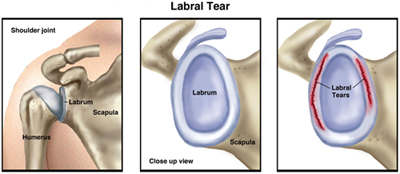
An unstable shoulder joint can be the cause or the result of a labral tear. "Labral" refers to the ring of cartilage (glenoid labrum) that surrounds the base of the shoulder joint. Injuries to the labrum are common, can cause a great deal of pain, and may make it hard to move your arm. A labral tear can occur from a fall or from repetitive work activities or sports that require you to use your arms raised above your head. Some labral tears can be managed with physical therapy; in severe cases, surgery may be required to repair the torn labrum.

The ring of cartilage called the glenoid labrum provides extra support for the shoulder joint, helping to keep it in place. A shoulder labral tear occurs when part of this ring is disrupted, frayed, or torn. Tears may lead to shoulder pain, an unstable shoulder joint, and, in severe cases, dislocation of the shoulder. Likewise, a shoulder dislocation can result in labral tears.

When you think of the shoulder joint, picture a golf ball (the head of the upper-arm bone, or humerus) resting on a golf tee (the glenoid fossa, a shallow cavity or socket located on the shoulder blade, or scapula). The labrum provides a rim for the socket (golf tee) so that the humerus (golf ball) does not easily fall off. If the labrum is torn, it is harder for the humerus to stay in the socket. The end result is that the shoulder joint becomes unstable and prone to injury.

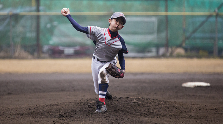
Because the biceps tendon attaches to the shoulder blade through the labrum, labral tears can occur when you put extra strain on the biceps muscle, such as when you throw a ball. Tears also can result from pinching or compressing the shoulder joint, when the arm is raised overhead.

There are 2 types of labral tears:

  • Traumatic labral tears usually occur because of a single incident, such as a shoulder dislocation or an injury from heavy lifting. People who use their arms raised over their heads—such as weight lifters, gymnasts, and construction workers—are more likely to experience traumatic labral tears. Activities where the force occurs at a distance from the shoulder, such as striking a hammer or swinging a racquet, can cause a traumatic labral tear. Falling on an outstretched arm also can cause this type of tear.

  • Nontraumatic labral tears most often occur because of muscle weakness or shoulder joint instability. When the muscles that stabilize the shoulder joint are weak, more stress is put on the labrum, leading to a tear. People with nontraumatic tears tend to have more "looseness" or greater mobility throughout all their joints, which might be a factor in the development of a tear.

LabralTear_SM.jpg

How Does it Feel?

A shoulder labral tear may cause you to feel:

  • Pain over the top of your shoulder

  • "Popping," "clunking," or "catching" with shoulder movement, because the torn labrum has "loose ends" that are flipped or rolled within the shoulder joint during arm movement, and may even become trapped between the upper arm and shoulder blade

  • Shoulder weakness, often on one side

  • A sensation that your shoulder joint will pop out of place

How Is It Diagnosed?

Not all shoulder labral tears cause symptoms. In fact, when tears are small, many people function without any symptoms. However, healing may be difficult due to the lack of blood supply available to a torn labrum. The shoulder with a labral tear may pop or click without being painful, but if the tear progresses, it is likely to lead to pain and weakness.

If your physical therapist suspects that you have a labral tear, your physical therapist will review your health history and perform an examination that is designed to test the condition of the glenoid labrum. The tests will place your shoulder in positions that may recreate some of your symptoms, such as "popping," "clicking," or mild pain, to help your physical therapist determine whether your shoulder joint is unstable. Magnetic resonance imaging (MRI) also may be used to complete the diagnosis. Some labral tears may be difficult to diagnose with certainty without arthroscopic surgery. Your physical therapist may consult with an orthopedic surgeon if necessary.

How Can a Physical Therapist Help?

When shoulder labral tears cause minor symptoms but don’t cause shoulder instability, they usually are treated with physical therapy. Your physical therapist will educate you about positions and activities to avoid, and tailor a treatment plan for your recovery. Your treatment may include:

Manual therapy. Your physical therapist may provide gentle manual (hands-on) therapy to decrease your pain and begin to restore movement in the shoulder area.

Strengthening exercises. Improving the strength of the muscles of the shoulder will help you decrease the stresses placed on the torn labrum and allow for better healing. Your physical therapist may design rotation exercises that target the muscles of the shoulder joint, and shoulder-blade (scapular) exercises to provide stability to the shoulder joint itself.

Stretching exercises. An imbalance in the muscles or a decrease in flexibility can result in poor posture or excessive stress within the shoulder joint. Your physical therapist may prescribe stretching exercises—such as gentle stretches of the chest (pectoralis) muscles—to improve the function of the muscles surrounding the shoulder. Your physical therapist also may introduce middle-back (thoracic) stretches to allow your body to rotate or twist to the side, so the shoulder joint doesn’t have to stretch further to perform tasks, such as swinging a racquet or golf club.

Postural exercises. Your physical therapist will assess your posture, and teach you specific exercises to ensure your shoulders are positioned properly for daily tasks. A forward-head and rounded-shoulder posture puts the shoulders at risk for injury.

Education. Education is an important part of any physical therapy treatment plan. Your physical therapist will help you understand your injury, the reasons for modifying your activities, and the importance of doing your exercises to decrease your risk of future injury.

Home-exercise program. A home-exercise program is an important companion to treatment in the physical therapy clinic. Your physical therapist will identify the stretching and strengthening exercises that will help you steadily improve your shoulder function and meet your work, home, and activity goals.

Following Surgery

In more severe cases, when conservative treatments are unable to completely relieve the symptoms of a labral tear, surgery may be required to reattach the torn labrum. Following surgery, your physical therapist will design a treatment program based on your specific needs and goals, and work with you to help you safely return to your daily activities.

A surgically repaired labrum takes 9 to 12 months to completely heal. Immediately following surgery, your physical therapist will teach you ways to avoid putting excessive stress or strain on the repaired labrum.

As the labrum heals, your physical therapist will introduce resistance and strengthening exercises, such as those listed above, to your treatment plan, to address your specific needs, and help you slowly and safely return to performing daily tasks that require force or lifting. Your physical therapist is trained to gradually introduce movements in a safe manner to allow you to return to your usual activities without re-injuring the repaired tissues.

Can this Injury or Condition be Prevented?

Forceful activities performed with the arms raised overhead may increase the likelihood of developing a labral tear. To avoid putting excessive stress on the labrum, you need to develop strength in the muscles that surround the shoulder and scapula. Your physical therapist can:

  • Design exercises to help you strengthen your shoulder and shoulder blade muscles

  • Show you how to avoid potentially harmful positions

  • Train you to properly control your shoulder movement and modify your activities to reduce your risk of sustaining a labral injury

  • Provide posture education to help you avoid placing unnecessary forces on the shoulder

  • Help you increase your shoulder and middle-back flexibility


What Kind of Physical Therapist Do I Need?

All physical therapists are prepared through education and experience to treat patients who have a shoulder labral tear, but you may want to consider:

  • A physical therapist who is experienced in treating people with musculoskeletal problems. Some physical therapists have a practice with an orthopedic focus.

  • A physical therapist who is a board-certified clinical specialist or who completed a residency or fellowship in orthopedics physical therapy has advanced knowledge, experience, and skills that may apply to your condition.

You can find physical therapists who have these and other credentials by using Find a PT, the online tool built by the American Physical Therapy Association to help you search for physical therapists with specific clinical expertise in your geographic area.

General tips when you're looking for a physical therapist (or any other health care provider):

  • Get recommendations from family and friends or from other health care providers.

  • When you contact a physical therapy clinic for an appointment, ask about the physical therapists' experience in helping people with labral tears.

  • During your first visit with the physical therapist, be prepared to describe your symptoms in as much detail as possible, and say what makes your symptoms worse.

Further Reading

The American Physical Therapy Association (APTA) believes that consumers should have access to information that could help them make health care decisions and also prepare them for their visit with their health care provider.

APTA has determined that the following articles provide some of the best scientific evidence for how to treat labral tears. The articles report recent research and give an overview of the standards of practice for treatment both in the United States and internationally. The article titles are linked either to a PubMed* abstract of the article or to free access of the full article, so that you can read it or print out a copy to bring with you to your health care provider.

Mazzocca AD, Cote MP, Solovyova O, et al. Traumatic shoulder instability involving anterior, inferior, and posterior labral injury: a prospective clinical evaluation of arthroscopic repair of 270° labral tears. Am J Sports Med. 2011;39:1687-1696.  Article Summary on PubMed.

Dodson CC, Altchek DW. SLAP lesions: an update on recognition and treatment. J Orthop Sports Phys Ther. 2009;39:71-80. Article Summary on PubMed.

Keener JD, Brophy RH. Superior labral tears of the shoulder: pathogenesis, evaluation, and treatment. J Am Acad Orthop Surg. 2009;17:627-637. Article Summary on PubMed.

 *PubMed is a free online resource developed by the National Center for Biotechnology Information (NCBI).  PubMed contains millions of citations to biomedical literature, including citations in the National Library of Medicine’s MEDLINE database.”

Authored by Charles Thigpen, PhD, PT, ATC and Lane Bailey, PT, DPT, CSCS. Reviewed by the MoveForwardPT.com editorial board.


Hip Bursitis

Hip bursitis is a painful condition that affects 15% of women and 8.5% of men of all ages in the United States. The condition tends to develop more in middle-aged and elderly individuals. Hip bursitis can have many causes, but the most common is a repetitive activity, such as walking or running on an uneven surface, which creates friction in the hip area. Athletes often develop hip bursitis after running up and down hills repetitively. The condition can also be caused by abnormal walking, such as limping, due to an uneven leg length, or arthritis in the back, hip, knee, or other joints in the leg. It can also occur without any specific cause. Physical therapy can be an effective treatment for hip bursitis to reduce pain, swelling, stiffness, and any associated weakness in the hip, back, or lower extremity.

Hip bursitis (also called trochanteric bursitis) occurs when one or both of the 2 bursae (fluid-filled sacs on the side of the hip) become damaged, irritated, or inflamed. Bursitis (“-itis” means “inflammation of”) means the bursa has become irritated and inflamed, usually causing pain. Normally, the bursa acts as a cushion for muscles and tendons that are close to it. Certain positions, motions, or disease processes can cause constant friction or stress on the bursa, leading to the development of bursitis. When the bursa becomes injured, those muscles and tendons don’t glide smoothly over it, and they can become painful.

Hip bursitis can be caused by:

  • Repetitive motions (running up and down hills or stairs)

  • Muscle weakness

  • Incorrect posture

  • Direct trauma (being hit or falling on the side of the hip)

  • Differences in the length of each leg

  • Hip surgery or replacement

  • Bone spurs in the hip

  • Infection

  • Diseases, such as rheumatoid arthritis, gout, psoriasis, or thyroid disease

  • Muscles or tendons in the hip area rubbing the bursa and causing irritation

How Does it Feel?

With hip bursitis, you may experience:

  • Pain on the outer side of the hip, thigh, buttock, or outside of the leg.

  • Pain when you push on the outer side of the hip with your fingers.

  • Pain when lying on the affected hip.

  • Pain when climbing stairs.

  • Pain that worsens when getting up from sitting, such as from a low chair or car seat.

  • Pain when walking or running.

  • Pain when lifting the leg up to the side.

  • Pain when sitting with legs crossed.

How Is It Diagnosed?

If you see your physical therapist first, the physical therapist will conduct a thorough evaluation that includes taking your health history. Your physical therapist also will ask you detailed questions about your condition, such as:

  • How and when did you notice the pain?

  • Have you been performing any repetitive activity?

  • Did you receive a direct hit to the hip or fall on it? 

Your physical therapist will perform special tests to help determine the likelihood that you have hip bursitis. Your physical therapist will gently press on the outer side of the hip to see if it is painful to the touch, and may use additional tests to determine if other parts of your hip are injured. Your physical therapist also will observe how you are walking.

Your physical therapist will test and screen for other, more serious conditions that could cause lateral hip pain. To provide a definitive diagnosis, your physical therapist may collaborate with an orthopedic physician or other health care provider, who may order further tests (eg, an x-ray) to confirm the diagnosis and rule out other damage to the hip, such as a fracture.

How Can a Physical Therapist Help?

Your physical therapist will work with you to design a specific treatment program that will speed your recovery, including exercises and treatments that you can do at home. Physical therapy will help you return to your normal lifestyle and activities. The time it takes to heal the condition varies, but results can be achieved in 2 to 8 weeks or less, when a proper stretching and strengthening program is implemented.

During the first 24 to 48 hours following your diagnosis, your physical therapist may advise you to:

  • Rest the area by avoiding walking or any activity that causes pain.

  • Apply ice packs to the area for 15 to 20 minutes every 2 hours.

  • Consult with a physician for further services, such as medication or diagnostic tests.

Your physical therapist will work with you to:

Reduce Pain and Swelling. If repetitive activities have caused the hip bursitis, your physical therapist will help you understand how to avoid or modify your activities to allow healing to begin. Your physical therapist may use different types of treatments and technologies to control and reduce your pain and swelling, including ice, heat, ultrasound, electrical stimulation, taping, exercises, and hands-on therapy, such as specialized massage.

Improve Motion. Your physical therapist will choose specific activities and treatments to help restore normal movement in the hip and leg. These might begin with "passive" motions that the physical therapist performs for you to gently move your hip joint, and progress to active exercises and stretches that you do yourself.

Improve Flexibility. Your physical therapist will determine if any leg or spinal muscles are tight, help you to stretch them, and teach you how to stretch them.

Improve Strength. Hip bursitis is often related to weak or injured hip muscles. Certain exercises will aid healing at each stage of recovery; your physical therapist will choose and teach you the correct exercises and equipment to steadily restore your strength and agility. These may include using cuff weights, stretch bands, weight-lifting equipment, and cardio-exercise equipment, such as treadmills or stationary bicycles.

Improve Balance. Regaining your sense of balance is important after an injury. Your physical therapist will teach you exercises to improve your balance skills.

Learn a Home-Exercise Program. Your physical therapist will teach you strengthening and stretching exercises to perform at home. These exercises will be specific for your needs; if you do them as prescribed by your physical therapist, you can speed your recovery.

Return to Activities. Your physical therapist will discuss your activity goals with you and use them to set your work, sport, and home-life recovery goals. Your treatment program will help you reach your goals in the safest, fastest, and most effective way possible. Your physical therapist will teach you exercises, work retraining activities, and sport-specific techniques and drills that match your lifestyle.

Speed Recovery Time. Your physical therapist is trained and experienced in choosing the best treatments and exercises to help you safely heal, return to your normal lifestyle, and reach your goals faster than you are likely to do on your own.

If Surgery Is Necessary

Surgery is not commonly required for hip bursitis. However, if surgery is needed, you will follow a recovery program over several weeks guided by your physical therapist. Your physical therapist will help you minimize pain, regain motion and strength, and return to normal activities in the safest and speediest manner possible.

Can this Injury or Condition be Prevented?

Your physical therapist can recommend a home-exercise program to strengthen and stretch the muscles around your knee, upper leg, and abdomen to help prevent future injury. These may include strength and flexibility exercises for the leg, knee, and core muscles.

To help prevent a recurrence of the injury, your physical therapist may advise you to:

  • Follow a consistent flexibility and strengthening exercise program, especially for the hip muscles, to maintain good physical conditioning, even in a sport's off-season.

  • Always warm up before starting a sport or heavy physical activity.

  • Gradually increase any athletic activity, rather than suddenly increasing the activity amount or intensity.

  • Obtain a professionally fitted shoe insert, if your legs are different lengths.

  • Learn and maintain correct posture.

Real Life Experiences

John is a 30-year-old dentist who enjoys running, biking, and triathlon training. He recently decided to add CrossFit training to his exercise routine, and joined an aggressive CrossFit gym. After running stairs intensely during 1 session, John felt pain developing on the outside of his right hip a few hours later. He realized that he was limping. By the end of the day, he was having trouble climbing stairs because of the hip pain. He contacted his physical therapist.

John's physical therapist asked him how and when he felt the pain. She observed him walking, and noted his limp. She gently touched the soft tissues on the outside of his hip, and noted the tenderness he felt when she pushed right over the bony spot on the outer side of the hip. She tested the strength and flexibility in his hip and back. She concluded that his hip bursa was irritated and inflamed, and that he had some tight and weak muscles in the area. She diagnosed hip bursitis. She asked John about his goals, which he said were to get rid of the pain, walk normally, and return to his previous level of exercising as well as his CrossFit club activities.

John's physical therapist began his treatment by gently stretching the muscles she had found were tight. Next, she applied ice and electrical stimulation to the area to help halt and reverse the inflammation process. She discussed with John the importance of “relative rest” for the hip area, meaning he should avoid activities like stair running, lunges, and squats for a few days, until his pain subsided.

When John returned for his next treatment, he was already feeling less pain. His physical therapist noted that his limp was almost gone. She started teaching John strengthening exercises for the muscle weakness she had found in his hip and back muscles. Only light weights were used to avoid causing new pain. As John improved, the weights were steadily increased.

Two weeks later, John's physical therapist added light jogging to John’s program, which he was able to do without increasing his pain. During the third week, John was improved enough to add lunges. By the fourth week, he was able to begin jogging up stairs in short sessions.

By the fifth week, John was able to rejoin his CrossFit club, although he was mindful to increase his workouts slowly and gradually, especially when it came to running stairs. He learned from his physical therapist to avoid any sudden increases in exercise. John also incorporated some of the special strengthening exercises he learned from his physical therapist into his warm-up and workout.

When he met his physical therapist at the store 6 months later, John was happy to report he was functioning at his top fitness level, and by following the advice he learned from her, he had prevented any recurrence of hip bursitis.

What Kind of Physical Therapist Do I Need?

All physical therapists are prepared through education and experience to treat hip bursitis. However, you may want to consider:

  • A physical therapist who is experienced in treating people with orthopedic injuries. Some physical therapists have a practice with an orthopedic focus.

  • A physical therapist who is a board-certified clinical specialist, or who completed a residency or fellowship in orthopedic or sports physical therapy. This therapist has advanced knowledge, experience, and skills that may apply to your condition.

You can find physical therapists who have these and other credentials by using Find a PT, the online tool built by the American Physical Therapy Association to help you search for physical therapists with specific clinical expertise in your geographic area.

General tips when you're looking for a physical therapist (or any other health care provider):

  • Get recommendations from family and friends or from other health care providers.

  • When you contact a physical therapy clinic for an appointment, ask about the physical therapists' experience in helping people who have your type of injury.

  • During your first visit with the physical therapist, be prepared to describe your symptoms in as much detail as possible, and say what makes your symptoms worse.

Further Reading

The American Physical Therapy Association (APTA) believes that consumers should have access to information that could help them make health care decisions and also prepare them for their visit with their health care provider. The following articles provide some of the best scientific evidence related to physical therapy treatment of hip bursitis. The articles report recent research and give an overview of the standards of practice both in the United States and internationally. The article titles are linked either to a PubMed* abstract of the article or to free full text, so that you can read it or print out a copy to bring with you to your health care provider.

Haviv B. Update on trochanteric bursitis of the hip. OA Orthopaedics. 2013;1(1):10. Article summary not available.

Lustenberger DP, Ng VY, Best TM, Ellis TJ. Efficacy of treatment of trochanteric bursitis: a systematic review. Clin J Sport Med. 2011;21(5):447–453. Free Article.

Segal NA, Felson DT, Torner JC, et al. Greater trochanteric pain syndrome: epidemiology and associated factors. Arch Phys Med Rehabil. 2007;88(8):988–992. Free Article.

Jones DL, Erhard RE. Diagnosis of trochanteric bursitis versus femoral neck stress fracture. Phys Ther. 1997;77(1):58–67. Free Article.

Butcher JD, Salzman KL, Lillegard WA. Lower extremity bursitis [erratum in: Am Fam Physician. 1996;54(2):468]. Am Fam Physician. 1996;53(7):2317–2324.

Hammer WI. The use of transverse friction massage in the management of chronic bursitis of the hip or shoulder. J Manipulative Physiol Ther. 1993;16(2):107–111. Article Summary in PubMed.

Sears B. Physical therapy for hip pain. About.com website. Accessed December 9, 2014.

Physical therapy management of trochanteric bursitis. Morphopedics website. Accessed December 9, 2014.

Trochanteric bursitis. Cleveland Clinic website. Accessed December 9, 2014.

University of Washington Orthopedics and Sports Medicine. Bursitis, tendinitis, and other soft tissue rheumatic syndromes. Accessed December 9, 2014.

*PubMed is a free online resource developed by the National Center for Biotechnology Information (NCBI). PubMed contains millions of citations to biomedical literature, including citations in the National Library of Medicine’s MEDLINE database.

Authored by Andrea Avruskin, PT, DPT. Reviewed by the MoveForwardPT.com editorial board.


What Is Vertigo?

Vertigo is the sensation of spinning—even when you're perfectly still, you might feel like you're moving or that the room is moving around you. Most causes of vertigo involve the inner ear ("vestibular system"). A number of conditions can produce vertigo, such as:

  • Inner ear infections or disorders

  • Migraines

  • Tumors, such as acoustic neuroma

  • Surgery that removes or injures the inner ear or its nerves

  • Head injury that results in injury to the inner ears

  • A hole in the inner ear

  • Stroke

You also might have:

  • Nausea

  • Vomiting

  • Sweating

  • Abnormal eye movements

One of the most common forms of vertigo is benign paroxysmal positional vertigo, an inner-ear problem that causes short periods of a spinning sensation when your head is moved in certain positions.

How Is It Diagnosed?

Your physical therapist will use your answers to the following questions to help identify the cause of your vertigo and to determine the best course of treatment:

  • When did you first have vertigo (the sensation of spinning)?

  • What are you doing when you have vertigo (turning your head, bending over, standing perfectly still, rolling in bed)?

  • How long does the vertigo last(seconds, minutes, hours, days)?

  • Have you had vertigo before?

  • Do you have hearing loss, ringing, or fullness in your ears?

  • Do you have nausea with the spinning?

  • Have you had any changes in your heart rate or breathing?

Your physical therapist will perform tests to determine the causes of your vertigo and also to assess your risk of falling. Depending on the results of the tests, your therapist may recommend further testing or consultation with your physician.

How Can a Physical Therapist Help?

Based on your physical therapist's evaluation and your goals for recovery, the therapist will customize a treatment plan for you. The specific treatments will depend on the cause of your vertigo. Your therapist's main focus is to help you get moving again and manage the vertigo at the same time. Treatment may include specialized head and neck movements or other exercises to help eliminate your symptoms. Conditions such as benign paroxysmal positional vertigo have very specific tests and treatments.

If you have dizziness and balance problems after your vertigo has stopped, your physical therapist can develop a treatment plan that targets those problems. Your physical therapist will teach you strategies to help you cope with your symptoms:

  • Do certain activities or chores around the house cause you to become dizzy? Your therapist will show you how to do those activities in a different way to help reduce the dizziness.

  • Have simple activities become difficult and cause fatigue and more dizziness? Your therapist will help you work through these symptoms right away so you can get moving again and return to your roles at home and at work more quickly.

Physical therapy treatments for dizziness can take many forms. The type of exercise that your therapist designs for you will depend on your unique problems and might include:

  • Exercises to improve your balance

  • Exercises to help the brain "correct" differences between your inner ears

  • Exercises to improve your ability to focus your eyes and vision

In addition, your physical therapist might prescribe exercises to improve your strength, your flexibility, and your heart health—with the goal of improving your overall physical health and well being.

What Kind of Physical Therapist Do I Need?

All physical therapists are prepared through education and experience to treat people with dizziness. You may want to consider:

  • A physical therapist who is experienced in treating people with neurological problems. Some physical therapists have a practice with a neurological vestibular rehabilitation focus.

  • A physical therapist who is a board-certified clinical specialist or who completed a residency or fellowship in neurological physical therapy. This therapist has advanced knowledge, experience, and skills that may apply to your condition.

You can find physical therapists who have these and other credentials by using Find a PT, the online tool built by the American Physical Therapy Association to help you search for physical therapists with specific clinical expertise in your geographic area.

General tips when you're looking for a physical therapist (or any other health care provider):

  • Get recommendations from family and friends or from other health care providers.

  • When you contact a physical therapy clinic for an appointment, ask about the physical therapists' experience in helping people with inner ear injury.

  • During your first visit with the physical therapist, be prepared to describe your symptoms in as much detail as possible, and say what makes your symptoms worse.

Further Reading

The American Physical Therapy Association (APTA) believes that consumers should have access to information that could help them make health care decisions and also prepare them for their visit with their health care provider.

The following articles provide some of the best scientific evidence about physical therapy treatments for vertigo. The articles report recent research and give an overview of the standards of practice for treatment both in the United States and internationally. The article titles are linked either to a PubMed abstract of the article or to free full text, so that you can read it or print out a copy to bring with you to your health care provider.

Hillier SL, McDonnell M. Vestibular rehabilitation for unilateral peripheral vestibular dysfunction. Cochrane Database Syst Rev. 2011;16;(2):CD005397. Systematic Review. Article Summary on PubMed.

Helminski JO, Zee DS, Janssen I, Hain TC. Effectiveness of particle repositioning maneuvers in the treatment of benign paroxysmal positional vertigo: a systematic review.Phys Ther. 2010;90:663–678. Free Article.

Scherer MR, Schubert MC. Traumatic brain injury and vestibular pathology as a comorbidity after blast exposure. Phys Ther. 2009;89:980-992. Free Article.

Cohen HS, Sangi-Haghpeykar H. Canalith repositioning variations for benign paroxysmal positional vertigo. Otolaryngol Head Neck Surg. 2010;143:405–412. Free Article.

*PubMed is a free online resource developed by the National Center for Biotechnology Information (NCBI). PubMed contains millions of citations to biomedical literature, including citations in the National Library of Medicine’s MEDLINE database.

Authored by Melissa S. Bloom, PT, DPT; Bob Wellmon, PT, PhD, NCS; and APTA's Neurology Section. Reviewed by the MoveForwardPT.com editorial board.


Degenerative Disk Disease

What Is Degenerative Disk Disease?

Your spine is made up of 33 vertebrae that are stacked on top of one another. Between each of these vertebrae is a rubbery piece of cartilage called an "intervertebral disk." (See images: Degenerative Disk Disease - Cervical | Lumbar.) Imagine the disk as a tire, with gelatin filling the hole in the tire. The tire is called the "annulus," and the gelatin is called the "nucleus." When we're young—under 30 years of age—the disk is made mostly of gelatin. As we age, and sometimes with injury, we start to lose some of that gelatin and the volume of the disk decreases, resulting in less space between the vertebrae. The disk becomes flatter and less flexible, leaving less space between each set of vertebrae. Sometimes bone spurs form in response to this degeneration of the disk, which could make the spine stiff. Often, this flattening and additional stiffness to the spine is not at all painful.  However, in some cases, when the rough surfaces of the vertebral joints rub together, pain and inflammation may result. The nerve root, the point where a spinal nerve exits the spine and extends to other parts of the body, may become irritated or compressed.

Disk degeneration may occur throughout several regions of the spine, or it may be limited to 1 disk. Degeneration does not always lead to pain. For some people, however, it can cause a great deal of pain and disability.

You are more likely to develop DDD if you:

  • Smoke
  • Are obese
  • Do heavy physical work
  • Don't get very much exercise
DDD_Cervical_md.jpg


How Does it Feel?

With DDD, you may experience mild to intense neck and back pain—or no pain at all:

  • A degenerative disk in the neck may cause pain in the arm, shoulder, or neck.
  • A degenerative disk in the low back may cause pain in the back, buttocks, or legs.

The pain is often made worse by sitting, bending, and reaching. It may be worse first thing in the morning and after staying in any one position for a long time.

In severe cases, when DDD results in pressure on the nerve root, it can lead to numbness, tingling, and even weakness in the arms or legs.

How Is It Diagnosed?

Your physical therapist will conduct a thorough evaluation that includes a review of your medical history, and will use screening tools to determine the likelihood of DDD. For example, your physical therapist may:

  • Ask you very specific questions about the location and behavior of your pain, weakness, and other symptoms
  • Ask you to fill out a body diagram to indicate specific areas of pain, numbness, and tingling
  • Perform tests of muscle strength and sensation to determine the presence of pressure on the nerve
  • Examine your posture and observe how you walk and perform other activities
  • Measure the range of motion of your spine and your arms and legs
  • Use manual (hands-on) therapy to evaluate the mobility of the joints and muscles in your spine
  • Test the strength of important muscle groups

If you have muscle weakness, loss of sensation, or severe pain, diagnostic tests, such as an X-ray or MRI, may be needed. However, X-ray evidence of wearing in the joints of the spine is found in people with back pain as well as in many who have never experienced back pain. Studies have shown that disc degeneration is present in 40% of individuals under the age of 30 and present in over 90% of those aged 50 to 55 years. Do not panic if your X-ray or MRI shows “problems” with your discs; these are normal changes that happen from the age of 20 years and onward. Physical therapists work closely with physicians and other health care providers to make certain that an accurate diagnosis is made and the appropriate treatment is provided.

Research shows that in all but the most extreme cases (usually involving muscle weakness or high levels of pain), conservative care, such as physical therapy, has better results than surgery. Research also shows that results from conservative care and surgery are the same after 18 months post operatively.

After the evaluation, if your physical therapist suspects you have DDD and there are no major medical problems, treatment can begin right away.

How Can a Physical Therapist Help?

Your physical therapist's overall purpose is to help you continue to participate in your daily activities and life roles. Your physical therapist will design a treatment program based on both the findings of the evaluation and your personal goals. The treatment program may include:

Stretching and flexibility exercises. Your physical therapist will teach you specific exercises to improve movement in the joints and muscles of your spine, arms, and legs. Improving motion in a joint is often the key to pain relief.

Strengthening exercises. Strong trunk muscles provide support for your spinal joints, and strong arm and leg muscles help take some of the workload off your spinal joints.

Aerobic exercise. Regular aerobic exercise, such as walking, swimming, or taking a low-impact aerobics class, has been shown to help relieve pain, promote a healthy body weight, and improve overall strength and mobility—all important factors in managing DDD.

This might sound like a lot of exercise, but don't worry, research shows that the more exercise you can handle, the quicker you'll get rid of your pain and other symptoms.

Your treatment program may also include:

Manual therapy. Your physical therapist may apply manual (hands-on) therapy, such as massage, to improve movement in stiff joints and tight muscles that may be contributing to your symptoms.

Posture and body mechanics education. Your physical therapist may show you how to make small changes in how you sit, stand, bend, and lift—even in how you sleep—to help relieve your pain and help you manage your condition on your own.

Note: Studies show that recurrence of neck and low back pain is common when a condition such as DDD is not properly treated. Regular performance of the exercises your physical therapist chooses for you is extremely important to make sure your pain does not return.

Can this Injury or Condition be Prevented?

DDD is a natural result of aging. However, you can make choices that lessen its impact on your life and slow its progression. Your local physical therapy clinic can offer you advice on staying strong and fit. Some physical therapy clinics conduct regular educational seminars to help people in the community learn to take care of their backs and necks. Your physical therapist can help you develop a fitness program that takes into account your DDD. There are some exercises that are better than others for people with DDD, and your physical therapist will choose the right ones for you. For instance:

  • Exercising in water can often be a great way to stay physically active when other forms of exercise are painful.
  • Exercises involving repetitive twisting and bending need to be performed with some caution. If you start to notice some aching or pain after exercising, consult with a health care professional, such as a physical therapist, who can improve the way you move—and help reduce or eliminate your back or neck symptoms.
  • Weight-training exercises, though important, need to be done with proper form to avoid stress to the back and neck. Your physical therapist will work with you to ensure your weight training is safe and effective.

What Kind of Physical Therapist Do I Need?

All physical therapists are prepared through education and experience to treat people who have DDD. You may want to consider:

  • A physical therapist who is experienced in treating people with pain, orthopedic, or musculoskeletal, diagnoses.
  • A physical therapist who is a board-certified clinical specialist or who completed a residency or fellowship in orthopaedic physical therapy. This therapist has advanced knowledge, experience, and skills that may apply to your condition.
  • You can find physical therapists who have these and other credentials by using Find a PT, the online tool built by the American Physical Therapy Association to help you search for physical therapists with specific clinical expertise in your geographic area.

General tips when you're looking for a physical therapist:

  • Get recommendations from family and friends or from other health care providers.
  • When you contact a physical therapy clinic for an appointment, ask about the physical therapist's experience in helping people with DDD.
  • During your first visit with the physical therapist, be prepared to describe your symptoms in as much detail as possible, and say what makes your symptoms worse.

Further Reading

The American Physical Therapy Association (APTA) believes that consumers should have access to information that could help them make health care decisions and also prepare them for their visit with their health care provider.

The following articles provide some of the best scientific evidence related to physical therapy treatment of degenerative disk disease. The articles report recent research and give an overview of the standards of practice for treatment of DDD both in the United States and internationally. The article titles are linked either to a PubMed abstract of the article or to free full text, so that you can read it or print out a copy to bring with you to your health care provider.

Macedo LG, Maher CG, Latimer J, McAuley JH. Motor control exercise for persistent, nonspecific low back pain: a systematic review. Phys Ther. 2009;89:9–25. Free Article.

Beattie PF. Current understanding of lumbar intervertebral disc degeneration: a review with emphasis upon etiology, pathophysiology, and lumbar magnetic resonance imaging findings. J Orthop Sports Phys Ther. 2008;38:329–340. Free Article.

Chou R, Qaseem A, Snow V, et al. Diagnosis and treatment of low back pain: a joint clinical practice guideline from the American College of Physicians and the American Pain Society [published correction appears in: Diagnosis and treatment of low back pain. Ann Intern Med. 2008]. Ann Intern Med. 2007;147:478–491. Free Article.

Roh JS, Teng AL, Yoo JU, et al. Degenerative disorders of the lumbar and cervical spine. Orthop Clin North Am. 2005:36:255–262. Article Summary on PubMed.

Authored by Chris Bise, PT, DPT. Reviewed by the MoveForwardPT.com editorial board.

Thoracic Outlet Syndrome

Thoracic Outlet Syndrome (TOS) is a potentially painful and disabling condition of the upper extremity. It results from the compression of structures in the thoracic outlet, a space just above the first rib, and behind the clavicle (collar bone). Due to the range of signs and symptoms that can lead to a diagnosis of TOS, the incidence rates of the condition currently are unknown. Physical therapists work with individuals who have TOS to ease their symptoms and restore their upper-body function.

How Does it Feel?

Because TOS generally is classified based upon the type of structures compressed, the symptoms experienced may vary. However, more than 90% of TOS cases are thought to be neurogenic (nerve compression) in nature (categories 3 and 4 below).

Arterial TOS

  • Pain in the hand; rarely in shoulder or neck
  • Coldness or cold intolerance
  • Numbness and tingling

Venous TOS

  • Pain in the arm
  • Swelling in the arm
  • Change in arm coloration (appears bluish)
  • Feeling of heaviness in the arm
  • Numbness and tingling in fingers and hands

True Neurogenic TOS

  • Pain, numbness, and tingling in the hand, arm, shoulder, and often the neck
  • Headaches
  • Numbness and tingling of the arm, often waking the individual up at night
  • Hand clumsiness
  • Intolerance to cold
  • Hand coldness and color changes

Disputed Neurogenic TOS

  • Pain, numbness and tingling in the hand, arm, shoulder and often the neck
  • Headaches
  • Numbness and tingling of the arm, often waking the individual up at night
  • Hand clumsiness
  • Intolerance to cold
  • Hand coldness and color changes
  • Symptoms greater at night vs day
  • Tests may come back normal (hence, the term "disputed").

How Is It Diagnosed?

Diagnosis of TOS begins with a thorough health history and clinical examination.

Your physical therapist will likely check for color changes in the affected area, and gently attempt to provoke symptoms by moving the affected limb in different directions.

You also may be referred for diagnostic testing, such as a Doppler ultrasound, which can confirm arterial and venous TOS, or nerve conduction velocity testing to help confirm a true neurogenic TOS.

Your physical therapist may be the first to recognize an onset of TOS, because of its effects on your physical function. Your physical therapist may ask you:

  • When did you begin experiencing these symptoms, and when are they the worst?
  • Have you noticed any change in your symptoms when the temperature changes?
  • Have you noticed any significant changes in your ability to perform physical tasks that require hand movements?
  • Have you noticed any changes in the appearance of your arm or hand?

In addition, your physical therapist will rule out other conditions, which may mimic this disorder. Your therapist may ask you to fill out a questionnaire in order to better understand your physical state, and to screen for the presence of other conditions.

How Can a Physical Therapist Help?

Once you have received a diagnosis of TOS, your physical therapist will work with you to develop a treatment plan to help ease the discomfort, and improve your ability to perform daily activities. Most research on this condition recommends a treatment plan that involves physical therapy to help ease your symptoms and improve function.

Physical therapy treatments may include:

Manual Therapy. Manual (hands-on) therapy may be applied to manipulate or mobilize the nerves of the arm to help reduce symptoms, such as pain and numbness/tingling. Your physical therapist also may attempt to gently mobilize your first rib and/or collar bone.

Movement and Strengthening Exercises. Your physical therapist will teach you muscle-strengthening exercises to improve movement and strength in the affected area.

Education. Your physical therapist will teach you strategies that can help minimize your symptoms while performing your daily functional activities.

Activity modification and postural strategies: Your physical therapist will teach you positions and strategies to place less stress on the structures involved with TOS.

Can this Injury or Condition be Prevented?

While some cases of TOS cannot be prevented, such as those due to anatomical variations, congenital conditions, trauma, or space-occupying lesions, others may be avoidable. Understanding risk factors that could make you more likely to develop this condition is the first step in prevention.

Your physical therapist will work with you to develop strategies to help you better understand and manage your risk factors and symptoms. As with many conditions, education is key. Understanding strategies, such as methods of reducing symptoms while performing activities, can help you live a full and functional life after the onset of TOS.

Real Life Experiences

George is a 45-year-old used-car salesman who takes potential customers on test drives in a crowded city area. Recently on a test drive, his customer ran a red light and the vehicle was struck by another motorist on the passenger side, where George was sitting. George was shaken up, although the police assured him the actual collision was minor.

George was taken to the emergency room following the accident, and received an X-ray, which was negative for a fracture of his clavicle. Fearing that he may be having a heart attack, he also followed up with his primary care physician, who was able to rule that out as well.

Over the next few days, George felt pain on his right side, and numbness and tingling down his right arm. His symptoms seemed to be worse at night. He called in sick to work, fearing he could be injured further on the job. He called his physical therapist.

George's physical therapist conducted a full physical examination. During the exam, George reported that he felt very anxious about the recent event, and wasn’t sure he'd be able to trust taking customers on test drives anymore.    

George's physical therapist noticed the presence of a rounded shoulder and forward-head posture, as she examined him. She was able to provoke George’s symptoms by gently placing his arm in particular positions, and gently pressing in the region of George’s first rib. She carefully checked for any other conditions that could be causing his symptoms. She told George that she suspected neurogenic TOS.

She developed a strategy for physical therapy that was best for him, consisting of activities and exercises to increase his strength, confidence, and function, while also easing his pain. She showed him postural activities to reduce compressive forces on his nerves. She also helped him with "nerve gliding" activities (encouraging his nerves to glide normally as his joints moved) to improve the function of the affected nerves.

Despite the complexity of the condition, George did well with his personalized course of physical therapy. Following several weeks of treatment and exercise, he was able to return to work without symptoms, and with a new-found confidence that he could ride in a car with customers again. With his physical therapist's ongoing help, George has returned to his normal activities of daily living.

This story highlights an individualized experience of TOS. Your case may be different. Your physical therapist will tailor a treatment program to your specific needs.

What Kind of Physical Therapist Do I Need?

All physical therapists are prepared through education and clinical experience to treat a variety of conditions or injuries.  You may want to consider:

  • A physical therapist who is experienced in treating people with TOS.
  • A physical therapist who is a board-certified clinical specialist or who completed a residency or fellowship in physical therapy. This therapist has advanced knowledge, experience, and skills that may apply to your condition.

You can find physical therapists who have these and other credentials by using Find a PT, the online tool built by the American Physical Therapy Association to help you search for physical therapists with specific clinical expertise in your geographic area.

General tips when you're looking for a physical therapist (or any other health care provider):

  • Get recommendations from family and friends or from other health care providers.
  • When you contact a physical therapy clinic for an appointment, ask about the physical therapists' experience in helping people with TOS.
  • During your first visit with the physical therapist, be prepared to describe your symptoms in as much detail as possible. Keeping a journal highlighting when you experience pain will help the physical therapist identify the best treatment approach.

Further Reading

The American Physical Therapy Association (APTA) believes that consumers should have access to information that could help them make health care decisions, and also prepare them for their visit with their health care provider.

The following articles provide some of the best scientific evidence related to physical therapy treatment of TOS. The articles report recent research and give an overview of the standards of practice both in the United States and internationally. The article titles are linked either to a PubMed* abstract of the article or to free full text, so that you can read it or print out a copy to bring with you to your health care provider.

Hooper TL, Denton J, McGalliard MK, Brismée JM, Sizer PS Jr. Thoracic outlet syndrome: a controversial clinical condition; part 1: anatomy and clinical examination/diagnosis. J Man Manip Ther. 2010:18(2):74–83. Free Article. Article Summary in PubMed.

Hooper TL, Denton J, McGalliard MK, Brismée JM, Sizer PS Jr. Thoracic outlet syndrome: a controversial clinical condition; part 2: non-surgical and surgical management. J Man Manip Ther. 2010;18z(3):132–138. Free Article. Article Summary in PubMed.

Watson LA, Pizzari T, Balster S. Thoracic outlet syndrome part 2: conservative management of thoracic outlet. Man Ther. 2010;15(4):305–314. Article Summary in PubMed.

Vanti C, Natalini L, Romeo A, Tosarelli D, Pillastrini P. Conservative treatment of thoracic outlet syndrome: a review of the literature. Eura Medicophys. 2007;43(1):55–70. Article Summary in PubMed.

*PubMed is a free online resource developed by the National Center for Biotechnology Information (NCBI). PubMed contains millions of citations to biomedical literature, including citations in the National Library of Medicine's MEDLINE database.

Authored by Joseph Brence, PT, DPT, FAAOMPT, COMT, DACReviewed by the MoveForwardPT.com editorial board.

Achilles Tendinopathy

Achilles tendinopathy is an irritation of the Achilles tendon. Achilles tendon pain is one of the most common types of pain felt behind the heel and up the back of the ankle when walking or running. Although Achilles tendinopathy affects both active and inactive individuals, 24% of athletes develop the condition, and an estimated 50% of runners will experience Achilles tendon pain in their running careers. In all individuals, Achilles tendinopathy can result in a limited ability to walk, climb stairs, or participate in recreational activities. Physical therapists help people with Achilles tendinopathy reduce pain in the affected area, and restore strength, flexibility, and mobility to the injured tendon and eliminate swelling when present.

Achilles Tendinopathy

Achilles tendinopathy is an irritation of the Achilles tendon, a thick band of tissue along the back of the lower leg that connects the calf muscles to the heel. The term “tendinopathy” refers to any problem with a tendon, either short- or long-term. The Achilles tendon transmits force from the calf muscles down to the foot when a person pushes the foot off the ground (eg, runs or jumps), and helps control the position of the ankle when the foot touches back down on the ground (eg, lands). Achilles tendinopathy results when the demand placed on the Achilles tendon is greater than its ability to function. The condition can occur after a single incident (acute injury) or after repetitive irritation or "microtrauma" (chronic injury). Most often, Achilles tendon pain is the result of repetitive trauma to the tendon that can result in chronic Achilles tendinopathy—a gradual breakdown of the tissue—and is most often treated with physical therapy.

Achilles tendinopathy is linked to several different factors, including:

  • Calf muscle tightness
  • Calf muscle weakness
  • Abnormal foot structure
  • Abnormal foot mechanics
  • Improper footwear
  • A change in an exercise routine or sport activity
  • Obesity

Pain can be present at any point along the tendon; the most common area to feel tenderness is just above the heel (known as midportion Achilles tendinopathy), although it may also be present where the tendon meets the heel (known as insertional Achilles tendinopathy).

AchilleTendonInjury-SM.jpg

How Does it Feel?

With Achilles tendinopathy, you may experience:

  • Tenderness in the heel or higher up in the Achilles tendon with manually applied pressure
  • Pain and stiffness with walking, at its worst with the first several steps
  • Tightness in the calf
  • Swelling in the back of the ankle

How Is It Diagnosed?

Your physical therapist will review your medical history and complete a thorough examination of your heel, ankle, and calf. Your physical therapist will assess your foot posture, strength, flexibility, and movement. This process may include watching you stand in a relaxed stance, walk, squat, step onto a stair, or do a heel raise. The motion and strength in other parts of your leg also will be assessed.

Your physical therapist may also ask questions regarding your daily activities, exercise regimens, and footwear, to identify other contributing factors to your condition.

Imaging techniques, such as X-ray or MRI, are often not needed to diagnose Achilles tendinopathy. Although it is unlikely that your condition will ultimately require surgery, your physical therapist will consult with other medical professionals, such as an orthopedist, to determine the best plan of treatment for your specific condition if it does not respond to conservative care.

How Can a Physical Therapist Help?

Physical therapy promotes recovery from Achilles tendinopathy by addressing issues such as pain or swelling of the affected area, and any lack of strength, flexibility, or body control. You and your physical therapist will work together to develop an individualized treatment program to help you achieve your specific goals in the safest and most effective way possible. Your treatment may include:

Education. Your physical therapist will work with you to identify any possible external factors causing your pain, such as faulty footwear or inappropriate movements or exercises. Your physical therapist will assess your footwear and recommend improvements, and develop a personalized exercise program to help ensure a pain-free return to your desired activities.

Pain management. Many pain-relief strategies may be implemented, such as applying ice to the area, putting the affected leg in a brace, using heel lifts, or using therapies such as iontophoresis (a medicated patch placed on the skin that is electrically charged and used to decrease pain and inflammation), or therapeutic ultrasound. These strategies can reduce the need for pain medication, including opioids.

Manual therapy. Your physical therapist may apply hands-on treatments to gently move your muscles and joints in order to improve their motion and function. These techniques often address areas that are difficult to treat on your own.

Range-of-motion treatments. Your ankle, foot, or knee joint may be moving improperly, causing increased strain on the Achilles tendon. Self-stretching and manual therapy techniques (massage and movement) applied to the lower body to help restore and normalize motion in the foot, ankle, knee, and hip can decrease this tension and restore full range of motion.

Gentle exercise. Loading of the tendon (applying weight or resistance to it) through exercise is beneficial for recovery from Achilles tendinopathy. You may begin by performing gentle strengthening exercises in a seated position (eg, pushing and pulling on a resistive band with your foot). You then may advance to exercises in a standing position (eg, standing heel raises).

Muscle-strengthening exercises. Muscle weakness or imbalance can result in excessive strain on the Achilles tendon. Based on your specific condition, your physical therapist will design an individualized, progressive, lower-extremity resistance program for you to help correct any weakness-associated movement errors that may be contributing to your pain.

Functional training. Once your pain eases and your strength and motion improve, you will need to safely transition back into more demanding activities. To minimize the tension placed on the Achilles tendon and your risk of repeated injury, it is important to teach your body safe, controlled movements. Based on your goals and movement assessment, your physical therapist will create a series of activities that will help you learn how to use and move your body correctly to safely perform the tasks required to achieve your goals.

If additional interventions are needed, such as injections, minimally invasive tendon procedures, or surgery, your physical therapist will work with you after your procedure to help you regain motion, strength, and function.

Can this Injury or Condition be Prevented?

Maintaining appropriate lower-extremity mobility and muscular strength, and paying particular attention to your exercise routine—especially changes in an exercise surface, the volume of exercises performed, and your footwear—are the best methods for preventing Achilles tendinopathy.

When you have experienced an injury, your physical therapist will help guide you through a process that will progressively reintegrate more demanding activities into your routine without aggravating your Achilles tendon. Keep in mind that returning to high-level activities too soon after injury can lead to another episode of pain.

What Kind of Physical Therapist Do I Need?

All physical therapists are prepared through education and experience to treat Achilles tendinopathy. However, you may want to consider:

  • A physical therapist who is experienced in treating people with Achilles tendinopathy. Some physical therapists have a practice with an orthopedic or musculoskeletal focus.
  • A physical therapist who is a board-certified orthopaedic clinical specialist or who completed a residency or fellowship in orthopedic or sports physical therapy. This physical therapist has advanced knowledge, experience, and skills that may apply to your condition.

You can find physical therapists who have these and other credentials by using Find a PT, the online tool built by the American Physical Therapy Association to help you search for physical therapists with specific clinical expertise in your geographic area.

General tips when you're looking for a physical therapist (or any other health care provider):

  • Get recommendations from family and friends or from other health care providers.
  • When you contact a physical therapy clinic for an appointment, ask about the physical therapists' experience in helping people who have Achilles tendinopathy.
  • During your first visit with the physical therapist, be prepared to describe your symptoms in as much detail as possible, and say what makes your symptoms worse.

Further Reading

The American Physical Therapy Association (APTA) believes that consumers should have access to information that could help them make health care decisions and also prepare them for their visit with their health care provider.

The following articles provide some of the best scientific evidence related to physical therapy treatment of Achilles tendinopathy. The articles report recent research and give an overview of the standards of practice both in the United States and internationally. The article titles are linked either to a PubMed* abstract of the article or to free full text, so that you can read it or print out a copy to bring with you to your health care provider.

Martin RL, Chimenti R, Cuddeford T, et al. Achilles pain, stiffness, and muscle power deficits: midportion Achilles tendinopathy revision 2018. J Orthop Sports Phys Ther. 2018;48:A1–A38. Free Article.

Maffulli N, Longo UG, Loppini M, Denaro V. Current treatment options for tendinopathy. Expert Opin Pharmacother. 2010;11:2177–2186. Article Summary on PubMed.

Jonsson P, Alfredson H, Sunding K, Fahistrom M, Cook J. New regimen for eccentric calf-muscle training in patients with chronic insertional Achilles tendinopathy: results of a pilot study. Br J Sports Med. 2008;42:746–749. Article Summary on PubMed.

Alfredson H, Cook J. A treatment algorithm for managing Achilles tendinopathy: new treatment options. Br J Sports Med. 2007;41:211–216. Free Article.

Maffulli N, Wong J, Almekinders LC. Types and epidemiology of tendinopathy. Clin Sports Med. 2003;22:675–692. Article Summary on PubMed.

*PubMed is a free online resource developed by the National Center for Biotechnology Information (NCBI). PubMed contains millions of citations to biomedical literature, including citations in the National Library of Medicine’s MEDLINE database.

Reviewed by Ruth Chimenti, PT, DPT, PhD. Authored by Allison Mumbleau, PT, DPT. Dr Mumbleau is a board-certified sports clinical specialist. Reviewed by the MoveForwardPT.com editorial board.

What is Pitcher's Elbow?

Pitcher's elbow, also known as medial epicondyle apophysitis, is a common injury that occurs among young baseball players. Caused by "overuse" and "repetitive motion," pitcher's elbow causes pain and swelling inside of the elbow, can limit one's ran…

Pitcher's elbow, also known as medial epicondyle apophysitis, is a common injury that occurs among young baseball players. Caused by "overuse" and "repetitive motion," pitcher's elbow causes pain and swelling inside of the elbow, can limit one's range of motion, and will limit or prevent the ability to throw a ball.

Causes

The forceful and repetitive nature of overhand throwing for baseball players (pitchers in particular) can cause inflammation of the growth plate inside the throwing elbow, resulting in pitcher's elbow. Adolescent baseball players are most likely to experience this injury because their elbow structure (ie, bones, growth plates, and ligaments) is not fully mature or developed.

The following risk factors contribute to pitcher's elbow:

Age. Young baseball players (particularly those between the ages of 9 and 14 years) are at greater risk because their elbow joints are not fully developed. Less mature bones, looseness of the ligaments, open growth plates, and undeveloped musculature are common in youth pitchers and predispose them to overuse injuries. 

Pitching too many games. The number of games pitched should be carefully monitored and the league's pitch count rules followed. Research has proven that overuse in baseball contributes to injuries such as pitcher's elbow. Specifically, there is evidence it can occur over the course of a game (pitching more than than 75 pitches per game), per season (pitching more than 1,000 pitches per season), or per year (pitching more than 3,000 pitches per year. Also, it is advised that pitching not occur for greater than 8 months of the year. If pain occurs before the pitch count limit is reached, the player should stop immediately. Additionally, pitching should be halted if fatigue is experienced during the game. Rotating pitchers within games is a good idea to ensure each pitcher gets adequate rest.

Curveballs and breaking pitches. Likely due to poor pitch mechanics, both of these types of pitches appear to put more stress on the growth plate than other pitches. These should be limited, especially in players between the ages of 9 and 14 years.

Improper mechanics. Improper throwing mechanics can put undue force on the elbow joint. Proper throwing mechanics can help a young player avoid unnecessary injury and develop proper technique that improves their game. Your coach or other qualified instructors can be used as a resource to ensure you have learned proper mechanics.

More about pitch count

No multiple pitching appearances in a single game. Your child should not make more than 1 pitching appearance in 1 game (ie, pitch, change positions, then pitch again).

No circumventing pitch count rules by pitching in multiple leagues. Most youths now play in multiple leagues and the number of pitches can only be tracked for each individual league. It is important to avoid violating the pitch count restrictions by pitching in multiple leagues.

No pitching at home after having pitched in a game. To limit the number and amount of overall pitches thrown, players are advised not to pitch at home after having pitched in a game.

Get appropriate rest between pitching performancesSee recommended pitch count rules.

How a Physical Therapist Can Help

Physical therapists are experts in restoring and improving mobility and motion in people's lives, and eliminating pain. For young baseball players, this means a physical therapist will work with you to help prevent pitcher's elbow, and recover safely if it does occur.

In addition to following the guidelines for pitch counts and recommendations for rest, a physical therapist will help baseball players prevent the occurrence of pitcher's elbow by teaching them stretching and strengthening exercises that are individualized to their specific needs. Everybody is different, which means pitcher's elbow may occur for different reasons for each person. A physical therapist will help a player recover by designing an individualized treatment plan to regain range of motion, flexibility, and strength.

Bibliography

MomsTeam.com. Protecting Young Pitching Arms. The Little League pitch count regulation guide for parents, coaches, and league officials. Updated February 27, 2017. Accessed March 7, 2018.

Fleisig GS, Andrews JR, Cutter GR, et al. Risk of serious injury for young baseball pitchers: a 10-year prospective study. Am J Sports Med. 2011;39(2):253–257. Free Article.

Nissen CW, Westwell M, Ounpuu S, Patel M, Solomito M, Tate J. A biomechanical comparison of the fastball and curveball in adolescent baseball pitchers. Am J Sports Med. 2009;37(8):1492–1498. Free Article.

Dun S, Loftice J. Fleisig GS, Kingsley D, and Andrews JR. A biomechanical comparison of youth baseball pitches: is the curveball potentially harmful? Am J Sports Med. 2008;36(4):686–692. Free Article.

Olsen SJ Jr, Fleisig GS, Dun S, Loftice J, Andrews JR. Risk factors for shoulder and elbow injuries in adolescent baseball pitchers. Am J Sports Med. 2006;34(6):905–912. Free Article.

USA Baseball Medical & Safety Advisory Committee. Position statement on youth baseball injuries. Updated May 2006. Accessed March 7, 2018.

Lyman S, Fleisig GS, Andrews JR, Osinski ED. Effect of pitch type, pitch count, and pitching mechanics on risk of elbow and shoulder pain in youth baseball pitchers. Am J Sports Med. 2002;30(4):463 –468. Free Article.

Lyman S, Fleisig GS, Waterbor JW, et al. Longitudinal study of elbow and shoulder pain in youth baseball pitchers. Med Sci Sports Exerc. 2001;33(11):1803–1810. Free Article.

Andrews JR, Fleisig GS. Preventing throwing injuries [editorial]. J Orthop Sports Phys Ther. 1998;27(3):187–188. Free Article.

Ireland ML, Hutchinson MR. Upper extremity injuries in young athletes. Clin Sports Med. 1995;14(3):533–569. Article Summary in PubMed.

Guide to Calf Strain

What is a Calf Strain?

The “calf muscle” consists of 9 different muscles. The gastrocnemius, soleus, and plantaris muscles attach onto the heel bone, and work together to produce the downward motion of the foot. The other 6 muscles cause knee, toe, and foot movements in different directions; these muscles are the popliteus, flexor digitorum longus, flexor hallucis longus, tibialis posterior, and the fibularis (or peroneal) longus and brevis. They extend from the lower leg bones around the sides of the ankle and attach to various parts of the foot and toes. Injuries to these 6 muscles are sometimes wrongly attributed to the first 3 muscles mentioned here, as the pain is felt in similar areas of the calf.

A calf strain is caused by overstretching or tearing any of the 9 muscles of the calf. Calf strains can occur suddenly or slowly over time, and activities, such as walking, climbing stairs, or running can be painful, difficult, or impossible.

A muscle strain is graded according to the amount of muscle damage that has occurred:

  • Grade 1. A mild or partial stretch or tearing of a few muscle fibers. The muscle is tender and painful, but maintains its normal strength. Use of the leg is not impaired, and walking is normal.
  • Grade 2. A moderate stretch or tearing of a greater percentage of the muscle fibers. A snapping or pulling sensation may occur at the time of the injury and after the injury. There is more tenderness and pain, noticeable loss of strength, and sometimes bruising. Use of the leg is visibly impaired, and limping when walking is common.
  • Grade 3. A severe tear of the muscle fibers, sometimes a complete muscle tear. A “popping” sound may be heard or felt when the injury occurs. Bruising is apparent, and sometimes a “dent” in the muscle where it is torn is visible beneath the skin. Use of the leg is extremely difficult, and putting weight on the leg is very painful.

When muscles are strained or torn, muscle fibers and other cells are disrupted and bleeding occurs, which causes bruising. Within a few hours of the injury, swelling can occur, causing the injured area to expand and feel tight and stiff.

After a severe calf strain, bruising may also be seen around the ankle or foot, as gravity pulls the escaped blood toward the lower part of the leg.


How Does it Feel?

If you strain your calf muscles, you may feel:

  • Sharp pain or weakness in the back of the lower leg. The pain can quickly resolve, or can persist.
  • A throbbing pain at rest with sharp stabs of pain occurring when you try to stand or walk.
  • A feeling of tightness or weakness in the calf area.
  • Spasms (a gripping or severe tightening feeling in the calf muscle).
  • Sharp pain in the back of the lower leg, when trying to stretch or move the ankle or knee.
  • A “pop” or hear a “pop” sound at the time of injury (with a Grade 3 calf strain).


Signs and Symptoms

With a calf strain, you may experience:

  • A snap or pull felt or heard at the time of injury (with a Grade 1 and 2 calf strain). A "pop" may be felt or heard at the time of injury of a Grade 3 calf strain.
  • Pain and weakness in the calf area.
  • Swelling in the area.
  • Tightness in the area.
  • Bruising.
  • Weakness in the calf when trying to walk, climb stairs, or stand.
  • Limping when walking.
  • Difficulty performing daily activities that require standing and walking.
  • An inability to run or jump on the affected leg.


How Is It Diagnosed?

If you see your physical therapist first, your physical therapist will conduct a thorough evaluation that includes taking your health history. Your physical therapist will ask you:

  • What were you doing when you first felt pain?
  • Where did you feel the pain?
  • Did you hear or feel a "pop" when it occurred?
  • Did you receive a direct hit to your calf area?
  • Did you see severe swelling in the first 2 to 3 hours following the injury? 
  • Do you feel pain when moving your ankle or knee, standing, or walking?

Your physical therapist will perform special tests to help determine whether you have a calf strain, such as:

  • Watch how you walk, and see if you can bear weight on the injured leg.
  • Test the different calf muscles for weakness.
  • Look for swelling or bruising.
  • Gently feel parts of the muscle to determine the specific location of the injury (palpation).

Your physical therapist may use additional tests to assess possible damage to specific muscles of the lower leg.

In certain cases, your physical therapist may collaborate with an orthopedist or other health care provider. The orthopedist may order further tests, such as an x-ray or magnetic resonance imaging (MRI), to confirm the diagnosis and to rule out other potential damage. These tests, however, are not commonly required for a calf strain.


How Can a Physical Therapist Help?

Your physical therapist will design a specific treatment program to speed your recovery, including exercises and treatments that you can do at home to help you return to your normal lifestyle and activities.

The First 24 to 48 Hours

Your physical therapist may advise you to:

  • Rest the area by avoiding walking or any activity that causes pain. Crutches or a brace may be recommended to reduce further strain on the muscles when walking.
  • Apply ice packs to the area for 15 to 20 minutes every 2 hours.
  • Compress the area with an elastic bandage wrap.
  • Insert heel lift pads into both of your shoes.
  • Consult with another health care provider for further services, such as medication or diagnostic tests.

Treatment Plan

Your physical therapist will provide treatments to:

Reduce Pain. Your physical therapist can use different types of treatments and technologies to control and reduce your pain, including ice, heat, ultrasound, electricity, taping, exercises, heel lifts, and hands-on therapy, such as massage.

Improve Motion. Your physical therapist will choose specific activities and treatments to help restore normal movement in the knee and ankle. These might begin with "passive" motions that the physical therapist performs for you to gently move your knee and ankle, and progress to active exercises and stretches that you perform yourself to increase muscle flexibility.

Improve Strength. Certain exercises will benefit healing at each stage of recovery; your physical therapist will choose the appropriate exercises, and teach you how to safely and steadily restore your strength and agility. These may include using cuff weights, stretchy bands, weight-lifting equipment, and cardio exercise equipment, such as treadmills or stationary bicycles.

Speed Recovery Time. Your physical therapist is trained and experienced in choosing the right treatments and exercises to help you safely heal, return to your normal lifestyle, and reach your goals faster than you are likely to do on your own.

Return to Activities. Your physical therapist will collaborate with you to decide on your recovery goals, including your return to work or sport, and will design your treatment program to help you reach those goals in the safest, fastest, and most effective way possible. Your physical therapist will apply hands-on therapy, such as massage, and teach you exercises, work retraining activities, and sport-specific techniques and drills to help you achieve your goals.

Prevent Future Reinjury. Your physical therapist can recommend a home-exercise program to strengthen and stretch the muscles around your ankle and knee to help prevent future reinjury of your calf. These may include strength and flexibility exercises for the calf, toe, knee, and ankle muscles.

If Surgery Is Necessary

Surgery is rarely necessary in the case of calf strain, but if a calf muscle fully tears and requires surgical repair, your physical therapist will help you minimize pain, restore motion and strength, and return to normal activities in the safest and speediest manner possible after surgery.


Can this Injury or Condition be Prevented?

Calf strains can be prevented by:

  • Increasing the intensity of any activity or sport gradually, not suddenly. Avoid pushing yourself too hard, too fast, too soon.
  • Always warming up before starting a sport or heavy physical activity.
  • Following a consistent strength and flexibility/stretching exercise program to maintain good physical conditioning, even in a sport's off-season.
  • Wearing shoes that are in good condition and fit well.


What Kind of Physical Therapist Do I Need?

All physical therapists are prepared through education and experience to treat calf strains. However, you may want to consider:

  • A physical therapist who is experienced in treating people with calf strains.
  • A physical therapist whose practice focus is in orthopedics or sports rehabilitation.
  • A physical therapist who is a board-certified clinical specialist, or who completed a residency or fellowship in sports physical therapy. This therapist has advanced knowledge, experience, and skills that may apply to your condition.

You can find physical therapists who have these and other credentials by using Find a PT, the online tool built by the American Physical Therapy Association to help you search for physical therapists with specific clinical expertise in your geographic area.

General tips when you're looking for a physical therapist (or any other health care provider):

  • Get recommendations from family and friends or from other health care providers.
  • When you contact a physical therapy clinic for an appointment, ask about the physical therapists' experience in helping people who have calf strains.
  • During your first visit with the physical therapist, be prepared to describe your symptoms in as much detail as possible, and describe what makes your symptoms worse.

Authored by Andrea Avruskin, PT, DPT. Reviewed by the MoveForwardPT.com editorial board.

Physical Therapist's Guide to Iliotibial Band Syndrome (ITBS or "IT Band Syndrome")

IliotibialBand_Small.jpg

Iliotibial band syndrome (ITBS) is one of the most common causes of knee pain, particularly in individuals involved in endurance sports. It accounts for up to 12% of running injuries and up to 24% of cycling injuries. ITBS is typically managed conservatively through physical therapy and temporary activity modification.

What is Iliotibial Band Syndrome (ITBS)?

Iliotibial band syndrome (ITBS) occurs when excessive irritation causes pain at the outside (or lateral) part of the knee. The iliotibial band (ITB), often referred to as the "IT band" is a type of soft tissue that runs along the side of the thigh from the pelvis to the knee. As it approaches the knee, its shape thickens as it crosses a prominent area of the thigh (femur) bone, called the lateral femoral condyle. Near the pelvis, it attaches to 2 important hip muscles, the tensor fascia latae (TFL) and the gluteus maximus.

Irritation and inflammation arise from friction between the ITB and underlying structures when an individual moves through repetitive straightening (extension) and bending (flexion) of the knee. Typically, ITBS pain occurs with overuse during activities such as running and cycling.

ITBS involves many lower extremity structures, including muscles, bones, and other soft tissues. Usually discomfort arises from:

  • Abnormal contact between the ITB and thigh (femur) bone
  • Poor alignment and/or muscular control of the lower body
  • Prolonged pinching (compression) or rubbing (shearing) forces during repetitive activities

The common structures involved in ITBS are:

  • Iliotibial band
  • Bursa (fluid-filled sack that sits between bones and soft tissues to limit friction)
  • Hip muscles

ITBS can occur in:

  • Athletes performing repetitive activities, such as squatting, and endurance sports such as running and cycling
  • Individuals who spend long periods of time in prolonged positions, such as sitting or standing for a long workday, climbing or squatting, or kneeling
  • Individuals who quickly start a new exercise regimen without proper warm-up or preparation

Back to Top


Signs and Symptoms

With ITBS, you may experience:

  • Stabbing or stinging pain along the outside of the knee
  • A feeling of the ITB “snapping” over the knee as it bends and straightens
  • Swelling near the outside of your knee
  • Occasionally, tightness and pain at the outside of the hip
  • Continuous pain following activity, particularly with walking, climbing, or descending stairs, or moving from a sitting to standing position

Pain is usually most intense when the knee is in a slightly bent position, either right before or right after the foot strikes the ground. This is the point where the ITB rubs the most over the femur.

Back to Top


How Is It Diagnosed?

Your physical therapist will ask you questions about your medical history and activity regimen. A physical examination will be performed so that your physical therapist can collect movement (range of motion), strength, and flexibility measurements at the hip, knee, and ankle.

When dealing with ITBS, it is also common for a physical therapist to use special tests and complete a movement analysis, which will provide information on the way that you move and how it might contribute to your injury. This could include assessment of walking/running mechanics, foot structure, and balance. Your therapist may have you repeat the activity that causes your pain to see firsthand how your body moves when you feel pain. If you are an athlete, your therapist might also ask you about your chosen sport, shoes, training routes, and exercise routine.

Typically, medical imaging tests, such as x-ray and MRI, are not needed to diagnosis ITBS.

Back to Top


How Can a Physical Therapist Help?

Your physical therapist will use treatment strategies to focus on:

Range of motion

Often, abnormal motion of the hip and knee and foot joint can cause ITBS because of how the band attaches to hip muscles. Your therapist will assess the motion of your injury leg compared with expected normal motion and the motion of the hip on your uninvolved leg.

Muscle strength

Hip and core weakness can contribute to ITBS. The "core" refers to the muscles of the abdomen, low back, and pelvis. Core strength is important, as a strong midsection will allow greater stability through the body as the arms and legs go through various motions. For athletes performing endurance sports, it is important to have a strong core to stabilize the hip and knee joints during repetitive leg motions. Your physical therapist will be able to determine which muscles are weak and provide specific exercises to target these areas.

Manual therapy

Many physical therapists are trained in manual therapy, which means they use their hands to move and manipulate muscles and joints to improve motion and strength. These techniques can target areas that are difficult to treat on your own.

Functional training

Even when an individual has normal motion and strength, it is important to teach the body how to perform controlled and coordinated movements so there is no longer excessive stress at the previously injured structures. Your physical therapist will develop a functional training program specific to your desired activity. This means creating exercises that will replicate your activities and challenge your body to learn the correct way to move.

Your physical therapist will also work with you to develop an individualized treatment program specific to your personal goals. He or she will offer tips to help you prevent your injury from reoccurring.

Back to Top


Can this Injury or Condition be Prevented?

Maintaining core and lower extremity strength and flexibility and monitoring your activity best prevents ITBS. It is important to modify your activity and contact your physical therapist soon after first feeling pain. Research indicates that when soft tissues are irritated and the offending activity is continued, the body does not have time to repair the injured area. This often leads to persistent pain, and the condition becomes more difficult to resolve.

Once you are involved in a rehabilitation program, your physical therapist will help you determine when you are ready to progress back to your previous activity level. He or she will make sure that your body is ready to handle the demands of your activities so that your injury does not return. You will also receive a program to perform at home that will help you maintain the improvements that you gained during rehabilitation.

Back to Top


Real Life Experiences

Sarah is a 31-year-old mother training for her first triathlon. With a young child at home, she has to squeeze in her training sessions early in the morning. She rarely has time to cool down or stretch after riding her bike or running because she has to get home before her child wakes up.

Sarah signs up for her first race and begins to increase her cycling and running in preparation. One day during the middle of a long run, she feels a sharp pain at the outside of her knee. It starts hurting with every step, and doesn't go away, even after she stops and stretches. Far from home, she has to finish her run despite the nagging pain. When she gets home, she puts ice on it, but for the rest of the day she has trouble going up and down stairs, or squatting to pick up her son, and feels pain when standing up after driving the car. The next day, she tries to ride her bike, but the knee pain is still there and feels worse.

Wisely, Sarah stops running and cycling and contacts her physical therapist.

Sarah's physical therapist conducts a comprehensive evaluation of her hip and knee motion, strength, balance, and running mechanics. She uses special tests and measures to determine if Sarah’s pain is related to her iliotibial band or if there are other problems occurring simultaneously. She talks with Sarah about her training routine, including equipment (shoes, position on the bike, etc), the routes she runs and their surfaces, and her stretching program. The therapist diagnoses Sarah with iliotibial band syndrome. She guides Sarah through specific exercises in the clinic, including manual stretching of the hip joint by the therapist, sidelying leg raises for hip strengthening, and single leg squats to promote integrated core, hip, knee, and ankle function. Sarah will also perform these exercises at home as a part of her daily exercise routine to maximize improvement and help ensure her sustainable success.

Sarah's physical therapist helps her develop strategies for training, taking into consideration her lifestyle as a busy mother, to help her stay injury-free. Together, they outline a 6-week rehabilitation program for iliotibial band syndrome. Sarah will come to the clinic 1-2 times each week, where her therapist will assess her progress, perform manual therapy techniques, and advance her exercise program as appropriate. Sarah will also have a daily exercise routine to perform independently at home, including stretching and strengthening activities.

In 6 weeks, Sarah has met all of her physical therapy goals and completes her rehabilitation in the clinic. After building her training gradually over the next month, she is able to train and successfully crosses the finish line just as planned!

Back to Top


What Kind of Physical Therapist Do I Need?

All physical therapists are prepared through education and clinical experience to treat a variety of conditions or injuries. You may want to consider:

  • A physical therapist who is experienced in treating people with orthopedic, or musculoskeletal, injuries.
  • A physical therapist who is a board-certified specialist or who has completed a residency in orthopedic or sports physical therapy, as he or she will have advanced knowledge, experience, and skills that apply to an athletic population.

You can find physical therapists that have these and other credentials by using Find a PT, the online tool built by the American Physical Therapy Association to help you search for physical therapists with specific clinical expertise in your geographic area.

General tips when you're looking for a physical therapist (or any other health care provider):

  • Get recommendations from family and friends or from other health care providers.
  • When you contact a physical therapy clinic for an appointment, ask about the physical therapists' experience in helping people with ITBS.
  • During your first visit with the physical therapist, be prepared to describe your symptoms in as much detail as possible, and report activities that make your symptoms worse.

Back to Top


Further Reading

The American Physical Therapy Association believes that consumers should have access to information that could help them make health care decisions and also prepare them for their visit with their health care provider.

The following articles provide some of the best scientific evidence related to physical therapy treatment of ITBS. The articles report recent research and give an overview of the standards of practice both in the United States and internationally. The article titles are linked either to a PubMed* abstract of the article or to free full text, so that you can read it or print out a copy to bring with you to your health care provider.

Strauss EJ, Kim S, Calcei JG, Park D. Iliotibial band syndrome: evaluation and management. J Am Acad Orthop Surg. 2011;19:726–736. Free Article.

Ellis R, Hing W, Reid D. Iliotibial band friction syndrome: a systematic review. Man Ther. 2007;12:200–208. Article Summary on PubMed.

Fredericson M, Weir A. Practical management of iliotibial band syndrome in runners. Clin J Sports Med. 2006;16:261–268. Article Summary on PubMed.

Fredericson M, Wolf C. Iliotibial band syndrome in runners: innovations in treatment. Sports Med. 2005;35:451–459. Article Summary on PubMed.

Levin J. Run down: battling IT band syndrome in long distance runners. Biomechanics. 2003;1:22–25. Article Summary Not Available.

Fredericson M, Cookingham CL, Chaudhari AM, et al. Hip abductor weakness in distance runners with iliotibial band syndrome. Clin J Sports Med. 2000;10:169–175. Article Summary on PubMed.

* PubMed is a free online resource developed by the National Center for Biotechnology Information (NCBI). PubMed contains millions of citations to biomedical literature, including citations in the National Library of Medicine’s MEDLINE database.

Authored by Laura Stanley, PT, DPT, SCS. Reviewed by the MoveForwardPT.com editorial board.

Physical Therapy vs Opioids: When to Choose Physical Therapy for Pain Management

According to the Centers for Disease Control and Prevention (CDC), sales of prescription opioids have quadrupled in the United States, even though "there has not been an overall change in the amount of pain that Americans report."

In response to a growing opioid epidemic, the CDC released opioid prescription guidelines in March 2016. The guidelines recognize that prescription opioids are appropriate in certain cases, including cancer treatment, palliative care, and end-of-life care, and also in certain acute care situations, if properly dosed.

But for other pain management, the CDC recommends nonopioid approaches including physical therapy.

Patients should choose physical therapy when ...

  • ... The risks of opioid use outweigh the rewards.
    Potential side effects of opioids include depression, overdose, and addiction, plus withdrawal symptoms when stopping opioid use. Because of these risks, "experts agreed that opioids should not be considered firstline or routine therapy for chronic pain," the CDC guidelines state. Even in cases when evidence on the long-term benefits of non-opioid therapies is limited, "risks are much lower" with non-opioid treatment plans.
  • ... Patients want to do more than mask the pain.
    Opioids reduce the sensation of pain by interrupting pain signals to the brain. Physical therapists treat pain through movement while partnering with patients to improve or maintain their mobility and quality of life.
  • ... Pain or function problems are related to low back painhip or knee osteoarthritis, or fibromyalgia.
    The CDC cites "high-quality evidence" supporting exercise as part of a physical therapy treatment plan for those familiar conditions.
  • ... Opioids are prescribed for pain. 
    Even in situations when opioids are prescribed, the CDC recommends that patients should receive "the lowest effective dosage," and opioids "should be combined" with nonopioid therapies, such as physical therapy.
  • ... Pain lasts 90 days.
    At this point, the pain is considered "chronic," and the risks for continued opioid use increase. An estimated 116 million Americans have chronic pain each year. The CDC guidelines note that nonopioid therapies are "preferred" for chronic pain and that "clinicians should consider opioid therapy only if expected benefits for both pain and function are anticipated to outweigh risks to the patient."

Before you agree to a prescription for opioids, consult with a physical therapist to discuss options for nonopioid treatment.

"Given the substantial evidence gaps on opioids, uncertain benefits of long-term use and potential for serious harm, patient education and discussion before starting opioid therapy are critical so that patient preferences and values can be understood and used to inform clinical decisions," the CDC states.

Physical therapists can play a valuable role in the patient education process, including setting realistic expectations for recovery with or without opioids.


Physical Therapy Journal (PTJ) Special Issue Highlights Effectiveness of Nonopioid Approaches to Pain

ALEXANDRIA, VA, April 16, 2018 — Early utilization of physical therapist treatment can reduce opioid use and downstream health care costs. That's just one important takeaway from the latest edition of Physical Therapy (PTJ), the official scientific journal of the American Physical Therapy Association (APTA), which today published a special issue devoted to nonpharmacological pain management. 

In five original research papers released online ahead of print for the May special issue:

  • Patients who received physical therapist treatment immediately following arthroscopic hip surgery were associated with lower downstream costs and lower opioid use.
  • People treated by a physical therapist within three days of the onset of low back pain were associated with lower total health care costs and lower opioid use.
  • Telehealth physical activity programs for older adults with low back pain improved physical function.
  • Analysis of patient screening suggested it may be possible to predict which patients are at risk for long-standing musculoskeletal pain.
  • Patient education about pain's link to the brain improved the participation of patients with chronic spinal pain in beneficial physical activity programs.

"This special issue adds new evidence to a growing body of evidence on the important role of nonpharmacological interventions for the management of chronic pain," said Editor in Chief Alan M. Jette, PT, PhD. "The need for this information has never been so urgent."

According to the American Academy of Pain Medicine, 100 million Americans live with chronic pain, and many of them turn to opioids to manage it. The Centers for Disease Control and Prevention (CDC) recommends pursuing nonopioid options like physical therapy for the safe management of chronic pain. Physical therapists treat pain through prescribed movement and exercise, hands-on care and patient education.

APTA's #ChoosePT campaign raises awareness about the risk of opioids for long-term pain management and physical therapy as a safe and effective alternative, consistent with CDC guidelines.

The American Physical Therapy Association represents more than 100,000 physical therapists, physical therapist assistants and students of physical therapy nationwide. Visit MoveForwardPT.com to learn more about the types of conditions physical therapists treat, and find a physical therapist in your area.

Source: APTA

    4 Tips to Stay Active and Independent As You Age


    Aging can have a bad reputation; however, there is no reason that growing older can’t be a journey full of adventure and opportunities to live life to the fullest. All you need is proper guidance, proactive steps, and a positive attitude.

    And there is proof. Research suggests that improvements in physical function are possible well into older adulthood, and supports that continued activity as you age helps fight cognitive decline.

    With the guidance of a physical therapist, you can improve mobility, maintain your independence, and continue participating in your favorite daily activities. As a movement expert, a physical therapist can provide an evaluation and design a treatment program to address any of your ailments and deficiencies, make modifications based on other preexisting conditions, and help you achieve your goals.

    The following tips can keep you active and independent as you age:

    1. Staying fit. Whether you are participate in regular exercise or just want to stay fit for daily activities, exercise is necessary. Exercise is proven to help improve balance, strengthen bones, and prevent heart and brain conditions. A prescribed strength training and aerobic exercise program will help you maintain and strengthen critical muscle groups needed for your life.
    2. Staying balanced. Maintaining balance and avoiding falls are imperative to maintaining a quality of life and living independently. A physical therapist can prescribe a customized program of static and dynamic balance activities and exercises to improve your balance and prevent dangerous falls.
    3. Assessing the terrain. A physical therapist can make recommendations that make your home and other environments safe by eliminating dangerous barriers. Typically this means removing throw rugs, loose carpets, clutter, and modifying entry thresholds with ramps.
    4. Staying engaged. Successful aging is not only impacted by one’s environment, but also how one interacts with their environment. Fear and social isolation are substantial factors that increase fall risk and jeopardize independence. To ensure active and successful aging, continue to engage with friends, family, and the community. Social experiences with friends and family help diminish fear and improve physical and mental capabilities.


    References

    Stephens C, Breheny M, Mansvelt J. Healthy ageing from the perspective of older people: a capability approach to resilience. Psychol Health. 2015;30(6):715–731. Article Summary in PubMed.

    Avin KG, Hanke TA, Kirk-Sanchez N, et al. Management of falls in community-dwelling older adults: clinical guidance statement from the Academy of Geriatric Physical Therapy of the American Physical Therapy Association. Phys Ther. 2015;95(6):815–834. Free Article.

    Young WR, Mark Williams A. How fear of falling can increase fall-risk in older adults: Applying psychological theory to practical observations. Gait Posture. 2015;41(1):7–12. Free Article.

    Holt-Lunstad J, Smith TB, Layton JB. Social relationships and mortality risk: a meta-analytic review. PLOS Med. 2010;7(7):e1000316. Free Article.

    Sherrington C, Whitney JC, Lord SR, Herbert RD, Cumming RG, Close JC. Effective exercise for the prevention of falls: a systematic review and meta-analysis. J Am Geriatr Soc. 2008;56(12):2234–2243. Article Summary in PubMed.

    Moreland J, Richardson J, Chan DH, et al. Evidence-based guidelines for the secondary prevention of falls in older adults. Gerontology. 2003;49(2):93–116. Article Summary in PubMed.

    House JS, Landis KR, Umberson D. Social relationships and health. Science. 1988;241(4865):540–545. Article Summary in PubMed.

     Authored by Joseph V Libera, PT, DPT, MBA, MPH, GCS

    Article Source:  Move Forward PT


    Role of a Physical Therapist

    Physical therapists (PTs) are health care professionals who diagnose and treat individuals of all ages, from newborns to the very oldest, who have medical problems or other health-related conditions that limit their abilities to move and perform functional activities in their daily lives.

    PTs examine each individual and develop a plan using treatment techniques to promote the ability to move, reduce pain, restore function, and prevent disability. In addition, PTs work with individuals to prevent the loss of mobility before it occurs by developing fitness- and wellness-oriented programs for healthier and more active lifestyles.

    Physical therapists provide care for people in a variety of settings, including hospitals, private practices, outpatient clinics, home health agencies, schools, sports and fitness facilities, work settings, and nursing homes. State licensure is required in each state in which a physical therapist practices.

    The Physical Therapy Profession

    Physical therapy is a dynamic profession with an established theoretical and scientific base and widespread clinical applications in the restoration, maintenance, and promotion of optimal physical function. Physical therapists:

    • Diagnose and manage movement dysfunction and enhance physical and functional abilities.
    • Restore, maintain, and promote not only optimal physical function but optimal wellness and fitness and optimal quality of life as it relates to movement and health.
    • Prevent the onset, symptoms, and progression of impairments, functional limitations, and disabilities that may result from diseases, disorders, conditions, or injuries.

    The terms "physical therapy" and "physiotherapy," and the terms "physical therapist" and "physiotherapist," are synonymous.

    As essential participants in the health care delivery system, physical therapists assume leadership roles in rehabilitation; in prevention, health maintenance, and programs that promote health, wellness, and fitness; and in professional and community organizations. Physical therapists also play important roles both in developing standards for physical therapist practice and in developing health care policy to ensure availability, accessibility, and optimal delivery of health care services. Physical therapy is covered by federal, state, and private insurance plans. Physical therapists' services have a positive impact on health-related quality of life.

    As clinicians, physical therapists engage in an examination process that includes:

    • taking the patient/client history,
    • conducting a systems review, and
    • performing tests and measures to identify potential and existing problems.

    To establish diagnoses, prognoses, and plans of care, physical therapists perform evaluations, synthesizing the examination data and determining whether the problems to be addressed are within the scope of physical therapist practice. Based on their judgments about diagnoses and prognoses and based on patient/client goals, physical therapists:

    • provide interventions (the interactions and procedures used in managing and instructing patients/clients),
    • conduct re-examinations,
    • modify interventions as necessary to achieve anticipated goals and expected outcomes, and
    • develop and implement discharge plans.

    Physical therapy can be provided only by qualified physical therapists (PTs) or by physical therapist assistants (PTAs) working under the supervision of a physical therapist.

    Source: Guide to Physical Therapist Practice, 2nd Edition (2003)

    Vision Statement for the Physical Therapy Profession

    "Transforming society by optimizing movement to improve the human experience." Learn more about APTA's plan for the future of the physical therapy profession.

    Courtesy of the American Physical Therapy Association and MoveForward PT

    Article source: http://www.apta.org/PTCareers/RoleofaPT/

    Pro Dynamic Physical Therapy Featured in Style Magazine

    2016 have been an incredible year for Pro Dynamic Physical Therapy. We have enjoyed serving the communities of Granite Bay, Folsom and Roseville since opening our doors in March. Check our our featured piece in Style Mag's 'Welcome to the Community'. Get an idea of why PDPT Inc. was started and a couple other random facts about the owner.


    Pro Dynamic Physical Therapy Ribbon Cutting Ceremony June 16th

    We invite you to come meet our physical therapy team, get a first-hand look at our newly renovated facility and enjoy refreshments throughout the event! See flier for more details! 

    Open House 3:30 - 5:30pm

    Ribbon Cutting 4:00pm

    6955 Douglas Blvd - Granite Bay, CA - 95746